. 2021 Jan 5;118(1):e2007526118.
doi: 10.1073/pnas.2007526118.

Evolutionarily related small viral fusogens hijack distinct but modular actin nucleation pathways to drive cell-cell fusion

Affiliations

Evolutionarily related small viral fusogens hijack distinct but modular actin nucleation pathways to drive cell-cell fusion

Ka Man Carmen Chan et al. Proc Natl Acad Sci U S A. .

Abstract

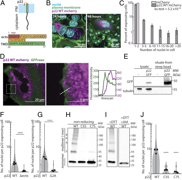
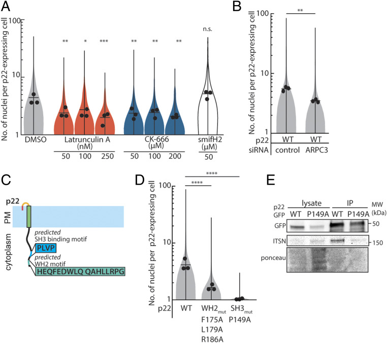
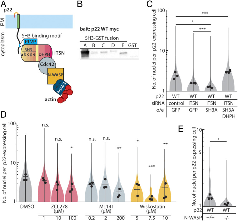
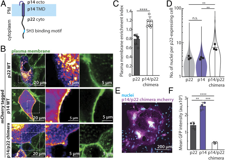
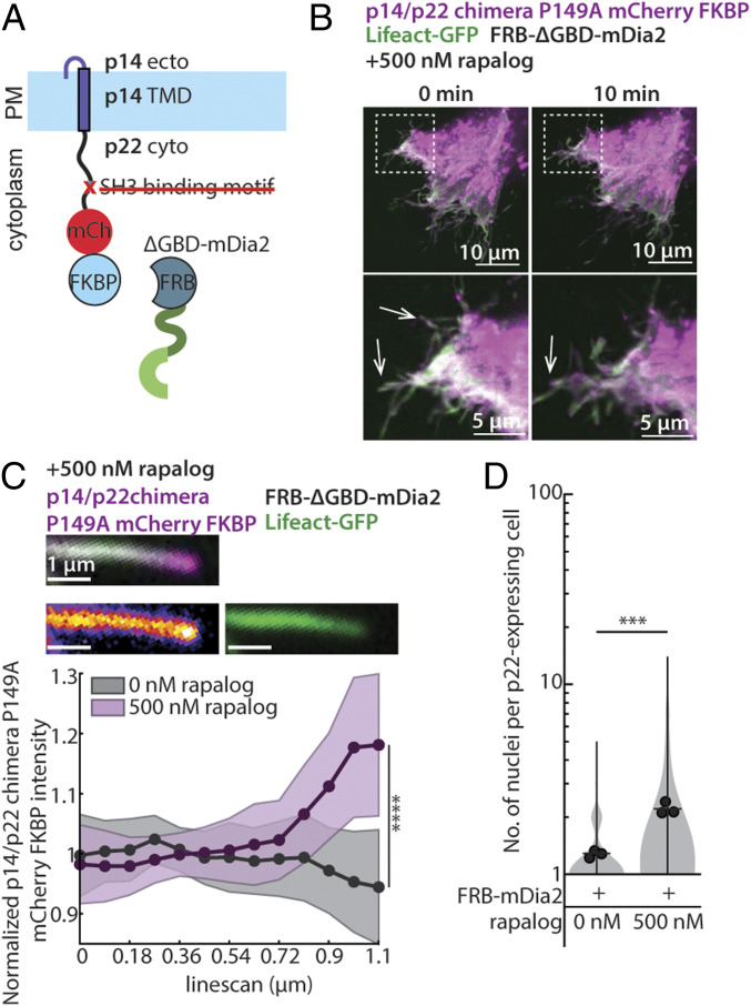
Fusion-associated small transmembrane (FAST) proteins are a diverse family of nonstructural viral proteins. Once expressed on the plasma membrane of infected cells, they drive fusion with neighboring cells, increasing viral spread and pathogenicity. Unlike viral fusogens with tall ectodomains that pull two membranes together through conformational changes, FAST proteins have short fusogenic ectodomains that cannot bridge the intermembrane gap between neighboring cells. One orthoreovirus FAST protein, p14, has been shown to hijack the actin cytoskeleton to drive cell-cell fusion, but the actin adaptor-binding motif identified in p14 is not found in any other FAST protein. Here, we report that an evolutionarily divergent FAST protein, p22 from aquareovirus, also hijacks the actin cytoskeleton but does so through different adaptor proteins, Intersectin-1 and Cdc42, that trigger N-WASP-mediated branched actin assembly. We show that despite using different pathways, the cytoplasmic tail of p22 can replace that of p14 to create a potent chimeric fusogen, suggesting they are modular and play similar functional roles. When we directly couple p22 with the parallel filament nucleator formin instead of the branched actin nucleation promoting factor N-WASP, its ability to drive fusion is maintained, suggesting that localized mechanical pressure on the plasma membrane coupled to a membrane-disruptive ectodomain is sufficient to drive cell-cell fusion. This work points to a common biophysical strategy used by FAST proteins to push rather than pull membranes together to drive fusion, one that may be harnessed by other short fusogens responsible for physiological cell-cell fusion.

Keywords: FAST proteins; actin cytoskeleton; cell-cell fusion; reovirus.


Conflict of interest statement

The authors declare no competing interest.

Figures

Fig. 1.
Fig. 1.
p22 is a membrane protein that multimerizes and drives cell-cell fusion. (A) Diagram of p22 topology on the plasma membrane and amino acid sequence of p22 ectodomain, predicted myristoylation, and transmembrane domain. (B) Expression of p22-mCherry (magenta) in Vero cells. Nuclei (Hoechst 33342; cyan) and plasma membrane (CellMaskDeepRed; green) are shown. (C) Nuclei distribution of cells expressing mCherry-tagged p22 and mCherry 36 h posttransfection, error bars indicate SD from three independent replicates, P value of ks-test between the two distributions is shown. (D) Representative confocal images of cells expressing p22-mCherry (magenta) and GFP-caax (plasma membrane marker; green). Contrast was adjusted in the magnified region. Magnified region and fluorescence intensity of line scan of dotted line is shown. (E) Western blot from surface biotinylation of p22-GFP–expressing cells and nontransfected cells. (F) Distribution of number of nuclei in p22-WT– and p22-Δecto–expressing cells from three independent transfections, with mean number of each replicate, average from three independent transfections shown. ****P < 0.0001 using two-tailed Student’s t test. (G) Distribution of number of nuclei in p22-WT– and p22-G2A–expressing cells from three independent transfections, mean number of each replicate, average from three independent transfections shown. ****P < 0.0001 using two-tailed Student’s t test. (H) Western blot of nonreducing SDS/PAGE of myc-tagged p22-WT, p22-C5S, p22-C7S probed with α-myc. (I) Western blot of SDS/PAGE of myc-tagged p22-WT, reduced with DTT and capped with iodoacetamide (IAA), and probed with α-myc. (J) Distribution of number of nuclei in p22-WT–, p22-C5S–, and p22-C7S–expressing cells from three independent transfections, mean number of each replicate, average from three independent transfections shown. ****P < 0.0001 using one-way ANOVA with Dunnett’s test.
Fig. 2.
Fig. 2.
Branched actin cytoskeleton plays a role in p22-mediated cell-cell fusion, and p22 binds to ITSN-1. (A) Distribution of number of nuclei in p22-WT–expressing cells treated with cytoskeletal drugs from three independent transfections, mean number of each replicate, average from three independent transfections shown. P values are one-way ANOVA with Dunnett’s test where not significant (n.s.). P > 0.05, *P < 0.05, **P < 0.01, ***P < 0.001. (B) Distribution of number of nuclei in p22-WT–expressing cells treated with control siRNA, siRNA targeting ARPC3 from three independent transfections, mean number of each replicate and average from three independent transfections shown. P values are two-way Student’s t test where **P < 0.01. (C) Diagram of predicted SH3 binding motif and predicted WH2 motif in p22 cytoplasmic tail. (D) Distribution of number of nuclei in p22-WT–, p22 WH2mut-, and p22 SH3mut-expressing cells from three independent transfections, mean number of each replicate and average from three independent transfections shown. P values are one-way ANOVA with Tukey’s test where ****P < 0.0001. (E) Western blot of coimmunoprecipitation of GFP-tagged p22-WT and p22-P149A with Intersectin-1.
Fig. 3.
Fig. 3.
Cdc42 and N-WASP signaling downstream of ITSN-1 is needed for p22-mediated cell-cell fusion. (A) Diagram of p22 binding to Intersectin-1 through a SH3 binding motif in its cytoplasmic tail. (B) In vitro binding assay with myc-tagged p22-WT as bait for purified GST-tagged SH3 domain A–E. (C) Distribution of number of nuclei in p22-WT–expressing cells treated with control siRNA or siRNA targeting ITSN-1 and/or overexpressing SH3A and SH3A-DHPH from three independent transfections, mean number of each replicate and average from three independent transfections shown. P values are two-way Student’s t test where *P < 0.05, ***P < 0.001. (D) Distribution of number of nuclei in p22-WT–expressing cells treated with drugs from three independent transfections, mean number of each replicate, average from three independent transfections shown. P values are one-way ANOVA with Dunnett’s test where not significant (n.s.) P > 0.05, *P < 0.05, **P < 0.01, ***P < 0.001. (E) Distribution of number of nuclei N-WASP null mouse embryonic fibroblasts and control cells expressing p22-WT from three independent transfections, with mean number of each replicate, average from three independent transfections shown. P values are two-tailed Student’s t test where *P < 0.05.
Fig. 4.
Fig. 4.
p14 and p22 are modular cell-cell fusogens, and their cytoplasmic tails can be swapped. (A) Diagram of chimeric fusogen with p14 ectodomain, p14 transmembrane domain, and p22 cytoplasmic tail. (B) Confocal images of mCherry-tagged p22-WT, p14-WT, and p14/p22 chimera with plasma membrane labeled with CellMaskDeepRed (green). Boxed regions are magnified. (C) Average plasma membrane enrichment index of mCherry-tagged p22-WT and p14/p22 chimera from three independent transfections, and error bars represent SD 24 h posttransfection. P values are two-tailed Student’s t test where ****P < 0.0001. (D) Distribution of number of nuclei in p22-WT, p14-WT, and p14/p22 chimera expressing cells 24 h posttransfection from three independent transfections, with mean number of each replicate, average from three independent transfections shown. P values are one-way ANOVA with Tukey’s test where not significant (n.s.) P > 0.05, **P < 0.01. (E) Representative confocal image of p14/p22 chimera mCherry (magenta) cell with nuclei labeled with Hoechst 33342 (cyan) at 24 h posttransfection. (F) Mean GFP intensity in each cell expressing GFP-tagged p22-WT, p14-WT, and p14/p22 chimera 24 h posttransfection from three independent transfection and error bars represent SDs. P values are two-tailed Student’s t test where **P < 0.01, ***P < 0.001, ****P < 0.0001.
Fig. 5.
Fig. 5.
Replacing the p14/p22 branched actin nucleator with a formin is sufficient to drive cell-cell fusion. (A) Diagram of p14/p22 chimera with FKBP at C terminus and P149A mutation with FRB-tagged ΔGBD-mDia2. (B) Confocal images of p14/p22 chimera P149A FKBP-mCherry (magenta) coexpressed with FRB-ΔGBD-mDia2 and Lifeact-GFP (green) cell at 0 min and 10 min after addition of 500 nM rapalog. Filopodia-like protrusions before and after rapalog addition is denoted with arrows. Box regions magnified. (C) Representative confocal merged image of a filopodia-like protrusion from cell expressing p14/p22 chimera-P149A-FKBP-mCherry (magenta), Lifeact-GFP (green), and FRB-ΔGBD-mDia2 10 min after addition of 500 nM rapalog. Each channel is shown with p14/p22 chimera-P149A-FKBP-mCherry (fire) and Lifeact-GFP (green). Average normalized fluorescence intensity of p14/p22 chimera-P149A-FKBP-mCherry along the length before (n = 33 filopodia-like protrusions) and 10 min after (n = 36 filopodia-like protrusions) addition of 500 nM rapalog. SD above and below the average are shown. ****P < 0.0001 by Student’s t test on the last datapoint. (D) Distribution of number of nuclei in p14/p22 chimera-P149A-FKBP– and FRB-ΔGBD-mDia2–expressing cells with and without rapalog from three independent transfections, with mean number of each replicate, average from three independent transfections shown. P values are two-tailed two-sample Student’s t test where ***P < 0.001.

References

    1. Ciechonska M., Duncan R., Reovirus FAST proteins: Virus-encoded cellular fusogens. Trends Microbiol. 22, 715–724 (2014). - PubMed
    1. Duncan R., Fusogenic reoviruses and their fusion-associated small transmembrane (FAST) proteins. Annu. Rev. Virol. 6, 341–363 (2019). - PubMed
    1. Kanai Y., et al. , Cell-cell fusion induced by reovirus FAST proteins enhances replication and pathogenicity of non-enveloped dsRNA viruses. PLoS Pathog. 15, e1007675 (2019). - PMC - PubMed
    1. Lefkowitz E. J.et al., Virus taxonomy: the database of the International Committee on Taxonomy of Viruses (ICTV). Nucleic Acids Res. 46, D708–D717 (2018). - PMC - PubMed
    1. Corcoran J. A., et al. , Myristoylation, a protruding loop, and structural plasticity are essential features of a nonenveloped virus fusion peptide motif. J. Biol. Chem. 279, 51386–51394 (2004). - PubMed

Publication types

MeSH terms