. 2021 Jan 12;118(2):e2021174118.
doi: 10.1073/pnas.2021174118. Epub 2020 Dec 21.

Molecular mechanism of the repressive phase of the mammalian circadian clock

Affiliations

Molecular mechanism of the repressive phase of the mammalian circadian clock

Xuemei Cao et al. Proc Natl Acad Sci U S A. .

Abstract

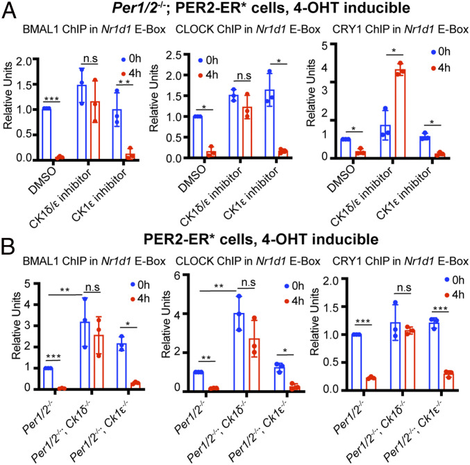
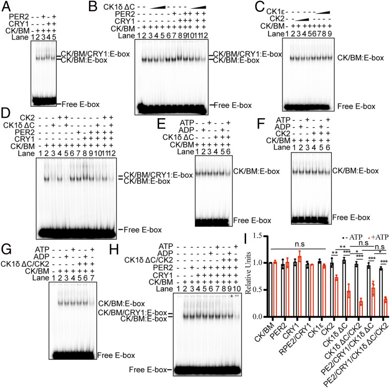
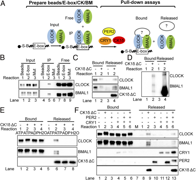
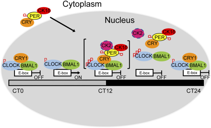
The mammalian circadian clock consists of a transcription-translation feedback loop (TTFL) composed of CLOCK-BMAL1 transcriptional activators and CRY-PER transcriptional repressors. Previous work showed that CRY inhibits CLOCK-BMAL1-activated transcription by a "blocking"-type mechanism and that CRY-PER inhibits CLOCK-BMAL1 by a "displacement"-type mechanism. While the mechanism of CRY-mediated repression was explained by both in vitro and in vivo experiments, the CRY-PER-mediated repression in vivo seemed in conflict with the in vitro data demonstrating PER removes CRY from the CLOCK-BMAL1-E-box complex. Here, we show that CRY-PER participates in the displacement-type repression by recruiting CK1δ to the nucleus and mediating an increased local concentration of CK1δ at CLOCK-BMAL1-bound promoters/enhancers and thus promoting the phosphorylation of CLOCK and dissociation of CLOCK-BMAL1 along with CRY from the E-box. Our findings bring clarity to the role of PER in the dynamic nature of the repressive phase of the TTFL.

Keywords: DNA binding proteins; casein kinase; circadian clock; cryptochrome; period.


Conflict of interest statement

The authors declare no competing interest.

Figures

Fig. 1.
Fig. 1.
PER2-mediated displacement of CLOCK, BMAL1, and CRY1 from an E-box in vivo is CK1δ dependent. (A) After treatment of Per1/2−/−; PER2–ER* cells for 24 h with 10 μM PF670462 (CK1δ/ε inhibitor) or 10 μM PF4800567 (CK1ε-selective inhibitor), 1 μM 4-OHT was added for 0 or 4 h to induce nuclear entry of PER2–ER*. CLOCK, BMAL1, and CRY1 binding to the Nr1d1 E-box was then measured by ChIP. (B) Experiments were then repeated (without inhibitors) to test cells with knocked-out CK1δ and CK1ε genes (Per1/2−/−; Ck1δ−/−; PER2–ER* and Per1/2−/−; Ck1ε−/−; PER2–ER* cells), using Per1/2−/−; PER2–ER* cells as a control. Results indicate a role of CK1δ but not CK1ε in removal of the CRY1–CLOCK–BMAL1 complex in vivo. Data for each panel were normalized to a value of 1 given to a control signal obtained with 0-h 4-OHT treatment (DMSO for A; PER2–ER* in B). Three biological repeats were used for quantification. Data are represented as dots for individual experiments and as columns for means. Error bars represent SDs. n.s, not significant; *P < 0.05, **P < 0.01, ***P < 0.001, as determined by t test between 0 and 4 h in same cell line and two-way ANOVA between different cell lines.
Fig. 2.
Fig. 2.
Mapping PER2 domains required for removing CRY1–CLOCK–BMAL1 from an E-box in vivo. (A) Illustration of the PER2–ER* constructs expressed in the Per1/2−/− cell line. The numbers indicate amino acid residues bordering deletions made in the full-length, 1,257-amino acid-long PER2. CBD, CRY-binding domain; CKBDa, casein kinase 2 binding domain; CKBDb, casein kinase 1ε binding domain; ER*, estrogen receptor “tag”; PAS, PER–ARNT–SIM domain. The dashed lines indicate regions deleted (Δ) in the constructs. Locations of the CK binding domains a and b (CKBDa and CKBDb) are shown. (BD) ChIP analyses of BMAL1, CLOCK, and CRY1 binding to an E-box in the Nr1d1 promoter in Per1/2−/− cell lines expressing different PER2–ER* proteins. Full-length PER2 (1–1,257), S659A PER2, and ΔCKBDb PER2 disrupt CRY1–CLOCK–BMAL1 binding to chromatin. Only the PER2 construct lacking both CKBDs (ΔCKBDa/b PER2) is unable to remove CRY1–CLOCK–BMAL1 from chromatin. S659A PER2, and ΔCKBDa PER2 appear to have only partial ability to remove CRY1 from the CLOCK–BMAL1–E-box complex. All data were normalized to a value of 1 for full-length PER2 (1–1,257) at 0-h 4-OHT. Three biological repeats were used for quantification. Data are represented as dots for individual experiments and as columns for means. Error bars represent SDs. n.s, not significant; *P < 0.05, **P < 0.01, ***P < 0.001, as determined by t test.
Fig. 3.
Fig. 3.
CLOCK hyperphosphorylation and CK1δ nuclear translocation are PER and CRY-dependent in vivo. Preliminary experiments were done in vitro to assess phosphorylation of core clock proteins by CKs. Purified proteins were incubated with [γ-32P]ATP and separated by SDS-PAGE (sodium dodecyl sulfate–polyacrylamide gel electrophoresis), and reaction products were visualized by autoradiography. Autoradiograms show that PER2 (A) and CLOCK–BMAL (B) can be directly phosphorylated by CK1δ and CK1ε, and marginally phosphorylated by CK2. (C and D) CLOCK–BMAL1 phosphorylation levels in WT, Per1/2−/−, and Cry1/2−/− mice as detected by bandshifts in immunoblots. Nuclear protein extracts from mouse livers were prepared at six CT points for analysis. In WT mice, when CLOCK–BMAL1 binding to E-boxes is high at CT4–8, CLOCK is hypophosphorylated. However, when CLOCK–BMAL1 binding to E-boxes is low at CT12–22, CLOCK is hyperphosphorylated. BMAL1 is hyperphosphorylated between CT8 and CT12, and BMAL1 is hypophosphorylated between CT16 and CT24. In Per1/2−/− and Cry1/2−/− mice, CLOCK is hypophosphorylated but BMAL1 is hyperphosphorylated at all time points. (E) Nuclear (“Nuc”) and cytoplasmic (“Cyto”) temporal expression of core clock proteins in WT and Per1/2−/− mouse liver. (F) Nuclear (Nuc) and cytoplasmic (Cyto) temporal expression of core clock proteins in WT and Cry1/2−/− mouse liver. Equal amounts of cytoplasmic protein (120 μg) and nuclear protein (6 μg) were loaded in each protein lane. α-Tubulin (cytoplasmic) and Lamin B1 (nuclear) were probed to provide loading controls. All the immunoblots were performed with at least two biological repeats and some had three technical repeats. All data yielded similar results. Representative images are shown.
Fig. 4.
Fig. 4.
CK1δ and CK2 reduce CLOCK/BMAL1 binding to an E-box in vitro. (A) Effect of CRY1 and PER2 on the mobility of the CLOCK–BMAL1–E-box complex. EMSA was performed with a 32P-labeled 30-bp duplex containing an E-box (1 nM) and CLOCK–BMAL1 (2 nM). A supershift was caused by CRY1 (15 nM) but not PER2 (15 nM) (lanes 3 and 4). PER2 removes only CRY1 from the CLOCK–BMAL1–CRY1–E-box complex; the CLOCK–BMAL1–E-box remains (lane 5). (B) Effect of CK1δ ΔC, CRY1, and PER2 on the mobility of the CLOCK–BMAL1–E-box complex. The E-box duplex (1 nM) was incubated with CLOCK–BMAL1 complex at 2 nM and increasing amounts of CK1δ ΔC (40, 100, and 200 nM). CRY1 and PER2 were added as in A. (C) Effect of CK2 and CK1ε on the amount of CLOCK–BMAL1–E-box complex. (D) Effect of CK1δ and CK2 on the entire circadian protein assembly binding to the E-box. MgCl2 and ATP were present in all reactions in Fig. 4 AD. EMSA showing the effect of CK1δ (E), CK2 (F), and CK1δ and CK2 together (G) on the amount of CLOCK–BMAL1–E-box complexes in the presence of ADP, ATP, or no nucleotide. (H) Effect of both CK1δ and CK2 on the entire circadian protein assembly binding to the E-box with/without ATP. The EMSAs data are representative of three independent experiments. (I) Quantitative analysis of the amount of CLOCK–BMAL1–E-box complex when adding core clock proteins with/without ATP. Three biological repeats were used for quantification. Data are represented as dots for individual experiments and as columns for means. Error bars are SDs of triplicate data. n.s, not significant; *P < 0.05, **P < 0.01, ***P < 0.001 were determined by two-way ANOVA.
Fig. 5.
Fig. 5.
Release of CLOCK–BMAL1 from an E-box by CK1δ in vitro. (A) Schematic showing the DNA pull-down assay to test the roles of PER2, CRY1, and CK1δ ΔC on CLOCK–BMAL1 release from an E-box. The E-box–containing duplex is tagged with biotin (“B”) and binds to and pellets with streptavidin (“S”) beads. CLOCK–BMAL1, bound to the E-box, also pellets with the beads. (In F, beads are prepared by adding CRY1 together with CLOCK–BMAL1 at this step, and in this case, the CLOCK–BMAL1–CRY1 complex pellets with the beads.) For pull-down assay, beads with CLOCK–BMAL1–E-box (or CLOCK–BMAL1–CRY1–E-box complexes) are then resuspended and incubated with PER2, CRY1, or CK1 δ, then pulled down to assess E-box bound and released proteins. (B) Preparation of CLOCK–BMAL1–E-box complexes (A, Left). CLOCK–BMAL1 (20 nM) binds to the DNA duplex (10 pmol) containing the E-box sequence and is pulled down (“IP”) by the streptavidin beads as shown in lane 5 of the immunoblot. When the duplex has a mutated E-box sequence, or in the absence of DNA, CLOCK–BMAL1 remain entirely in the “Free” fraction following pull-down, demonstrating specificity of binding. (C) For pull-down assay (A, Right), CLOCK–BMAL1–E-box complexes immunoprecipitated as in B, lane 5, were resuspended, incubated with CK1δ ΔC, and then pulled down again to separate Bound and Released fractions. The immunoblot analysis shows that CLOCK–BMAL1 is released from the E-box after adding 200 nM CK1δ ΔC. (D) SDS-PAGE autoradiogram showing that the bound and released CLOCK is hyperphosphorylated, but only bound BMAL1 is hyperphosphorylated after addition of 200 nM CK1δ ΔC. Proteins were radiolabeled by adding [γ-32P]ATP to the binding reactions. (E) Release of CLOCK–BMAL1 from the E-box by 200 nM CK1δ ΔC is ATP dependent in the pull-down assay. (F) Effect of 15 nM PER2, and 200 nM CK1δ ΔC on the CLOCK–BMAL1–CRY1–E-box complex. Note that the CRY1 amount associated with CLOCK–BMAL1 in the bound fraction (lanes 4 to 6) is lower than that observed in the EMSAs (Fig. 4 A, B, and D) because the multiple washes in the pull-downs causes dissociation of CRY from the CLOCK–BMAL1 complex. The DNA pull-down data are representative of three independent experiments.
Fig. 6.
Fig. 6.
Analysis of circadian complexes by glycerol gradient sedimentation. Mouse nuclear extract prepared from a mouse harvested at ZT19 and reference proteins were mixed together, and the extract and reference proteins were sedimented together through a 10 to 30% (wt/wt) glycerol gradient. Reference proteins included bovine thyroglobulin (669 kDa, 19 S), sweet potato β-amylase (222 kDa, 8.9 S), and chicken ovalbumin (43 kDa, 3.55 S). Fractions were collected starting at the bottom of the gradient and analyzed by loading samples to two SDS-PAGE gels. Gels showing gradient profiles for extracts from WT (A), Per1/2−/− (B), and Cry1/2−/− mice (C) at ZT19 were developed by immunoblot with PER1, PER2, CRY1, CRY2, CK1δ, CLOCK, and BMAL1 antibodies. “P” stands for pellet and indicates the sample obtained by washing the empty gradient tube with a small volume (240 μL) after collecting all fractions. The purpose of the P sample was to discover whether an insoluble pellet existed following centrifugation and to examine its composition. Three percent of the original sample was loaded directly to the SDS-PAGE gel as “Input.” The arrows indicate positions of the peak fraction of each reference protein as determined from the Coomassie Blue-stained SDS-PAGE gel (see SI Appendix, Fig. S9 for additional information related to this figure). Quantification of relative intensity is shown beside the immunoblot. Data for each protein were normalized to a value of 1 given to the peak fraction. Glycerol gradient experiments were performed with three biological repeats for WT and Cry1/2−/−, and two biological repeats for Per1/2−/− mice, and all data yielded essentially identical results. Representative images are shown in the figure.
Fig. 7.
Fig. 7.
Model showing the role of blocking-type and displacement-type repression in the mammalian circadian clock. At around CT4–8, CLOCK–BMAL1 binds to E-boxes to drive clock-controlled gene transcription. After protein synthesis in the cytoplasm, CRY recruits PER and PER recruits CK1δ/CK2 through its CKBD and then enters the nucleus and phosphorylates CLOCK, leading to CLOCK–BMAL1 dissociation from the E-box (CT12–22 displacement-type repression). At around CT0–4, PER levels are too low to be detected and only CRY1 binds to the CLOCK–BMAL1–E-box to block CLOCK–BMAL1 activity (blocking-type repression), which maintains CLOCK–BMAL1 in a repressed state until the next TTFL cycle begins. Note the CK2 is shown a circle with discontinuous circumference to indicate its partial contribution relative to CK1δ. In addition, the entire “repressor–activator complex” is shown in brackets to indicate that it must exist as a kinetic intermediate and not a stable megadalton complex.

References

    1. Reppert S. M., Weaver D. R., Coordination of circadian timing in mammals. Nature 418, 935–941 (2002). - PubMed
    1. Hastings M. H., Reddy A. B., Maywood E. S., A clockwork web: Circadian timing in brain and periphery, in health and disease. Nat. Rev. Neurosci. 4, 649–661 (2003). - PubMed
    1. Partch C. L., Green C. B., Takahashi J. S., Molecular architecture of the mammalian circadian clock. Trends Cell Biol. 24, 90–99 (2014). - PMC - PubMed
    1. Patke A., Young M. W., Axelrod S., Molecular mechanisms and physiological importance of circadian rhythms. Nat. Rev. Mol. Cell Biol. 21, 67–84 (2020). - PubMed
    1. Sancar A., Mechanisms of DNA repair by photolyase and excision nuclease (Nobel lecture). Angew. Chem. Int. Ed. Engl. 55, 8502–8527 (2016). - PubMed

Publication types

MeSH terms

LinkOut - more resources