. 2021 Aug 1;6(8):945-950.
doi: 10.1001/jamacardio.2020.7444.

Evaluation for Myocarditis in Competitive Student Athletes Recovering From Coronavirus Disease 2019 With Cardiac Magnetic Resonance Imaging

Affiliations

Evaluation for Myocarditis in Competitive Student Athletes Recovering From Coronavirus Disease 2019 With Cardiac Magnetic Resonance Imaging

Jitka Starekova et al. JAMA Cardiol. .

Abstract

Importance: The utility of cardiac magnetic resonance imaging (MRI) as a screening tool for myocarditis in competitive student athletes returning to training after recovering from coronavirus disease 2019 (COVID-19) infection is unknown.

Objective: To describe the prevalence and severity of cardiac MRI findings of myocarditis in a population of competitive student athletes recovering from COVID-19.

Design, setting, and participants: In this case series, an electronic health record search was performed at our institution (University of Wisconsin) to identify all competitive athletes (a consecutive sample) recovering from COVID-19, who underwent gadolinium-enhanced cardiac MRI between January 1, 2020, and November 29, 2020. The MRI findings were reviewed by 2 radiologists experienced in cardiac imaging, using the updated Lake Louise criteria. Serum markers of myocardial injury and inflammation (troponin-I, B-type natriuretic peptide, C-reactive protein, and erythrocyte sedimentation rate), an electrocardiogram, transthoracic echocardiography, and relevant clinical data were obtained.

Exposures: COVID-19 infection, confirmed using reverse transcription-polymerase chain reaction testing.

Main outcomes and measures: Prevalence and severity of MRI findings consistent with myocarditis among young competitive athletes recovering from COVID-19.

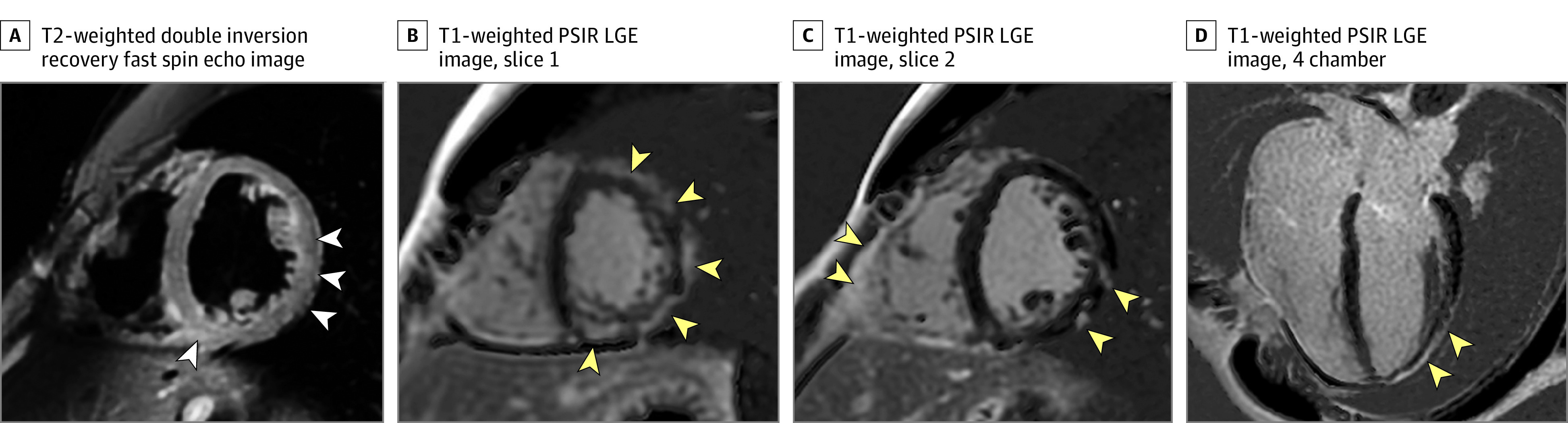
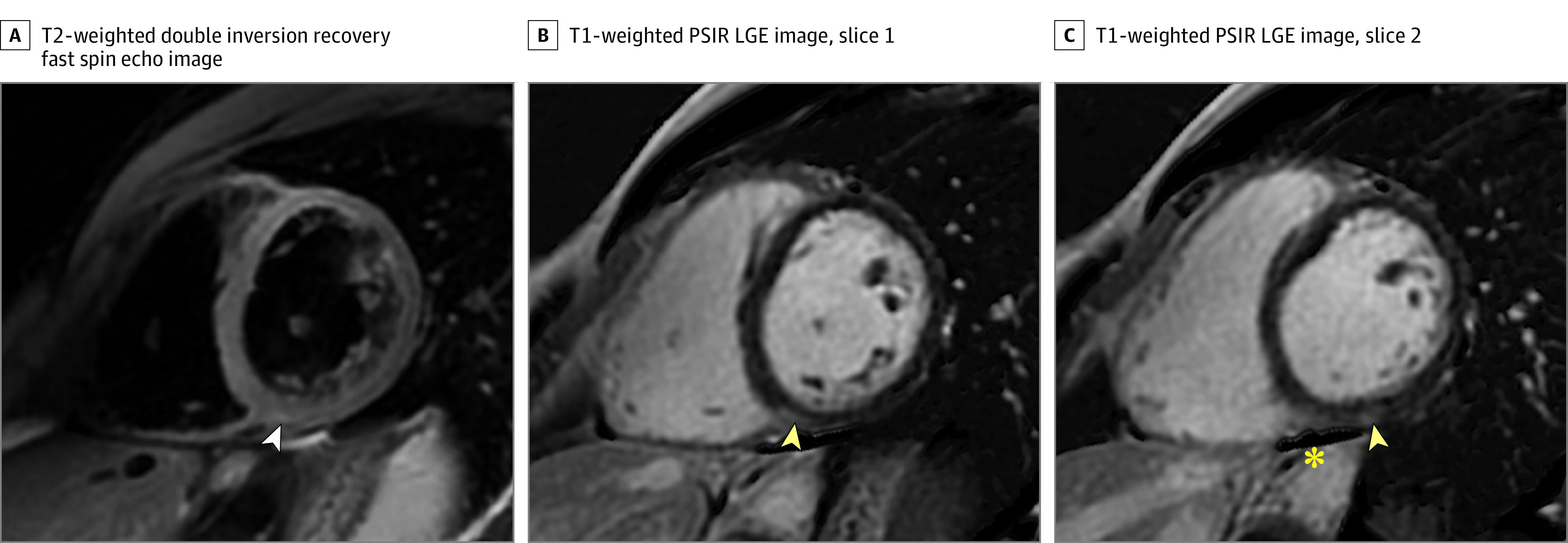
Results: A total of 145 competitive student athletes (108 male and 37 female individuals; mean age, 20 years; range, 17-23 years) recovering from COVID-19 were included. Most patients had mild (71 [49.0%]) or moderate (40 [27.6%]) symptoms during the acute infection or were asymptomatic (24 [16.6%]). Symptoms were not specified or documented in 10 patients (6.9%). No patients required hospitalization. Cardiac MRIs were performed a median of 15 days (range, 11-194 days) after patients tested positive for COVID-19. Two patients had MRI findings consistent with myocarditis (1.4% [95% CI, 0.4%-4.9%]). Of these, 1 patient had marked nonischemic late gadolinium enhancement and T2-weighted signal abnormalities over multiple segments, along with an abnormal serum troponin-I level; the second patient had 1-cm nonischemic mild late gadolinium enhancement and mild T2-weighted signal abnormalities, with normal laboratory values.

Conclusions and relevance: In this case series study, based on MRI findings, there was a low prevalence of myocarditis (1.4%) among student athletes recovering from COVID-19 with no or mild to moderate symptoms. Thus, the utility of cardiac MRI as a screening tool for myocarditis in this patient population is questionable.


Conflict of interest statement

Conflict of Interest Disclosures: Dr Reeder is a Romnes Faculty Fellow and has received an award provided by the University of Wisconsin–Madison Office of the Vice Chancellor for Research and Graduate Education with funding from the Wisconsin Alumni Research Foundation; reported that the University of Wisconsin receives research support from GE Healthcare and Bracco Diagnostics, outside the submitted work; and has ownership interests in Calimetrix, Reveal Pharmaceuticals, Cellectar Biosciences, Elucent Medical, and HeartVista and has received investigator-initiated grant support from Bayer Healthcare for research, outside the submitted work. Dr Grist has ownership interests in Elucent Medical, Shine Medical Technologies, OnLume, and Histosonics, outside the submitted work. Dr Grist also reported grants from Bracco Diagnostics Inc, research support to institution and grants from GE HealthCare Research support to institution during the conduct of the study, and personal fees from Bracco Diagnostics Inc advisory board, outside the submitted work. Dr Schiebler has ownership interests in Healthmyne Inc, Stemina Biomarker Discovery Inc, and X-Vax Inc, outside the submitted work. Dr Eckhardt is funded in part by the Gary and Marie Weiner Professorship in Cardiovascular Medicine Research. No other disclosures were reported.

Figures

Figure 1.
Figure 1.. Magnetic Resonance Imaging (MRI) Findings Consistent With Acute Myopericarditis in a Patient Recovering From Initially Asymptomatic Coronavirus Disease 2019
Shown are MRI findings consistent with acute inflammatory changes, with patchy midmyocardial and epicardial late gadolinium enhancement as well as pericardial enhancement (yellow arrowheads) on T1-weighted phase-sensitive inversion recovery late gadolinium–enhanced (PSIR LGE) short-axis images (B and C) and 4-chamber image (D), and corresponding elevated T2-weighted signal on fat-saturated double inversion recovery fast spin echo image (A) (white arrowheads). The troponin-I level was normal 1 day prior to the cardiac MRI (0.03 ng/mL; to convert to micrograms per liter, multiply by 1.0) but increased to 0.04 ng/mL and 0.09 ng/mL 2 and 4 days after the cardiac MRI examination, respectively.
Figure 2.
Figure 2.. Mild Magnetic Resonance Imaging Findings Consistent With Lake Louise Criteria for Myocarditis in an Athlete Recovering From Coronavirus Disease 2019
A, T2-weighted fat-saturated double inversion recovery fast spin echo short-axis image. B and C, T1-weighted phase-sensitive inversion recovery late gadolinium–enhanced (PSIR LGE) short-axis images. Yellow arrowheads demonstrate a 1-cm focus of epicardial LGE in the inferior wall at the base and the inferior right ventricular insertion point. A mildly elevated T2-weighted signal was also noted (white arrowhead), as well as a small amount of adjacent pericardial fluid (asterisk). Serum troponin levels were normal in this patient.

Comment in

References

    1. Puntmann VO, Carerj ML, Wieters I, et al. . Outcomes of cardiovascular magnetic resonance imaging in patients recently recovered from coronavirus disease 2019 (COVID-19). JAMA Cardiol. 2020;5(11):1265-1273. doi:10.1001/jamacardio.2020.3557 - DOI - PMC - PubMed
    1. Knight DS, Kotecha T, Razvi Y, et al. . COVID-19: myocardial injury in survivors. circulation. 2020;142(11):1120-1122. doi:10.1161/CIRCULATIONAHA.120.049252 - DOI - PMC - PubMed
    1. Beşler MS, Arslan H. Acute myocarditis associated with COVID-19 infection. Am J Emerg Med. 2020;38(11):2489.e1-2489.e2. doi:10.1016/j.ajem.2020.05.100 - DOI - PMC - PubMed
    1. Trogen B, Gonzalez FJ, Shust GF. COVID-19-associated myocarditis in an adolescent. Pediatr Infect Dis J. 2020;39(8):e204-e205. doi:10.1097/INF.0000000000002788 - DOI - PubMed
    1. Ho JS, Sia C-H, Chan MY, Lin W, Wong RC. Coronavirus-induced myocarditis: a meta-summary of cases. Heart Lung. 2020;49(6):681-685. doi:10.1016/j.hrtlng.2020.08.013 - DOI - PMC - PubMed

Publication types