Review
. 2021 Jul;41(7):1501-1516.
doi: 10.1177/0271678X20982383. Epub 2021 Jan 14.

Causes and consequences of baseline cerebral blood flow reductions in Alzheimer's disease

Affiliations
Review

Causes and consequences of baseline cerebral blood flow reductions in Alzheimer's disease

Oliver Bracko et al. J Cereb Blood Flow Metab. 2021 Jul.

Abstract

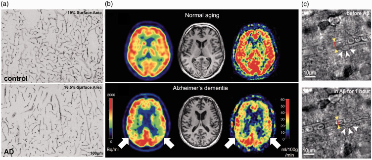
Reductions of baseline cerebral blood flow (CBF) of ∼10-20% are a common symptom of Alzheimer's disease (AD) that appear early in disease progression and correlate with the severity of cognitive impairment. These CBF deficits are replicated in mouse models of AD and recent work shows that increasing baseline CBF can rapidly improve the performance of AD mice on short term memory tasks. Despite the potential role these data suggest for CBF reductions in causing cognitive symptoms and contributing to brain pathology in AD, there remains a poor understanding of the molecular and cellular mechanisms causing them. This review compiles data on CBF reductions and on the correlation of AD-related CBF deficits with disease comorbidities (e.g. cardiovascular and genetic risk factors) and outcomes (e.g. cognitive performance and brain pathology) from studies in both patients and mouse models, and discusses several potential mechanisms proposed to contribute to CBF reductions, based primarily on work in AD mouse models. Future research aimed at improving our understanding of the importance of and interplay between different mechanisms for CBF reduction, as well as at determining the role these mechanisms play in AD patients could guide the development of future therapies that target CBF reductions in AD.

Keywords: Alzheimer’s disease; cerebral blood flow; cognitive impairment; in vivo 2-photon imaging; magnetic resonance imaging.


Conflict of interest statement

Declaration of conflicting interests: The author(s) declared no potential conflicts of interest with respect to the research, authorship, and/or publication of this article.

ORCID iDs: Nozomi Nishimura https://orcid.org/0000-0003-4342-9416

Figures

Figure 1.
Figure 1.
Vascular alterations and brain blood flow reductions in AD patients. (a) Heparan sulfate proteoglycan mmunohistochemistry in the cortex of a control subject (upper panel) and an AD patient (lower panel), showing reduced vascular density with AD. (b) Images of brain perfusion using ASL-MRI from a normal aged subject (top images), and an AD patient (bottom images). Red colors represent higher perfusion, while blue colors represent lower perfusion. (c) Bright field images of a capillary segment from a human brain slice before (left) and after (right) application of 72 nM Aβ1-42 onto the slice, which triggered activation of a pericyte (white arrows) and constriction of the capillary (red line and yellow arrowheads).
Figure 2.
Figure 2.
Mechanisms contributing to CBF reductions in mouse models of Alzheimer’s disease. (a) Images from 15 month old WT (left) and APP/PS1-Tg4510 mice (right; overexpresses mutant APP and mutant tau), showing clearly increased vascular volume in this AD mouse model containing both APP and tau-related mutations (Scale bar, 20 μm). (b) Fragmentation of smooth muscle cells (arrows) along a cortical arteriole in Tg2576 mice (left image), which is attenuated by CD36 deletion (right image) (Scale bar, 10 μm; anti-α-actin: green, anti-Aβ: red). (c) Example image of a pericyte (red) constricting around a cortical capillary (blood plasma shown in green), with blood cell flow blocked, from an AD mouse. (d) Cerebral perfusion was maintained in TgCRND8 mice treated with dabigatran. (e) Image sequences showing stalled (left panels) and flowing (right panels) capillaries, where the darker spots in vessels are due to unlabeled blood cells within the fluorescently-labeled blood plasma. Stalled capillaries, where blood cells do not move frame-to-frame, occurred at higher incidence in APP/PS1 mice, as compared to controls.

References

    1. Roher AE, Debbins JP, Malek-Ahmadi M, et al. Cerebral blood flow in Alzheimer’s disease. Vasc Health Risk Manag 2012; 8: 599–611. - PMC - PubMed
    1. Hays CC, Zlatar ZZ, Wierenga CE.The utility of cerebral blood flow as a biomarker of preclinical Alzheimer’s disease. Cell Mol Neurobiol 2016; 36: 167–179. - PMC - PubMed
    1. Zhang N, Gordon ML, Goldberg TE.Cerebral blood flow measured by arterial spin labeling MRI at resting state in normal aging and Alzheimer’s disease. Neurosci Biobehav Rev 2017; 72: 168–175. - PubMed
    1. Sweeney MD, Kisler K, Montagne A, et al. The role of brain vasculature in neurodegenerative disorders. Nat Neurosci 2018; 21: 1318–1331. - PMC - PubMed
    1. Sweeney MD, Montagne A, Sagare AP, et al. Vascular dysfunction—the disregarded partner of Alzheimer’s disease. Alzheimers Dement 2019; 15: 158–167. - PMC - PubMed

Publication types

LinkOut - more resources