

Regional and depth-dependence of cortical blood-flow assessed with high-resolution Arterial Spin Labeling (ASL)
- PMID: 33444098
- PMCID: PMC8327107
- DOI: 10.1177/0271678X20982382
Regional and depth-dependence of cortical blood-flow assessed with high-resolution Arterial Spin Labeling (ASL)
Abstract
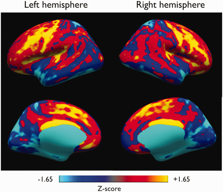
Methods for imaging of cerebral blood flow do not typically resolve the cortex and thus underestimate flow. However, recent work with high-resolution MRI has emphasized the regional and depth-dependent structural, functional and relaxation times variations within the cortex. Using high-resolution Arterial Spin Labeling (ASL) and T1 mapping acquisitions, we sought to probe the effects of spatial resolution and tissue heterogeneity on cortical cerebral blood flow (CBF) measurements with ASL. We acquired high-resolution (1.6mm)3 whole brain ASL data in a cohort of 10 volunteers at 3T, along with T1 and transit-time (ATT) mapping, followed by group cortical surface-based analysis using FreeSurfer of the different measured parameters. Fully resolved regional analysis showed higher than average mid-thickness CBF in primary motor areas (+15%,p<0.002), frontal regions (+17%,p<0.01) and auditory cortex, while occipital regions had lower average CBF (-20%,p<10-5). ASL signal was higher towards the pial surface but correction for the shorter T1 near the white matter surface reverses this gradient, at least when using the low-resolution ATT map. Similar to structural measures, fully-resolved ASL CBF measures show significant differences across cortical regions. Depth-dependent variation of T1 in the cortex complicates interpretation of depth-dependent ASL signal and may have implications for the accurate CBF quantification at lower resolutions.
Keywords: ASL; cerebral blood-flow; cortical perfusion; perfusion; surface-based analysis.
Conflict of interest statement
Figures


References
-
- Iida H, Law I, Pakkenberg B, et al. Quantitation of regional cerebral blood flow corrected for partial volume effect using O-15 water and PET: I. theory, error analysis, and stereologic comparison. J Cereb Blood Flow Metab 2000; 20:1237–1251. - PubMed
-
- Koopmans PJ, Yacoub E.Strategies and prospects for cortical depth dependent T2 and T2* weighted BOLD fMRI studies. NeuroImage 2019; 197: 668–676. - PubMed
-
- Petr J, Mutsaerts HJMM, De Vita E, et al. Effects of systematic partial volume errors on the estimation of gray matter cerebral blood flow with arterial spin labeling MRI. Magn Reson Mater Phys 2018; 31: 725–734. - PubMed
-
- Zhao MY, Mezue M, Segerdahl AR, et al. A systematic study of the sensitivity of partial volume correction methods for the quantification of perfusion from pseudo-continuous arterial spin labeling MRI. NeuroImage 2017; 162: 384–397. - PubMed
MeSH terms
Substances
LinkOut - more resources
Full Text Sources
Other Literature Sources
