. 2021 Jan 14;17(1):e1009193.
doi: 10.1371/journal.ppat.1009193. eCollection 2021 Jan.

A novel mode of control of nickel uptake by a multifunctional metallochaperone

Affiliations

A novel mode of control of nickel uptake by a multifunctional metallochaperone

Milica Denic et al. PLoS Pathog. .

Abstract

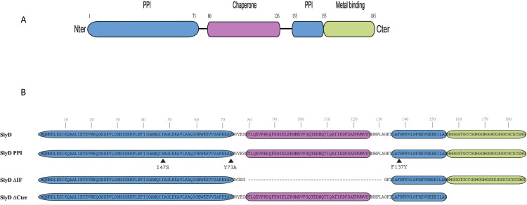
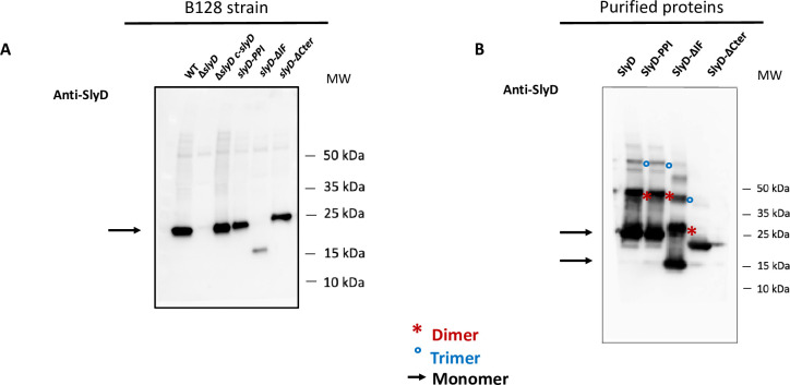
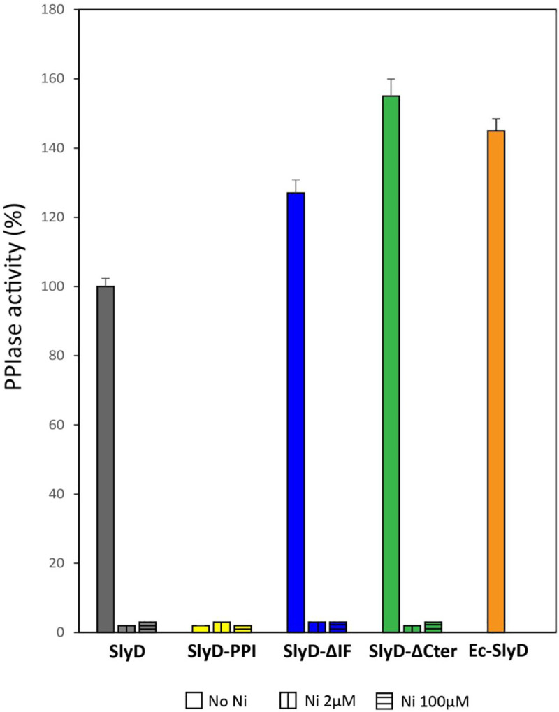
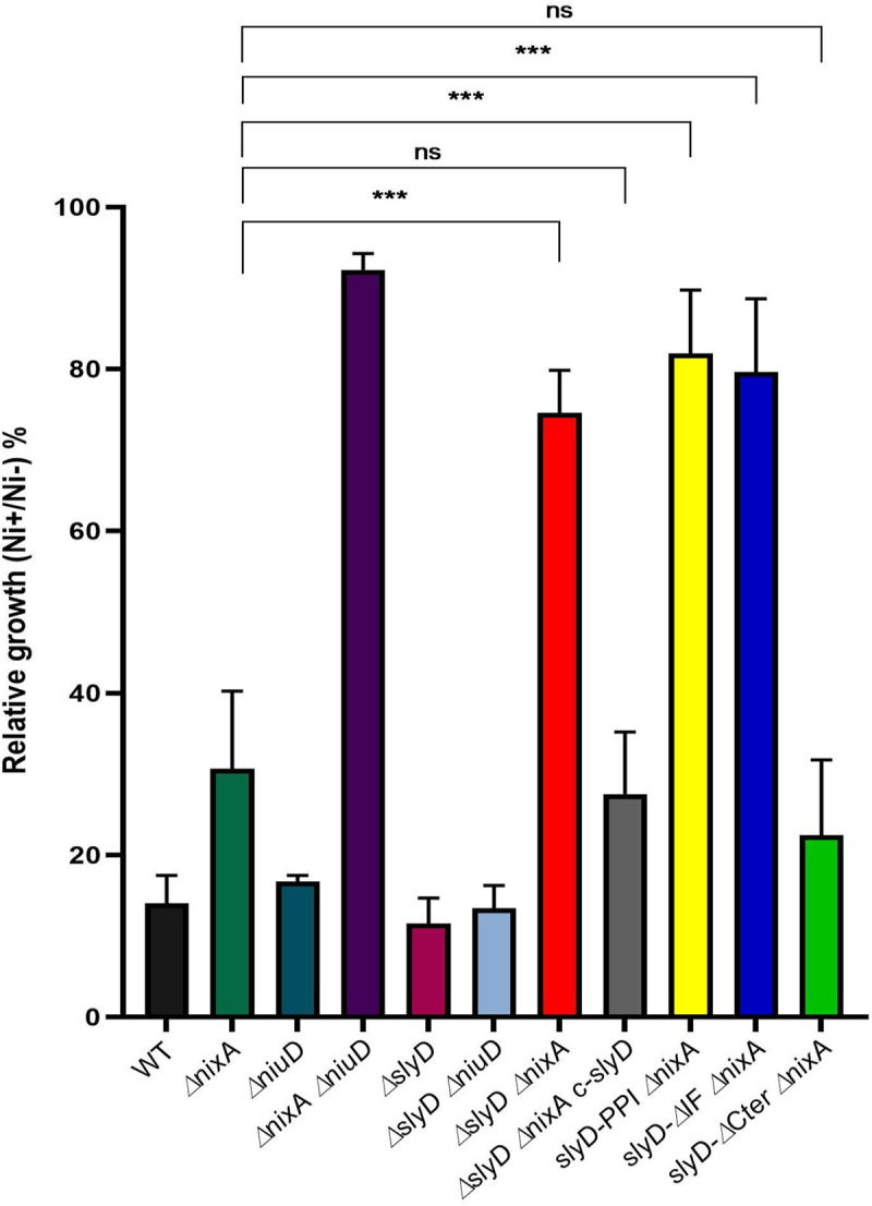
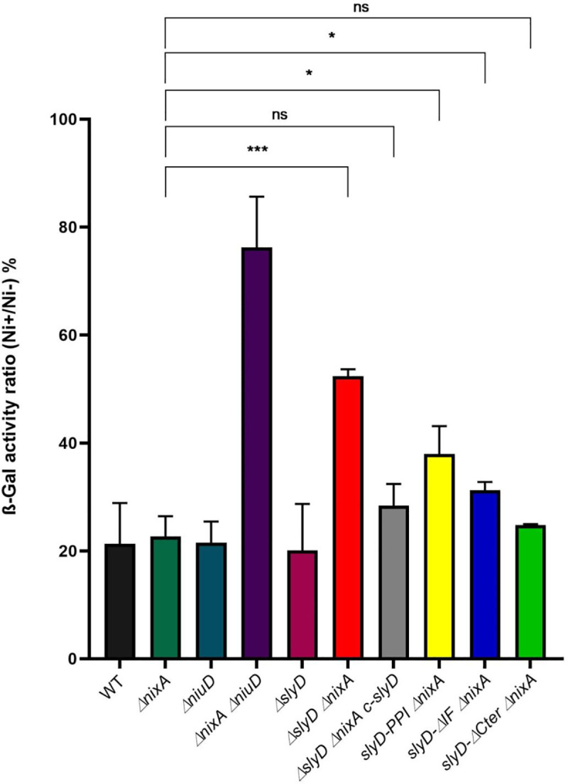
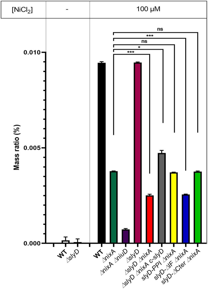
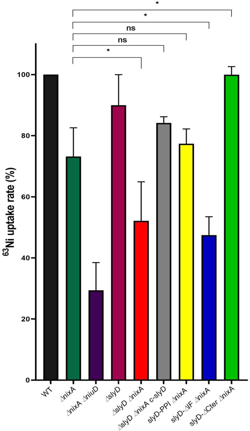
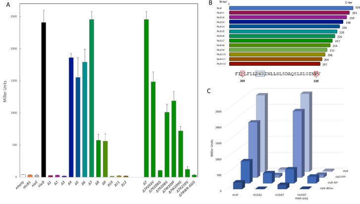
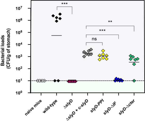
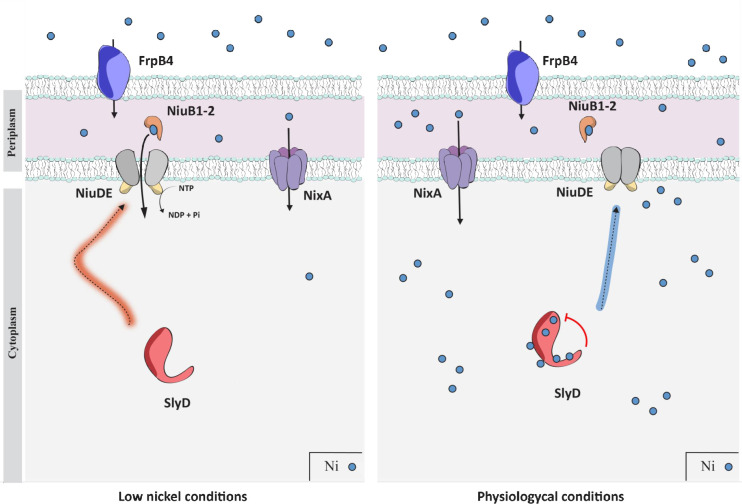
Cellular metal homeostasis is a critical process for all organisms, requiring tight regulation. In the major pathogen Helicobacter pylori, the acquisition of nickel is an essential virulence determinant as this metal is a cofactor for the acid-resistance enzyme, urease. Nickel uptake relies on the NixA permease and the NiuBDE ABC transporter. Till now, bacterial metal transporters were reported to be controlled at their transcriptional level. Here we uncovered post-translational regulation of the essential Niu transporter in H. pylori. Indeed, we demonstrate that SlyD, a protein combining peptidyl-prolyl isomerase (PPIase), chaperone, and metal-binding properties, is required for the activity of the Niu transporter. Using two-hybrid assays, we found that SlyD directly interacts with the NiuD permease subunit and identified a motif critical for this contact. Mutants of the different SlyD functional domains were constructed and used to perform in vitro PPIase activity assays and four different in vivo tests measuring nickel intracellular accumulation or transport in H. pylori. In vitro, SlyD PPIase activity is down-regulated by nickel, independently of its C-terminal region reported to bind metals. In vivo, a role of SlyD PPIase function was only revealed upon exposure to high nickel concentrations. Most importantly, the IF chaperone domain of SlyD was shown to be mandatory for Niu activation under all in vivo conditions. These data suggest that SlyD is required for the active functional conformation of the Niu permease and regulates its activity through a novel mechanism implying direct protein interaction, thereby acting as a gatekeeper of nickel uptake. Finally, in agreement with a central role of SlyD, this protein is essential for the colonization of the mouse model by H. pylori.


Conflict of interest statement

The authors have declared that no competing interests exist. Author Deborah Zamble was unable to confirm their authorship contributions. On their behalf, the corresponding author has reported their contributions to the best of their knowledge.

Figures

Fig 1
Fig 1. Illustration of the H. pylori SlyD wild type and mutant proteins.
A. Schematic representation of the functional domains of the SlyD protein of H. pylori strain B128, with indication of the corresponding encompassing residues. The regions required for the peptidyl-prolyl isomerase activity (PPIase) are colored in blue, the "inserted in Flap" IF chaperone domain is colored in pink and the C-terminal metal-binding region is colored in green. B. Illustration of the different SlyD mutants, SlyD-PPI, SlyD-ΔIF and SlyD-ΔCter. The three residues (I47S, Y73A and F137Y) changed in the SlyD-PPI mutant are marked by a black arrow.
Fig 2
Fig 2. Analysis of the production of SlyD wild type and mutant proteins in H. pylori.
A. Western blot of equal amounts of total extracts, under reducing conditions, from H. pylori B128 WT strain and B128-derived mutants carrying the following mutations ΔslyD, ΔslyD c-slyD (complemented mutant), slyD-PPI, slyD-ΔIF, and slyD-ΔCter strain, that were probed with specific anti-SlyD polyclonal antibodies prepared during this study. An arrow shows the position of the SlyD protein. B. Western blot of purified recombinant SlyD proteins (WT, SlyD-PPI, SlyD-ΔIF and SlyD-ΔCter) probed with specific anti-SlyD polyclonal antibodies. An arrow shows the position of the monomeric SlyD proteins, red stars and blue circles highlight SlyD dimers and trimers, respectively.
Fig 3
Fig 3. In vitro PPIase activity and nickel regulation of H. pylori wild type and mutant SlyD proteins.
PPIase activities of WT and mutant SlyD proteins, measured without or with 2 μM or 100 μM NiSO4. The PPIase activity of purified E. coli SlyD (EcSlyD) is also presented as a control. The data (S3B Fig) were fit to second-order rate equations and the PPIase activities are expressed as a percentage of the activity of the wild type H. pylori SlyD protein. Grey bars correspond to the wild type protein, yellow to the SlyD-PPI mutant, blue to the SlyD-ΔIF mutant and green to the SlyD-ΔCter mutant. The orange bar corresponds to the E. coli SlyD protein. The values are the averages from three replicates and error bars represent the standard deviation.
Fig 4
Fig 4. Tolerance of H. pylori wild type and mutant strains to nickel exposure.
Growth of H. pylori B128 wild type strain, isogenic slyD mutants and a complemented strain (c-slyD) was measured after 24h in the presence of 1.5 mM NiCl2 or without added metal. The results are presented as the percentage of growth in the presence of nickel relative to growth in its absence. In this figure as well as in Figs 5, 6 and 7, the same color codes were used for the bars corresponding to each strain or mutant which name is indicated below each bar. Black bars correspond to the wild type strain, dark green to the ΔnixA mutant, dark blue to the ΔniuD mutant, violet to the ΔnixA ΔniuD mutant, dark red to the ΔslyD mutant, light blue to the ΔslyD ΔniuD mutant, bright red to ΔslyD ΔnixA mutant, light grey to the ΔslyD ΔnixA c-slyD mutant, yellow to the slyD-PPI ΔnixA mutant, bright blue to the slyD-ΔIF ΔnixA mutant and light green to the slyD-ΔCter ΔnixA mutant. The data correspond to the mean value of three independent experiments. Error bars represent the standard deviation. Statistics are presented only for the comparison with the ΔnixA mutant: *** corresponds to p<0.001 and "ns" for non-significant. S9 Fig presents the complete statistical analysis of these data.
Fig 5
Fig 5. Evaluation of intracellular nickel availability of H. pylori wild type strain and isogenic mutants with a PfecA3::lacZ reporter fusion.
ß-galactosidase activity of a PfecA3::lacZ reporter fusion expressed from a plasmid in different H. pylori B128-derived strains, after 24H exposure to 100 μM NiCl2. The expression of the fusion decreases in a NikR-dependent manner with increasing intracellular nickel concentration. In medium without added nickel, the ß-galactosidase activities of the different strains were found to be comparable (about 6,000 miller units). ß-galactosidase activities are presented as the ratio of activity measured in strains grown in the presence of 100 μM NiCl2 or in the absence of nickel supplementation, expressed as a percentage. Color codes of the bars are as in Fig 4. The data correspond to the mean value of three independent experiments (S5 Table). Error bars represent the standard deviation. Statistics are presented only for the comparison with the ΔnixA mutant. * corresponds to p<0.05, *** to p<0.001 and "ns" for non-significant. S9 Fig presents the complete statistical analysis of these data.
Fig 6
Fig 6. Measurement of the intracellular nickel content of H. pylori wild type strain and isogenic mutants by ICP-OES.
Nickel amounts were measured by Inductively Coupled Plasma Optical Emission Spectrometry (ICP-OES) and are expressed as the percentage of the ratio of nickel mass versus total sample mass. Strains were grown either without added nickel or with 100 μM NiCl2. Color codes of the bars are as in Fig 4. The measurement of each strain under each condition was performed in triplicates in two experiments. Statistics are presented only for the comparison with the ΔnixA mutant. * corresponds to p<0.05, *** to p<0.001 and "ns" for non-significant. S9 Fig presents the complete statistical analysis of these data.
Fig 7
Fig 7. SlyD is required for the uptake of radioactive nickel by the Niu nickel transporter.
Measurements of radioactive nickel uptake rates in H. pylori B128 wild type strain and isogenic mutant strains in the presence of 10 μM of 63NiCl2. Uptake rates are normalized to the rate of wild type H. pylori strain. Color codes of the bars are as in Fig 4. Error bars represent the standard deviation. Statistics are presented only for the comparison with the ΔnixA mutant. * corresponds to p<0.05 and "ns" for non-significant. S9 Fig presents the complete statistical analysis of these data.
Fig 8
Fig 8. SlyD interacts with NiuD, the membrane permease of the Niu nickel uptake system.
Bacterial two-hybrid BACTH was used to analyze, in E. coli strain BTH101, the interaction between SlyD and the NiuD permease. The values and standard deviation for each strain and controls are available in S6 Table, and each measurement was performed three times. A. ß-galactosidase activities of pairwise combinations of WT SlyD with different truncated and mutant versions of the NiuD protein. B. Illustration of the truncated NiuD protein versions and sequence of the NiuD region surrounding the RWR motif in the region that is required for the interaction with SlyD. C. ß-galactosidase activities of pairwise combinations of wild type and mutant SlyD proteins (PPI, ΔIF and ΔCter) with wild type and mutant NiuD proteins.
Fig 9
Fig 9. SlyD is essential for mouse colonization by H. pylori SS1 strain.
Each diamond corresponds to the colonization load of one mouse one month after infection with the H. pylori strain indicated below. Each strain was tested in a group of seven mice. The color codes for the different strains are as in Fig 4. Horizontal bars represent the geometric means of the colonization load for the wild type bacteria, each mutant and the chromosomally complemented ΔslyD mutant (designated c-slyD). A dashed line shows the detection limit. The ΔslyD and isogenic mutants exhibited a statistically significant colonization defect when compared to the WT or ΔslyD c-slyD strains, respectively (**p<0.01, ***p<0.001 and "ns" non-significant).
Fig 10
Fig 10. Model for the regulation of nickel uptake by the SlyD protein.
In H. pylori, nickel ions (small blue dots) are transported across the outer membrane by FrpB4 (blue), a TonB-dependent transporter. Once in the periplasm, uptake of nickel through the inner membrane can be performed by the NixA permease (violet) or the ABC-transporter, NiuBDE (orange, grey and yellow). NiuB1-2 (orange) are the periplasmic nickel shuttles that deliver the metal to the NiuD permease, which activity is energized by the NiuE NTPase. The function of the Niu transporter requires activation by SlyD (red), a regulation that relies on direct interaction between SlyD and the NiuD permease. This is illustrated on the left panel, presenting conditions of low nickel availability. On the right panel, when nickel is available, binding of nickel to SlyD regulates its PPIase activity. This allows SlyD to sense the intracellular nickel concentration and to act as a gate keeper to control nickel entry.

Similar articles

Cited by

References

    1. Chandrangsu P, Rensing C, Helmann JD. Metal homeostasis and resistance in bacteria. Nat Rev Microbiol. 2017;15(6):338–50. 10.1038/nrmicro.2017.15 - DOI - PMC - PubMed
    1. Waldron KJ, Rutherford JC, Ford D, Robinson NJ. Metalloproteins and metal sensing. Nature. 2009;460(7257):823–30. 10.1038/nature08300 - DOI - PubMed
    1. Palmer LD, Skaar EP. Transition metals and virulence in bacteria. Annu Rev Genet. 2016;50(1):67–91. - PMC - PubMed
    1. Marchetti M, De Bei O, Bettati S, Campanini B, Kovachka S, Gianquinto E, et al. Iron metabolism at the interface between host and pathogen: From nutritional immunity to antibacterial development. Int J Mol Sci. 2020;21(6). 10.3390/ijms21062145 - DOI - PMC - PubMed
    1. Begg SL. The role of metal ions in the virulence and viability of bacterial pathogens. Biochem Soc Trans. 2019;47(1):77–87. 10.1042/BST20180275 - DOI - PubMed

Publication types

MeSH terms

Grants and funding