. 2021 Feb 9;54(2):247-258.e7.
doi: 10.1016/j.immuni.2020.11.020. Epub 2021 Jan 13.

A class of viral inducer of degradation of the necroptosis adaptor RIPK3 regulates virus-induced inflammation

Affiliations

A class of viral inducer of degradation of the necroptosis adaptor RIPK3 regulates virus-induced inflammation

Zhijun Liu et al. Immunity. .

Abstract

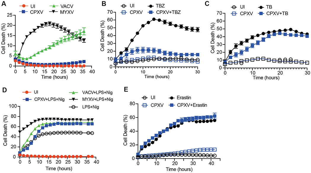
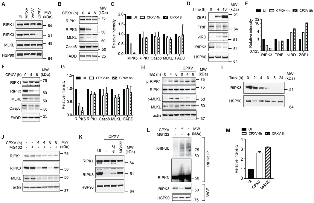
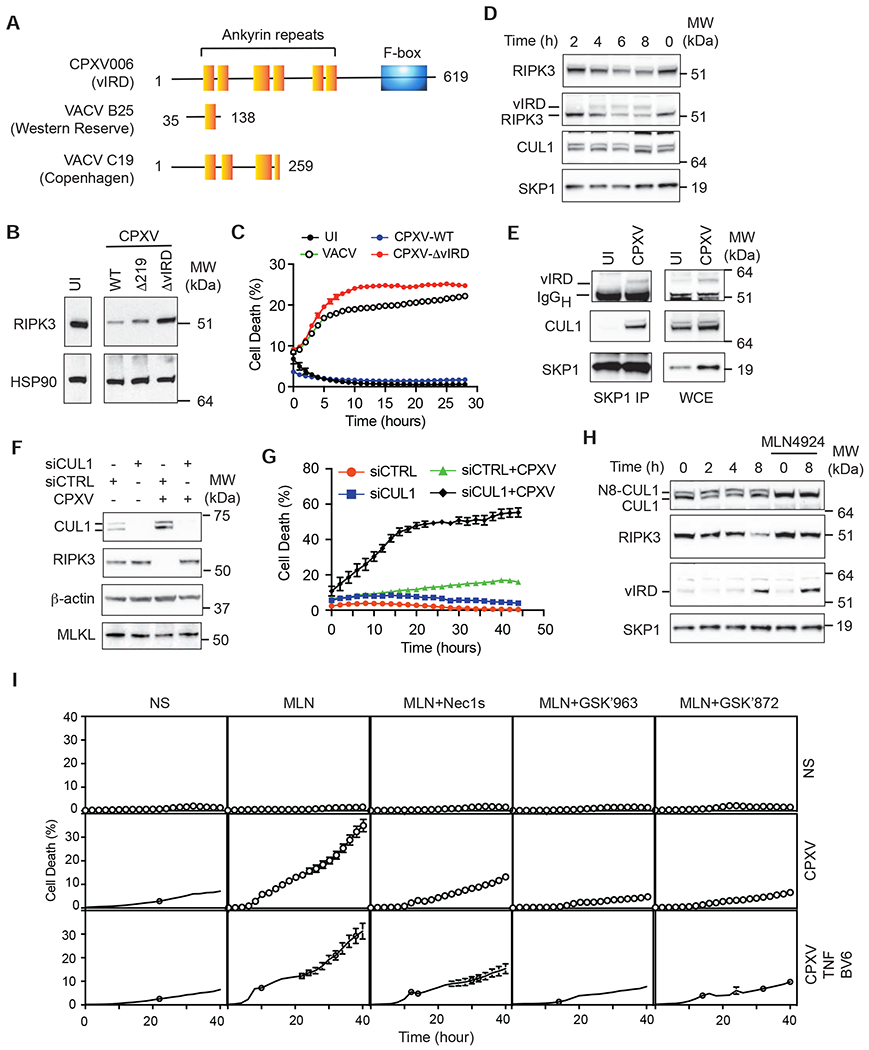
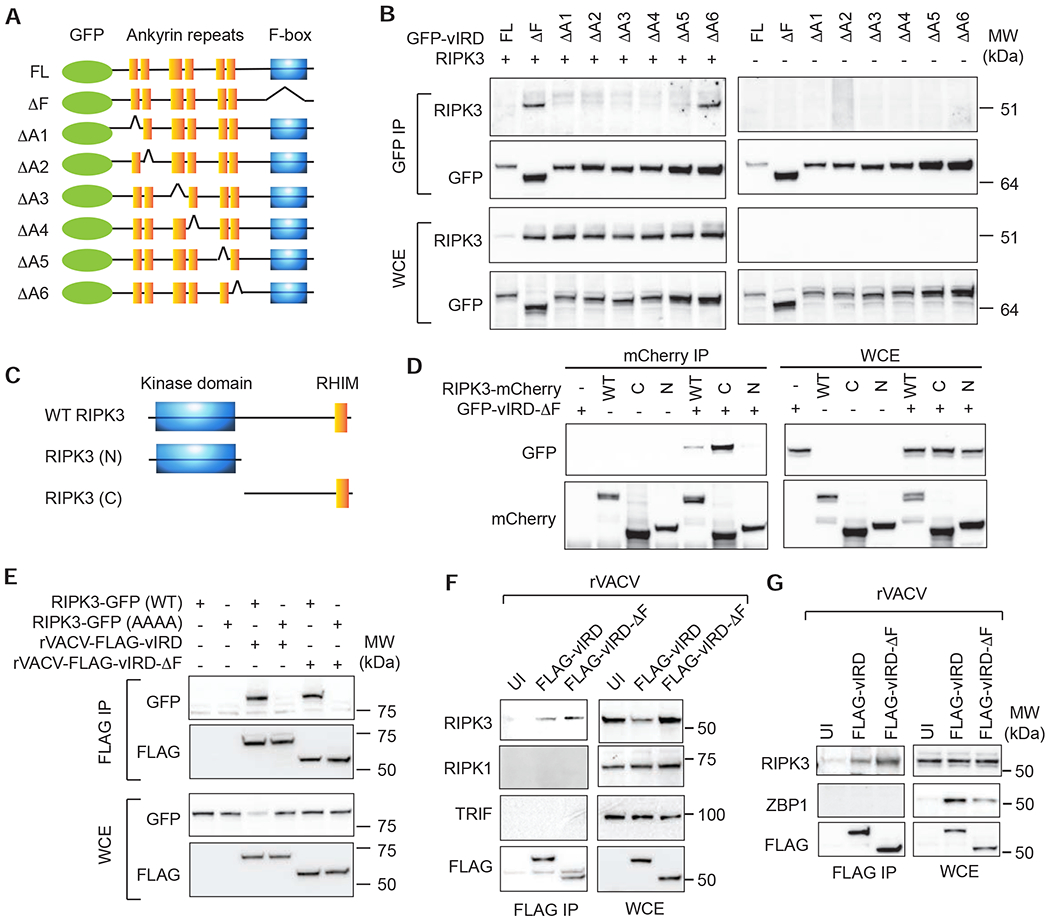
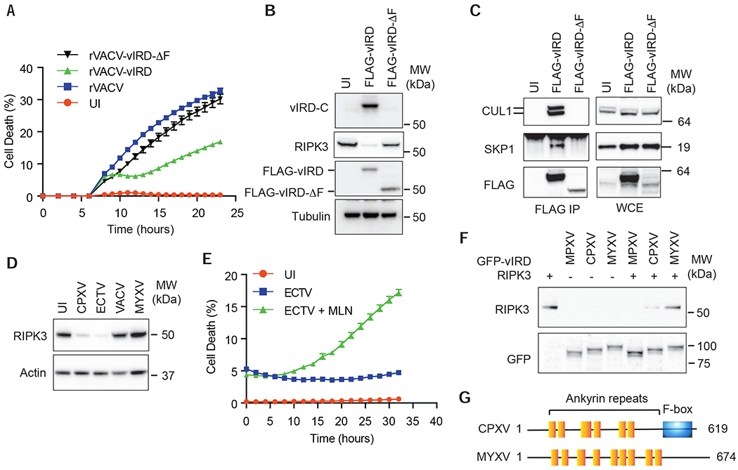
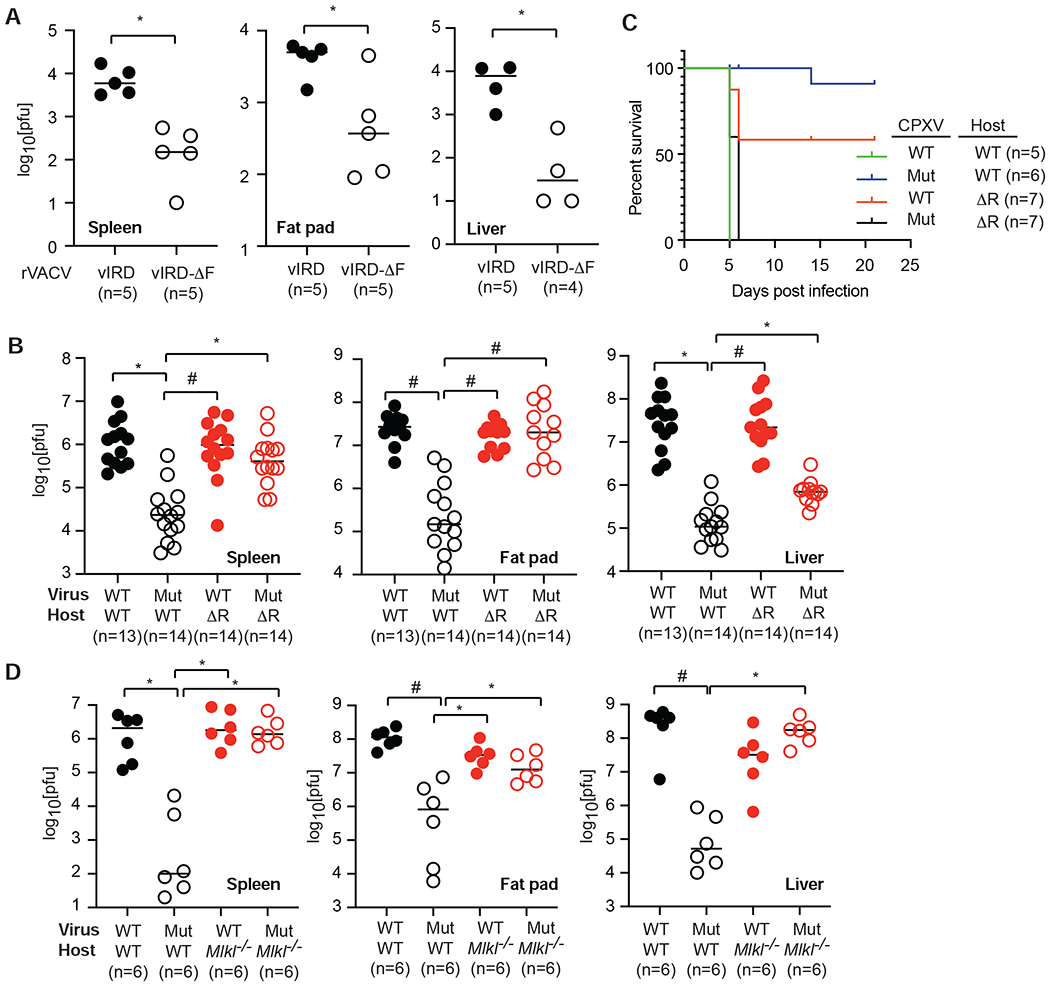
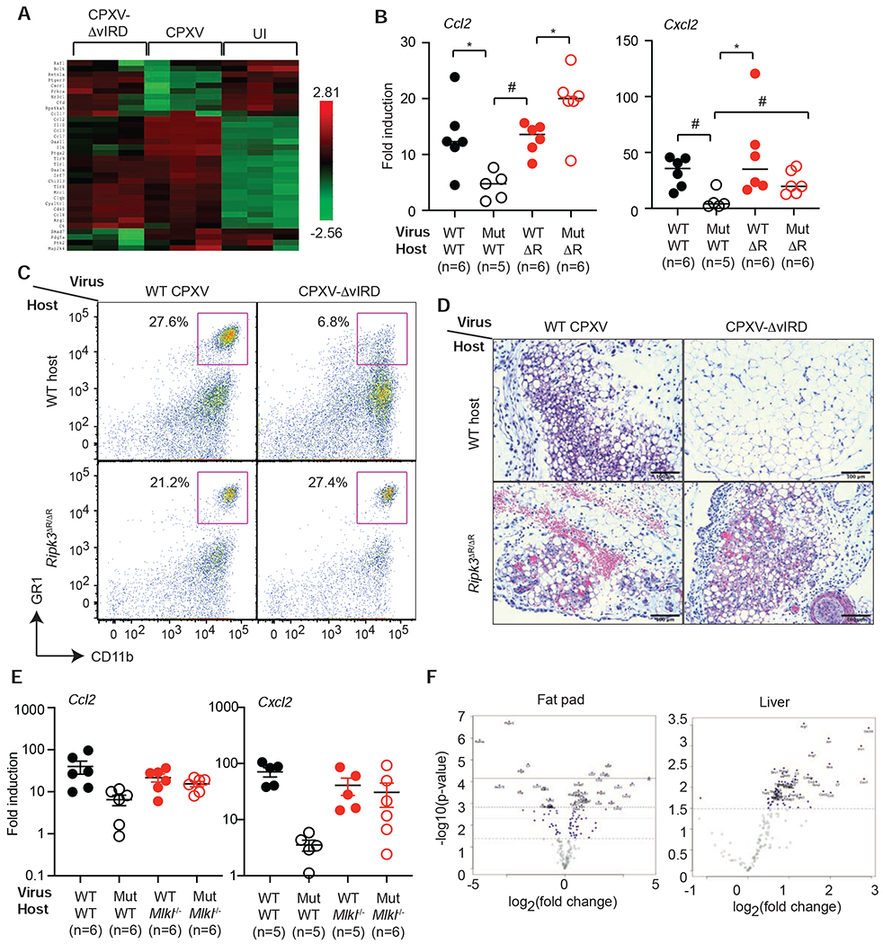
The vaccine strain against smallpox, vaccinia virus (VACV), is highly immunogenic yet causes relatively benign disease. These attributes are believed to be caused by gene loss in VACV. Using a targeted small interfering RNA (siRNA) screen, we identified a viral inhibitor found in cowpox virus (CPXV) and other orthopoxviruses that bound to the host SKP1-Cullin1-F-box (SCF) machinery and the essential necroptosis kinase receptor interacting protein kinase 3 (RIPK3). This "viral inducer of RIPK3 degradation" (vIRD) triggered ubiquitination and proteasome-mediated degradation of RIPK3 and inhibited necroptosis. In contrast to orthopoxviruses, the distantly related leporipoxvirus myxoma virus (MYXV), which infects RIPK3-deficient hosts, lacks a functional vIRD. Introduction of vIRD into VACV, which encodes a truncated and defective vIRD, enhanced viral replication in mice. Deletion of vIRD reduced CPXV-induced inflammation, viral replication, and mortality, which were reversed in RIPK3- and MLKL-deficient mice. Hence, vIRD-RIPK3 drives pathogen-host evolution and regulates virus-induced inflammation and pathogenesis.

Keywords: F-box; RIPK3; TNF; ankyrin repeats; cowpox virus; inflammation; necroptosis; poxvirus; ubiquitination; vaccinia virus.


Conflict of interest statement

Declaration of interests A U.S. provisional patent application (no. 62/848,255) has been filed for this study.

Figures

Figure 1.
Figure 1.. CPXV confers resistance to RIPK3-dependent necroptosis.
(A) L929 cells were infected with the indicated GFP-expressing recombinant virus. Cell death was determined by Incucyte live cell imaging. UI: uninfected cells. (B) CPXV-infected or uninfected (UI) L929 cells were stimulated with TNF, BV6 and zVAD-fmk. Necroptosis was measured by Incucyte. (C) CPXV-infected or uninfected (UI) L929 cells were treated with TNF and BV6. Cell death was measured by Incucyte. (D) Uninfected J2 virus-transformed macrophages or J2 macrophages infected with the indicated poxviruses were stimulated with LPS (20 μg/ml) and Nigericin (Nig, 10 μM). Cell death was detected using Incucyte. (E) Uninfected or CPXV-infected human colorectal carcinoma Colo205 were treated with Erastin (10 μM) and cell death was measured using Incucyte. See also Figure S1.
Figure 2.
Figure 2.. CPXV causes proteasome-mediated degradation of RIPK3
(A-G) Effect of CPXV on necroptosis signal adaptor expression. (A) L929 cells were infected with the indicated viruses for 18 hours and protein expression was determined by Western blot. (B-C) L929 (D-E) J2 virus-transformed macrophages and (F-G) MEFs were infected with CPXV for the indicated amount of time and protein expression was determined by Western blot. (C, E, G) Quantification of CPXV-induced changes of protein expression. Protein expression in uninfected cells at T=0 was normalized to “1”. (H) CPXV inhibited MLKL phosphorylation. Uninfected or CPXV-infected MEFs were treated with TNF, BV6 and zVAD for the indicated times. RIPK1 phosphorylation at S166 and MLKL phosphorylation at S345 were examined by Western blot. (I) Kinetics of RIPK3 protein degradation in CPXV-infected L929 cells. (J) The proteasome inhibitor MG132 inhibited CPXV-induced RIPK3 degradation. L929 cells were infected with CPXV for 2h, followed by MG132 (10 μM). Protein expression was determined by Western blot. (K) CPXV-infected L929 cells were treated with AraC or MG132 and their effect on RIPK3 degradation was determined by Western blot. (L, M) CPXV induces K48-linked RIPK3 ubiquitination. Cell lysates from CPXV-infected or MG132-treated L929 cells were denatured in 6M urea prior to immunoprecipitation with anti-RIPK3 antibody. Western blot was performed to determine the extent of RIPK3 K48-linked ubiquitination. The extent of RIPK3 ubiquitination was quantified in (M). See also Figure S2.
Figure 3.
Figure 3.. vIRD interacts with cellular SCF complex components to promote RIPK3 degradation and necroptosis resistance.
(A) Schematic diagram of the domain organization of vIRD from CPXV and the truncated vIRD orthologues from VACV Western Reserve and Copenhagen strains. (B) L929 cells infected with wild type or the indicated mutant CPXV were analyzed for RIPK3 expression by Western blot. (C) vIRD is essential for necroptosis resistance of CPXV-infected cells. L929 cells infected with VACV, vIRD-deleted CPXV (CPXV-ΔvIRD) or vIRD revertant (CPXV-WT) were monitored for cell death using Incucyte. (D) Kinetics of vIRD expression and RIPK3 degradation were determined by Western blot at the indicated times after CPXV infection. (E) Formation of the viral SCF complex. SKP1 was immunoprecipitated from CPXV-infected and uninfected L929 cells. vIRD and CUL1 recruitment upon CPXV infection was determined by Western blot. (F) CUL1 is essential for CPXV-induced RIPK3 degradation. L929 cells transfected with CUL1-specific or control scrambled siRNAs were monitored for RIPK3 degradation by Western blot. (G) CUL1 is essential for CPXV-mediated resistance to necroptosis. L929 cells transfected with the indicated siRNA were monitored for cell death by Incucyte live cell imaging. (H) Effect of CUL1 neddylation on CPXV-induced RIPK3 degradation. Uninfected or CPXV-infected L929 cells were treated with MLN4924 (1 μM) as indicated. The expression of vIRD and RIPK3 was determined by Western blot. (I) The effect of CUL1 neddylation on CPXV-induced necroptosis. Uninfected and CPXV-infected L929 cells were treated with the indicated inhibitors and/or stimulated with TNF and BV6. Cell death was monitored by Incucyte. See also Figure S3 and Tabls S1.
Figure 4.
Figure 4.. The ankyrin repeats of vIRD mediate binding to RIPK3.
(A) Schematic diagram of the vIRD mutants used. (B) HEK293T cells were transfected with the indicated GFP-tagged vIRD and RIPK3. Immunoprecipitation and Western blot were performed using the indicated antibodies. (C) Schematic diagram of the RIPK3 domain structure and truncation mutants used. (D) An intact RHIM is required for RIPK3 binding to vIRD. HEK293T cells were transfected with the indicated plasmids. Interaction between RIPK3 mutants and vIRD was determined by immunoprecipitation and Western blot as indicated. (E) The RHIM is essential for RIPK3 binding to vIRD. HEK293T cells transfected with the indicated plasmids were infected with rVACV-vIRD or rVACV-vIRD-ΔF. Binding with vIRD was determined by immunoprecipitation and Western blot as indicated. (F) Specific interaction between vIRD and RIPK3. L929 cells were infected with rVACV-FLAG-vIRD or rVACV-FLAG-vIRD-ΔF. vIRD was pulled down with anti-FLAG antibody followed by Western blot detection of the indicated RHIM adaptors. (G) vIRD does not interact with ZBP1. J2 virus transformed macrophages were infected with the indicated rVACV. Immunoprecipitation and Western blot were performed as indicated.
Figure 5.
Figure 5.. vIRD is conserved in other orthopoxviruses.
(A) Expression of vIRD inhibits rVACV-induced necroptosis. L929 cells infected with the indicated rVACV were monitored for cell death by Incucyte. (B) Expression of vIRD by VACV triggers RIPK3 degradation. Western blot analysis of RIPK3 expression in L929 cells infected with the indicated rVACV. vIRD-C is a rabbit polyclonal antibody specific for the C-terminus of vIRD. (C) Assembly of vIRD with SKP1 and CUL1 in L929 cells infected with the indicated rVACV expressing wild type or F-box deleted vIRD. (D) RIPK3 degradation by ECTV. L929 cells infected with the indicated viruses were examined for RIPK3 degradation by Western blot. (E) Resistance of ECTV-infected L929 cells to necroptosis is reversed by the neddylation inhibitor MLN4924. (F) The vIRD orthologues from MPXV and ECTV, but not MYXV, triggered RIPK3 degradation. HEK293T cells transfected with the indicated plasmids were determined for RIPK3 expression by Western blot. (G) Schematic diagram of the domain structure of MYXV-m148R compared to that of vIRD from CPXV. See also Figure S4 and S5.
Figure 6.
Figure 6.. vIRD promotes viral replication through RIPK3 and necroptosis inhibition.
(A) Expression of vIRD enhanced VACV replication in mice. Mice were infected with rVACV-vIRD or rVACV-vIRD-ΔF. Three and a half days later, viral titers in the indicated tissues were determined by Vero cell plaque assay. (B) Ripk3gfpfl/fl (WT) or Ripk3ΔR/ΔR (ΔR) mice were infected with wild type (WT) or CPXV-ΔvIRD (Mut) via the intraperitoneal route as indicated. Viral load in the indicated tissues was determined by Vero cell plaque assay 3.5 days post-infection. (C) Survival of Ripk3gfpfl/fl (WT) or Ripk3ΔR/ΔR (ΔR) mice infected with the indicated wild type or ΔvIRD CPXV. (D) MLKL deficiency rescues defective viral replication of CPXV-ΔvIRD. Mlkl−/− and wild type littermates were infected by the indicated virus. Viral load in the indicated tissues was determined by Vero cell plaque assay 3.5 days post-infection. * p < 0.05, # p < 0.01. See also Figure S6.
Figure 7.
Figure 7.. vIRD regulates virus-induced tissue inflammation.
(A) Heat map of the expression of the top 20 DE genes in WT CPXV vs UI and CPXV-ΔvIRD vs UI groups were shown. (B) Q-PCR analysis of Ccl2 and Cxcl2 expression in infected mouse visceral fat pad of the indicated groups. (C) FACS analysis of CD11b+GR1+ neutrophils isolated from the visceral fat pad of the infected mice. (D) H&E staining of infected visceral fat pad from mice in the different groups. Scale bars represent 100 μm. (E) Chemokine expression in Mlkl−/− mice. Wild type or Mlkl−/− littermates were infected with wild type CPXV or CPXV-ΔvIRD and Ccl2 and Cxcl2 expression was determined 3.5 days post-infection. (F) Tissue-specific effects of vIRD on inflammatory gene expression. Wild type mice infected with wild type CPXV or CPXV-ΔvIRD were analyzed for inflammatory gene expression by Nanostring. The top DE genes in visceral fat pad and liver were shown as Volcano plots. * p < 0.05, # p < 0.01. See also Figure S7 and Table S2.

Similar articles

Cited by

References

    1. Blasco R, and Moss B (1995). Selection of recombinant vaccinia viruses on the basis of plaque formation. Gene 158, 157–162. - PubMed
    1. Bratke KA, McLysaght A, and Rothenburg S (2013). A survey of host range genes in poxvirus genomes. Infection, genetics and evolution : journal of molecular epidemiology and evolutionary genetics in infectious diseases 14, 406–425. - PMC - PubMed
    1. Carpenter EA, Ruby J, and Ramshaw IA (1994). IFN-gamma, TNF, and IL-6 production by vaccinia virus immune spleen cells. An in vitro study. J Immunol 152, 2652–2659. - PubMed
    1. Chan FK, Luz NF, and Moriwaki K (2015). Programmed necrosis in the cross talk of cell death and inflammation. Annual review of immunology 33, 79–106. - PMC - PubMed
    1. Chan FK, Shisler J, Bixby JG, Felices M, Zheng L, Appel M, Orenstein J, Moss B, and Lenardo MJ (2003). A role for tumor necrosis factor receptor-2 and receptor-interacting protein in programmed necrosis and antiviral responses. J Biol Chem 278, 51613–51621. - PubMed

Publication types

MeSH terms

Substances