Review
. 2021 Jun:68:66-73.
doi: 10.1016/j.sbi.2020.12.009. Epub 2021 Jan 11.

Protein N-glycosylation and O-mannosylation are catalyzed by two evolutionarily related GT-C glycosyltransferases

Affiliations
Review

Protein N-glycosylation and O-mannosylation are catalyzed by two evolutionarily related GT-C glycosyltransferases

Lin Bai et al. Curr Opin Struct Biol. 2021 Jun.

Abstract

The structural folds of glycosyltransferases are categorized into three superfamilies: GT-A, GT-B, and GT-C. Few structures of GT-C fold existed in the Protein Data Bank prior to the recent advent of high-resolution cryo-EM, because the glycosyltransferases are large membrane proteins that are difficult to crystallize. The use of cryo-EM has resulted in the structures of several key GT-C glycosyltransferases. Here we summarize the latest structural features of and mechanistic insights into these membrane enzyme complexes.


Conflict of interest statement

Conflict of interest statement

Nothing declared.

Figures

Figure 1.
Figure 1.. N-glycosylation and O-mannosylation pathways in the ER of Saccharomyces cerevisiae.
Both OST and PMT complexes can directly associate with the Sec61 translocon to catalyze co-translational protein glycosylation. The OST donor substrate dolichol-PP-OS is sequentially synthesized by 14 enzymes (ALG1–14). The Pmt1/2 donor substrate dolichol-P-man is synthesized in the cytosol and flipped into the ER lumen for protein O-mannosylation. Post-translational protein glycosylation also occurs in eukaryotes but is not illustrated here for simplicity.
Figure 2.
Figure 2.. The cryo-EM structures of the yeast and human OSTs.
(A) Cryo-EM 3D map of the yeast OST complex (EMD-7336) in a frontside and a backside view (viewed within the membrane plane). (B) Cryo-EM 3D map of the human OST-A complex (EMD-10110). (C) Cryo-EM 3D map of the human OST-B complex (EMD-10112). (D) Cryo-EM 3D map of a mammalian OST–translocon–ribosome complex at 4.7 Å overall resolution (EMD-4317).
Figure 3.
Figure 3.. Cryo-EM structure of the yeast Pmt1-Pmt2 complex.
(A) Cryo-EM 3D map of yeast Pmt1–Pmt2 complex (EMD-20236) viewed from the side (left, from within the membrane plane) and from the top (right, from ER lumen). (B) The Pmt1–Pmt2 structure (PDB code 6P25) is shown in cartoons; colors are the same as in panel A.
Figure 4.
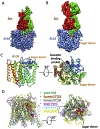
Figure 4.. Cryo-EM structure of the yeast ALG6 and its structural alignment with other GT-C glycosyltransferases.
(A) Cryo-EM 3D map of the yeast ALG6-Fab complex at 3.0 Å resolution (EMD-10258). (B) Cryo-EM 3D map of the yeast ALG6-Fab complex bound to a sugar donor at 3.9 Å average resolution (EMD-10257). (C) A side (left) and a top view (right) of the ALG6 structure (PDB code 6SNH), shown in cartoons and colored in rainbow from N-terminal blue to C-terminal red. (D) A side view (left) and a lumenal view (right) of the structural alignment of ALG6 (PDB code 6SNH) with yeast Stt3 (PDB code 6C26), human STT3A (PDB code 6S7O), STT3B (PDB code 6S7T), and yeast Pmt1 and Pmt2 (PDB code 6P25). The names of enzymes whose structures contain a sugar donor are underlined.

References

    1. Helenius A, Aebi M: Roles of N-linked glycans in the endoplasmic reticulum. Annu Rev Biochem 2004, 73:1019–1049. - PubMed
    1. Lairson LL, Henrissat B, Davies GJ, Withers SG: Glycosyltransferases: structures, functions, and mechanisms. Annu Rev Biochem 2008, 77:521–555. - PubMed
    1. Spiro RG: Protein glycosylation: nature, distribution, enzymatic formation, and disease implications of glycopeptide bonds. Glycobiology 2002, 12:43r–56r. - PubMed
    1. Bai XC, McMullan G, Scheres SH: How cryo-EM is revolutionizing structural biology. Trends Biochem Sci 2015, 40:49–57. - PubMed
    1. Cheng Y: Single-particle cryo-EM at crystallographic resolution. Cell 2015, 161:450–457. - PMC - PubMed

Publication types

LinkOut - more resources