

Role of Innate Inflammation in the Regulation of Tissue Remodeling during Tooth Eruption
- PMID: 33445432
- PMCID: PMC7827943
- DOI: 10.3390/dj9010007
Role of Innate Inflammation in the Regulation of Tissue Remodeling during Tooth Eruption
Abstract
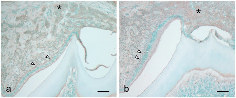
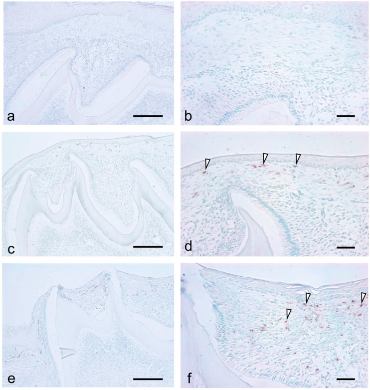
Tooth eruption is characterized by a coordinated complex cascade of cellular and molecular events that promote tooth movement through the eruptive pathway. During tooth eruption, the stratum intermedium structurally changes to the papillary layer with tooth organ development. We previously reported intercellular adhesion molecule-1 (ICAM-1) expression on the papillary layer, which is the origin of the ICAM-1-positive junctional epithelium. ICAM-1 expression is induced by proinflammatory cytokines, including interleukin-1 and tumor necrosis factor. Inflammatory reactions induce tissue degradation. Therefore, this study aimed to examine whether inflammatory reactions are involved in tooth eruption. Reverse transcription-polymerase chain reaction (RT-PCR) analysis revealed sequential expression of hypoxia-induced factor-1α, interleukin-1β, and chemotactic factors, including keratinocyte-derived chemokine (KC) and macrophage inflammatory protein-2 (MIP-2), during tooth eruption. Consistent with the RT-PCR results, immunohistochemical analysis revealed KC and MIP-2 expression in the papillary layer cells of the enamel organ from the ameloblast maturation stage. Moreover, there was massive macrophage and neutrophil infiltration in the connective tissue between the tooth organ and oral epithelium during tooth eruption. These findings suggest that inflammatory reactions might be involved in the degradation of tissue overlying the tooth organ. Further, these reactions might be induced by hypoxia in the tissue overlying the tooth organ, which results from decreased capillaries in the tissue. Our findings indicate that bacterial infections are not associated with the eruption process. Therefore, tooth eruption might be regulated by innate inflammatory mechanisms.
Keywords: HIF-1; IL-1; KC; neutrophil; tooth eruption.
Conflict of interest statement
The authors declare no conflict of interest.
Figures


Similar articles
-
Daughters of the Enamel Organ: Development, Fate, and Function of the Stratum Intermedium, Stellate Reticulum, and Outer Enamel Epithelium.Stem Cells Dev. 2016 Oct;25(20):1580-1590. doi: 10.1089/scd.2016.0267. Epub 2016 Sep 9. Stem Cells Dev. 2016. PMID: 27611344 Free PMC article.
-
Increased expression of cytokines and adhesion molecules in rat chronic esophagitis.Digestion. 2003;68(4):189-97. doi: 10.1159/000075698. Epub 2003 Dec 19. Digestion. 2003. PMID: 14691346
-
Role of the junctional epithelium in periodontal innate defense and homeostasis.J Periodontal Res. 2012 Dec;47(6):750-7. doi: 10.1111/j.1600-0765.2012.01490.x. Epub 2012 May 15. J Periodontal Res. 2012. PMID: 22587460
-
Cellular, molecular, and genetic determinants of tooth eruption.Crit Rev Oral Biol Med. 2002;13(4):323-34. doi: 10.1177/154411130201300403. Crit Rev Oral Biol Med. 2002. PMID: 12191959 Review.
-
Histological and immunological characteristics of the junctional epithelium.Jpn Dent Sci Rev. 2018 May;54(2):59-65. doi: 10.1016/j.jdsr.2017.11.004. Epub 2017 Dec 7. Jpn Dent Sci Rev. 2018. PMID: 29755616 Free PMC article. Review.
Cited by
-
The Role and Mechanism of Metformin in Inflammatory Diseases.J Inflamm Res. 2023 Nov 23;16:5545-5564. doi: 10.2147/JIR.S436147. eCollection 2023. J Inflamm Res. 2023. PMID: 38026260 Free PMC article. Review.
-
The radiological and histological investigation of the dental follicle of asymptomatic impacted mandibular third molars.BMC Oral Health. 2022 Dec 26;22(1):642. doi: 10.1186/s12903-022-02681-6. BMC Oral Health. 2022. PMID: 36567318 Free PMC article.
References
LinkOut - more resources
Full Text Sources
Other Literature Sources
Miscellaneous
