. 2021 Jan 12;13(1):90.
doi: 10.3390/pharmaceutics13010090.

Ultra-Small Iron Nanoparticles Target Mitochondria Inducing Autophagy, Acting on Mitochondrial DNA and Reducing Respiration

Affiliations

Ultra-Small Iron Nanoparticles Target Mitochondria Inducing Autophagy, Acting on Mitochondrial DNA and Reducing Respiration

Lorenzo Rivas-García et al. Pharmaceutics. .

Abstract

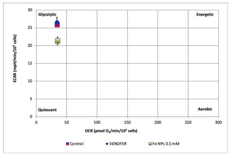
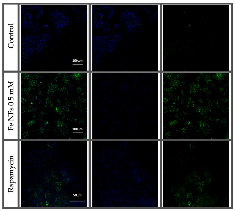
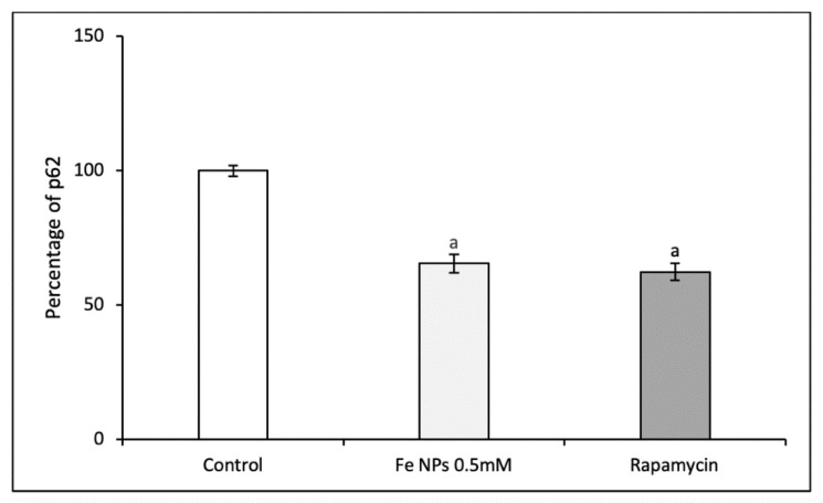
The application of metallic nanoparticles (materials with size at least in one dimension ranging from 1 to 100 nm) as a new therapeutic tool will improve the diagnosis and treatment of diseases. The mitochondria could be a therapeutic target to treat pathologies whose origin lies in mitochondrial dysfunctions or whose progression is dependent on mitochondrial function. We aimed to study the subcellular distribution of 2-4 nm iron nanoparticles and its effect on mitochondrial DNA (mtDNA), mitochondrial function, and autophagy in colorectal cell lines (HT-29). Results showed that when cells were exposed to ultra-small iron nanoparticles, their subcellular fate was mainly mitochondria, affecting its respiratory and glycolytic parameters, inducing the migration of the cellular state towards quiescence, and promoting and triggering the autophagic process. These effects support the potential use of nanoparticles as therapeutic agents using mitochondria as a target for cancer and other treatments for mitochondria-dependent pathologies.

Keywords: copy number; metals; mitochondria; mtDNA deletions; nanotechnology; respiration.


Conflict of interest statement

The authors declare no conflict of interest.

Figures

Figure 1
Figure 1
TEM images of cultured cells HT-29 exposed to (A) 0.5 mmol L−1 4 nm iron nanoparticles (FeNPs) and (B) 0.5 mmol L−1 Venofer®. Ellipses highlight the presence of mitophagy processes. Arrows highlight the presence of nanoparticles.
Figure 1
Figure 1
TEM images of cultured cells HT-29 exposed to (A) 0.5 mmol L−1 4 nm iron nanoparticles (FeNPs) and (B) 0.5 mmol L−1 Venofer®. Ellipses highlight the presence of mitophagy processes. Arrows highlight the presence of nanoparticles.
Figure 2
Figure 2
ND1/ND4 mtDNA deletion. Results expressed as mean ± SEM. (p < 0.05). (a) vs. control (p < 0.05).
Figure 3
Figure 3
ND1/ND4 mtDNA copy number. Results expressed as mean ± SEM. (a) vs. control (p < 0.05).
Figure 4
Figure 4
Modulation of mitochondrial respiration by iron nanoparticles in HT-29 cells. Cells were treated with DMEM (ctrl), iron nanoparticles Fe NPs or Venofer® for 24 h. OCR was determined by using the Seahorse XF-24 Extracellular Flux Analyzer after the sequential injections of oligomycin (1 μg mL−1), 2,4-DNP (1 mmol L−1), and rotenone/antimycin (1 μmol L−1/10 μmol L−1). (A) shows OCR. (B) shows maximal respiratory capacity. (C) shows the spare capacity. Data are indicated as the mean ± SEM (n = 3). (a) vs. control; (b) vs. Fe NPs 0.5 mM (p < 0.05).
Figure 5
Figure 5
Effects on Mitochondrial glycolysis by iron nanoparticles in HT-29 cells. Cells were treated with DMEM (ctrl), iron nanoparticles Fe NPs or Venofer® for 24 h. ECAR was determined by using the Seahorse XF-24 Extracellular Flux Analyzer after the injections of rotenone (1 μmol L−1), glucose (30 mmol L−1) and 2-DG (100 mmol L−1). Glycolytic capacity was calculated from the XF glycolysis stress test profile. (A) shows ECAR. (B) shows glycolysis. (C) shows the glycolitic capacity. (D) shows the glycolysis reserve. Data are indicated as the mean ± SEM (n = 3). (a) vs. control; (b) vs. Fe NPs 0.5 mmol L−1 (p < 0.05).
Figure 6
Figure 6
ECAR:OCR ratio plot showing the metabolic phenotype of HT-29 cells treated or not with iron nanoparticles FeNPs or Venofer®. Data represent the mean ± SEM (n = 3).
Figure 7
Figure 7
Autophagy induction evaluated by fluorescence using a microplate reader in HT-29 normalized to control cells (untreated). 5 mmol L−1, rapamycin 0.5 µmol L−1 and control cells (untreated). Results expressed as mean ± SEM. (a) vs. control; (b) vs. Fe NPs 0.5 mmol L−1 (p < 0.05).
Figure 8
Figure 8
Confocal microscopy images of HT-29 cells exposed to Fe NPs 0.5 mmol L−1, rapamycin 0.5 µmol L−1 and control cells (untreated).
Figure 9
Figure 9
Percentage of p62 (respect control cells) in HT-29 cells exposed to Fe NPs 0.5 mmol L−1, rapamycin 0.5 µmol L−1. Results expressed as mean ± SEM. (a) vs. control (p < 0.05).
Figure 10
Figure 10
Evaluation of apoptosis/necrosis by flow cytometry. Results expressed as mean ± SEM. (a) vs. control (p < 0.05).

References

    1. Zdrojewicz Z., Waracki M., Bugaj B., Pypno D., Cabała K. Medical applications of nanotechnology. Postepy Hig. Med. Dosw. 2015;69:1196–1204. doi: 10.5604/17322693.1177169. - DOI - PubMed
    1. Amendoeira A., Rivas García L., Fernandes A.R., Baptista P.V. Light Irradiation of Gold Nanoparticles Toward Advanced Cancer Therapeutics. Adv. Ther. 2020;3:1900153. doi: 10.1002/adtp.201900153. - DOI
    1. Lopez-Chaves C., Soto-Alvaredo J., Montes-Bayon M., Bettmer J., Llopis J., Sanchez-Gonzalez C. Gold nanoparticles: Distribution, bioaccumulation and toxicity. In vitro and in vivo studies. Nanomed. Nanotechnol. Biol. Med. 2018;14:1–12. doi: 10.1016/j.nano.2017.08.011. - DOI - PubMed
    1. Cordani M., Somoza Á. Targeting autophagy using metallic nanoparticles: A promising strategy for cancer treatment. Cell. Mol. Life Sci. 2019;76:1215–1242. doi: 10.1007/s00018-018-2973-y. - DOI - PMC - PubMed
    1. Alphandéry E. Biodistribution and targeting properties of iron oxide nanoparticles for treatments of cancer and iron anemia disease. Nanotoxicology. 2019;13:573–596. doi: 10.1080/17435390.2019.1572809. - DOI - PubMed

LinkOut - more resources