. 2021 Jan 12;22(2):678.
doi: 10.3390/ijms22020678.

JAZF1, A Novel p400/TIP60/NuA4 Complex Member, Regulates H2A.Z Acetylation at Regulatory Regions

Affiliations

JAZF1, A Novel p400/TIP60/NuA4 Complex Member, Regulates H2A.Z Acetylation at Regulatory Regions

Tara Procida et al. Int J Mol Sci. .

Abstract

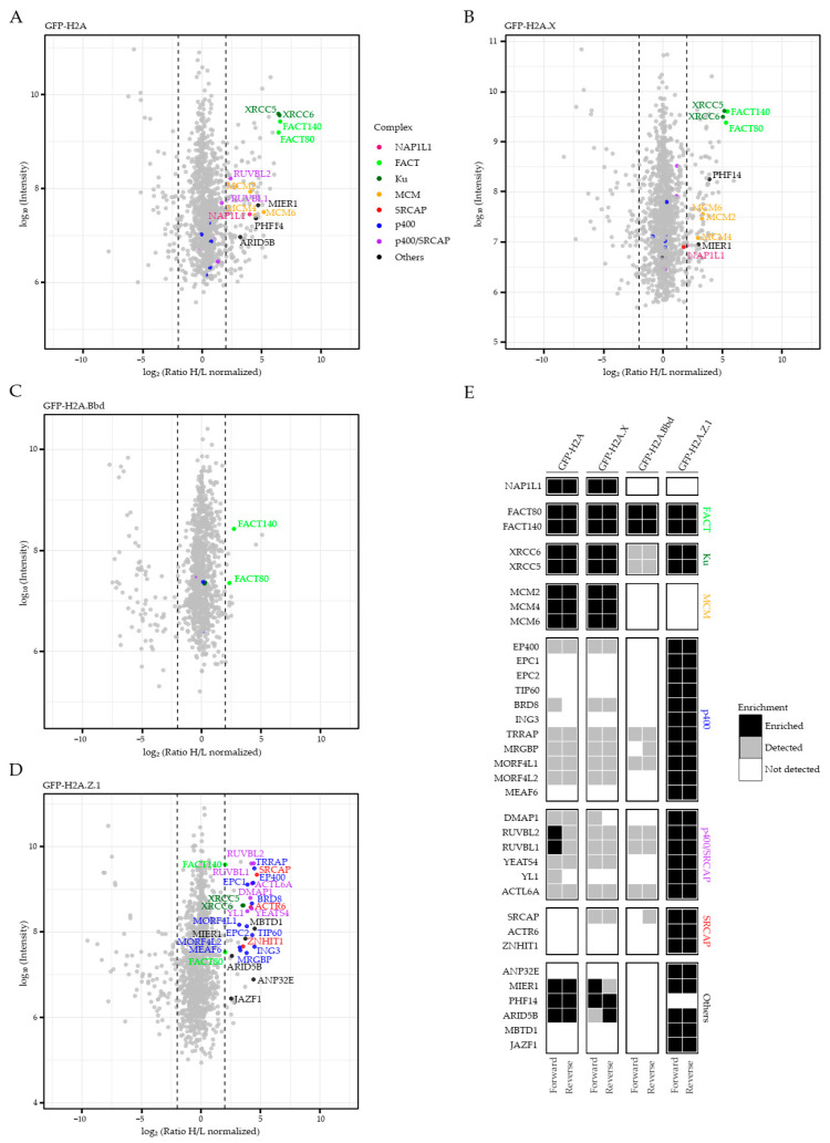
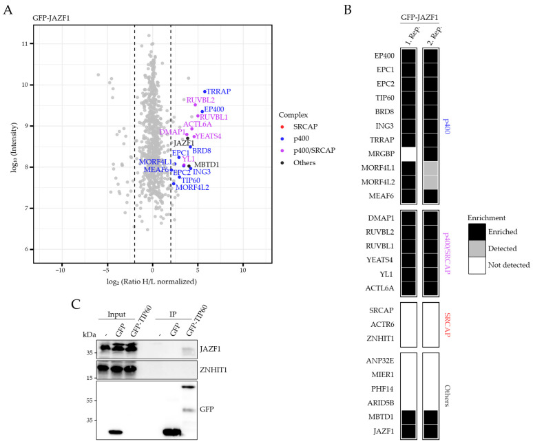
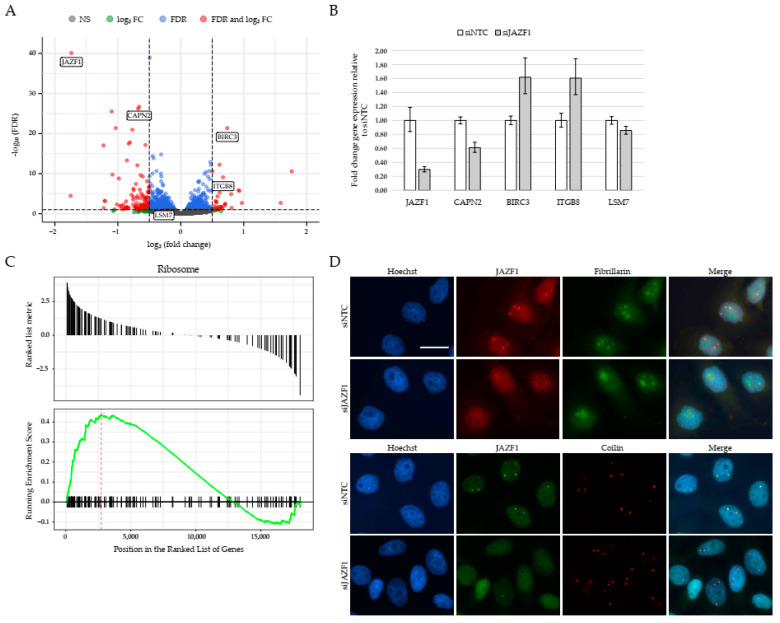
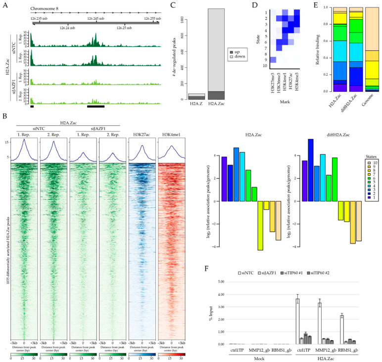
Histone variants differ in amino acid sequence, expression timing and genomic localization sites from canonical histones and convey unique functions to eukaryotic cells. Their tightly controlled spatial and temporal deposition into specific chromatin regions is accomplished by dedicated chaperone and/or remodeling complexes. While quantitatively identifying the chaperone complexes of many human H2A variants by using mass spectrometry, we also found additional members of the known H2A.Z chaperone complexes p400/TIP60/NuA4 and SRCAP. We discovered JAZF1, a nuclear/nucleolar protein, as a member of a p400 sub-complex containing MBTD1 but excluding ANP32E. Depletion of JAZF1 results in transcriptome changes that affect, among other pathways, ribosome biogenesis. To identify the underlying molecular mechanism contributing to JAZF1's function in gene regulation, we performed genome-wide ChIP-seq analyses. Interestingly, depletion of JAZF1 leads to reduced H2A.Z acetylation levels at > 1000 regulatory sites without affecting H2A.Z nucleosome positioning. Since JAZF1 associates with the histone acetyltransferase TIP60, whose depletion causes a correlated H2A.Z deacetylation of several JAZF1-targeted enhancer regions, we speculate that JAZF1 acts as chromatin modulator by recruiting TIP60's enzymatic activity. Altogether, this study uncovers JAZF1 as a member of a TIP60-containing p400 chaperone complex orchestrating H2A.Z acetylation at regulatory regions controlling the expression of genes, many of which are involved in ribosome biogenesis.

Keywords: H2A.Z; JAZF1; TIP60; acetylation; enhancer; gene regulation; histone variants; ribosome.


Conflict of interest statement

All authors declare no conflict of interest.

Figures

Figure 1
Figure 1
JAZF1 is member of H2A.Z chaperone complex(es). (A–D) Scatter plots of Green Fluorescent Protein (GFP) pull-downs for GFP–H2A (A), GFP–H2A.X (B), GFP–H2A.Bbd (C) and GFP–H2A.Z.1 (D). HeLa Kyoto cells stably expressing GFP–H2A, –H2A.X, -H2A.Bbd or GFP–H2A.Z.1 were SILAC-labeled and subjected to single-step affinity purifications of soluble nuclear proteins, using GFP nanotrap beads. In each panel, the ratio of the identified proteins after MS is plotted. Nucleosome-assembly protein 1-like 1 (NAP1L1) is plotted in pink, members of the facilitates chromatin transcription (FACT) complex are depicted in bright green, members of the Ku complex in dark green, mini chromosome maintenance (MCM) proteins in orange, members of the SRCAP complex in red, of the p400 complex in blue and shared members of both complexes in purple. Other proteins are highlighted in black. Significantly enriched proteins of the abovementioned categories are labeled with names, non-significantly enriched but detected proteins are only highlighted with the respective color within the plot. Dotted lines represent selected ratio cutoff of 4 and 0.25. Shown is one out of two similar replicates. See Supplementary Materials Table S1 for a list of all identified proteins. (E) Heatmap of all identified chaperone proteins described in (AD). Shown are enriched (black), detected but not enriched (gray) and not detected (white) proteins after forward or reverse SILAC–MS identifications. In forward and reverse experiments, selected ratio cutoff is 4 and 0.25, respectively. Shown is one of two similar biological replicates.
Figure 2
Figure 2
JAZF1 is a member of a TIP60- and MBTD1-containing p400 complex. (A) Scatter plot of GFP pull-downs for GFP–JAZF1. HeLa Kyoto cells transiently expressing GFP or GFP–JAZF1 were SILAC-labeled and subjected to single-step affinity purifications of soluble nuclear proteins, using GFP nanotrap beads. In each panel, the ratio of the identified proteins after MS is plotted. Members of the SRCAP complex are depicted in red, members of the p400 complex are in blue and shared members of both complexes are in purple. JAZF1 and MBTD1 are highlighted in black. Dotted lines represent selected ratio cutoff of 4. Shown is one of two biological replicates. See Supplementary Materials Table S1 for a list of all identified proteins. (B) Heatmap of p400, SRCAP and shared complex members identified in (A) and an additional replicate SILAC experiment. Shown are enriched (black), detected but not enriched (gray) and not detected (white) proteins after MS identifications. Selected ratio cutoff is 4. (C) Immunoblotting of JAZF1, SRCAP member ZNHIT1 and GFP upon transient transfection of HeLa Kyoto cells with GFP and GFP–TIP60, followed by GFP-trap pull-downs of soluble nuclear extracts.
Figure 3
Figure 3
JAZF1 depletion leads to deregulation of genes involved in ribosome biogenesis. (A) Volcano-plot representation of differential expression analysis of genes upon JAZF1 depletion (siJAZF1 vs. siNTC), showing 162 statistically significant deregulated genes, 130 downregulated and 32 upregulated. Red dots represent statistically significant deregulated genes (log2FC > 0.5 or <−0.5 and False Discovery Rate (FDR) < 0.1); blue dots represent genes with significant statistical values (FDR < 0.1) that not reach our fold change threshold; green dots represent genes with significant expression changes (log2FC > 0.5 or <−0.5) without providing enough statistical evidence; gray dots represent genes with no statistical or fold-change significance. Genes that were validated by using RT-qPCR (B) are highlighted with boxes. See also Supplementary Materials Figure S1B for PCA plot of deregulated genes upon JAZF1 knockdown. (B) RT-qPCR verification of two downregulated (JAZF1 and CAPN2) and two upregulated (BIRC3 and ITGB8) genes and one in its expression unchanged gene (LSM7) upon JAZF1 depletion. Shown is the fold change of three replicates normalized to HPRT1 expression. Error bars depict the SD of three replicates. Shown is one out of three biological replicates showing similar results. (C) Gene set enrichment analysis (GSEA) plot showing statistically significant and consistent differences of genes associated with the KEGG (Kyoto Encyclopedia of Genes and Genomes) pathway “Ribosome” upon JAFZ1 depletion. See also Supplementary Materials Figure S1C,D for further GSEA analyses. (D) Immunofluorescence microscopy analysis of HeLa Kyoto cells co-stained with antibodies against JAZF1 (top, red; bottom, green), Fibrillarin (green, top) or Coilin (red, bottom) and Hoechst (DNA, blue) after control (siNTC) or JAZF1 (siJAZF1) knockdowns. Scale bar for all pictures = 20 µm.
Figure 4
Figure 4
JAZF1 depletion leads to decrease in H2A.Z acetylation at regulatory regions. (A) Genome browser snap shot of human chromosome 8 locus as representative region displaying differential H2A.Zac ChIP-seq signals in two replicates of control (siNTC; non-target control siRNA; dark green) and JAZF1 knockdown (siJAZF1; light green). (B) ChIP-seq density heatmap of H2A.Zac upon control (siNTC) or JAZF1 (siJAZF1) knockdowns (two replicates, green), as well as visualization of the intensities of H3K27ac (blue, ENCODE) and H3K4me1 (red, ENCODE) at these differentially H2A.Z acetylated regions. Color intensities represents normalized and globally scaled tag counts. (C) Bar-plots visualizing deregulated H2A.Z or H2A.Zac peaks upon JAZF1-depletion. Notice that no major change is observed in H2A.Z peaks, but > 1000 H2A.Zac peaks are downregulated upon loss of JAZF1. (D) ChromHMM-based characterization of chromatin states [56,57]. The heatmap depicts the emission parameters of the HMM and describes the combinatorial occurrence of the individual histone modifications (mark) in different chromatin states (1–10). (E) Chromatin-state enrichment of global H2A.Zac and differentially deregulated H2A.Zac peaks (diffH2A.Zac) upon JAZF1 depletion in specific states (see (D)) calculated to frequency in complete genome (right). Notice enrichment of diffH2A.Zac peaks in states 2 and 6 that resemble H3K36me3-positive enhancer regions. See also Supplementary Materials Figure S2F for further characterizations. (F) ChIP–qPCR verification of three diffH2A.Zac sites (gb: gene body) upon JAZF1 (light gray) and TIP60 depletion (two independent knockdowns: siTIP60 #1 and siTIP60 #2, dark gray, see also Supplementary Materials Figure S2G for knockdown efficiency), compared to control knockdown (siNTC, white) using no (mock) or H2A.Zac antibodies. Shown is the respective enrichment as percentage of input signals. Error bars represent the SD of three replicates. Notice that TIP60 knockdown also leads to a similar reduction in H2A.Zac at these three diffH2A.Zac sites, compared to JAZF1 depletion.

Similar articles

Cited by

References

    1. Van Holde K.E. Chromatin. Springer; New York, NY, USA: 1988.
    1. Bonisch C., Nieratschker S.M., Orfanos N.K., Hake S.B. Chromatin proteomics and epigenetic regulatory circuits. Expert Rev. Proteom. 2008;5:105–119. doi: 10.1586/14789450.5.1.105. - DOI - PubMed
    1. Buschbeck M., Hake S.B. Variants of core histones and their roles in cell fate decisions, development and cancer. Nat. Rev. Mol. Cell Biol. 2017;18:299–314. doi: 10.1038/nrm.2016.166. - DOI - PubMed
    1. Zink L.M., Hake S.B. Histone variants: Nuclear function and disease. Curr. Opin. Genet. Dev. 2016;37:82–89. doi: 10.1016/j.gde.2015.12.002. - DOI - PubMed
    1. Iouzalen N., Moreau J., Mechali M. H2A.ZI, a new variant histone expressed during Xenopus early development exhibits several distinct features from the core histone H2A. Nucleic Acids Res. 1996;24:3947–3952. doi: 10.1093/nar/24.20.3947. - DOI - PMC - PubMed

MeSH terms

Grants and funding