. 2021 Jan 12;18(2):577.
doi: 10.3390/ijerph18020577.

Sex-Specific Alterations in Cardiac DNA Methylation in Adult Mice by Perinatal Lead Exposure

Affiliations

Sex-Specific Alterations in Cardiac DNA Methylation in Adult Mice by Perinatal Lead Exposure

Laurie K Svoboda et al. Int J Environ Res Public Health. .

Abstract

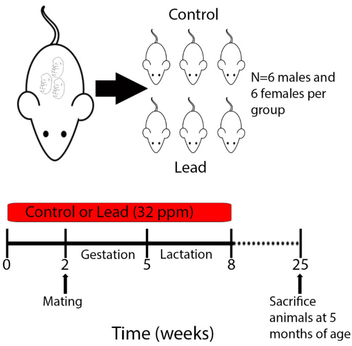
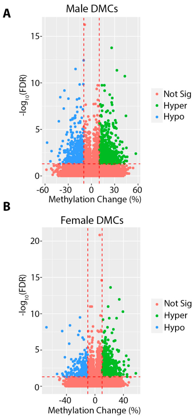
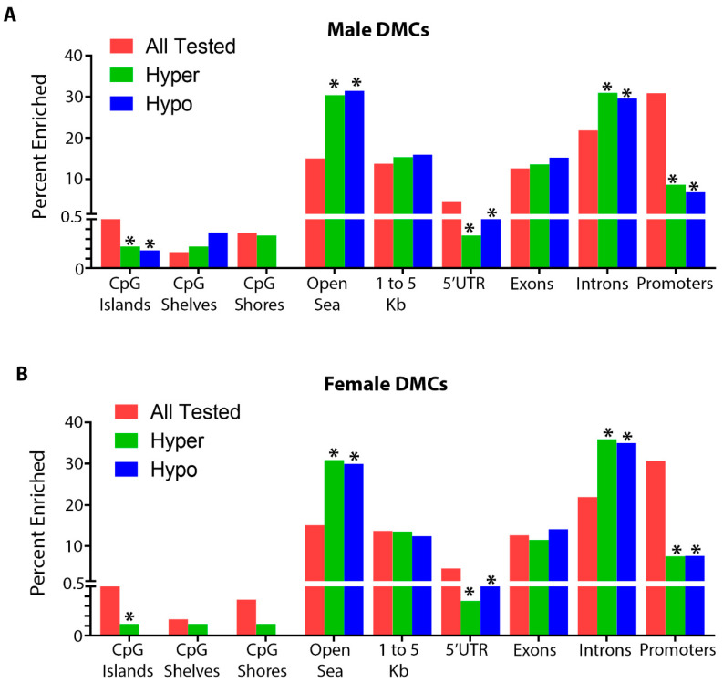
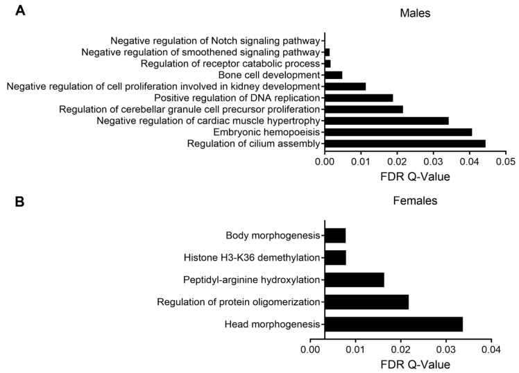
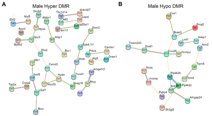
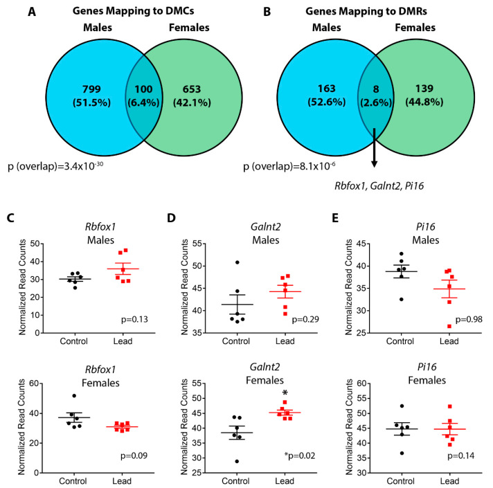
Environmental factors play an important role in the etiology of cardiovascular diseases. Cardiovascular diseases exhibit marked sexual dimorphism; however, the sex-specific effects of environmental exposures on cardiac health are incompletely understood. Perinatal and adult exposures to the metal lead (Pb) are linked to several adverse cardiovascular outcomes, but the sex-specific effects of this toxicant on the heart have received little attention. Perinatal environmental exposures can lead to disease through disruption of the normal epigenetic programming that occurs during early development. Using a mouse model of human-relevant perinatal environmental exposure, we investigated the effects of exposure to Pb during gestation and lactation on DNA methylation in the hearts of adult offspring mice (n = 6 per sex). Two weeks prior to mating, dams were assigned to control or Pb acetate (32 ppm) water, and exposure continued until offspring were weaned at three weeks of age. Enhanced reduced-representation bisulfite sequencing was used to measure DNA methylation in the hearts of offspring at five months of age. Although Pb exposure stopped at three weeks of age, we discovered hundreds of differentially methylated cytosines (DMCs) and regions (DMRs) in males and females at five months of age. DMCs/DMRs and their associated genes were sex-specific, with a small, but statistically significant subset overlapping between sexes. Pathway analysis revealed altered methylation of genes important for cardiac and other tissue development in males, and histone demethylation in females. Together, these data demonstrate that perinatal exposure to Pb induces sex-specific changes in cardiac DNA methylation that are present long after cessation of exposure, and highlight the importance of considering sex in environmental epigenetics and mechanistic toxicology studies.

Keywords: DNA methylation; Developmental Origins of Health and Disease (DOHaD); cardiovascular disease; heavy metals; sex differences; toxicoepigenetics.


Conflict of interest statement

The authors declare no conflict of interest.

Figures

Figure 1
Figure 1
Schematic of experimental design and sample collection. Dams were exposed to Pb 2 weeks prior to mating via drinking water. Maternal (and, in turn, offspring) exposure continued until weaning, when offspring reached 3 weeks of age. 6 males and 6 females per exposure were sacrificed at 5 months of age for tissue collection and ERRBS analysis.
Figure 2
Figure 2
Heart weights, expressed as a percentage of body weight, were assessed for males (A) and females (B) at 5 months of age. Data were analyzed using linear mixed-effects regression with litter-specific random effects to account for within-litter correlation. Black dots represent control animals and red squares depict Pb-treated animals. There were no statistically significant differences in hearts from Pb-exposed animals compared to controls.
Figure 3
Figure 3
Volcano plots depicting differentially methylated cytosines (DMCs) for Pb exposed compared to control in males (A) and females (B). DMCs in red did not meet the criteria for significance (at least a 10% change in methylation and FDR < 0.05). Significantly hypomethylated DMCs are shown in blue, and significantly hypermethylated DMCs are shown in green.
Figure 4
Figure 4
Annotation summary plots depicting the total number of CpGs tested in pink, hypermethylated differentially methylated cytosines (DMCs) in green, and hypomethylated DMCs in blue for each genomic annotation using the R annotatr package for males (A) and females (B). * Denotes statistically significance, p value < 0.05.
Figure 5
Figure 5
GREAT pathway analysis of DMRs in males (A) and females (B). Pathways are arranged from most significant (top) to least significant (bottom).
Figure 6
Figure 6
Genes associated with differentially methylated regions (DMRs) in males and females were placed into the STRING network interaction tool. DMR-associated genes in females showed no significant network interactions and are not shown in the figure. Panel (A) depicts genes associated with the top 100 most hypermethylated DMRs based on percentage methylation change. Panel (B) depicts all hypomethylated DMRs, since there were fewer than 100. To aid in visualization, only the genes that showed significant network interactivity are shown.
Figure 7
Figure 7
Venn Diagrams showing overlap in differentially methylated cytosine (DMC) (A) and differentially methylated regions (DMR) (B)-associated genes between males and females. Statistical significance of overlap was determined using a hypergeometric test. In panel B, three DMR-associated genes (Rbfox1, Galnt2, Pi16) that overlap between males and females are highlighted due to their association with cardiovascular disease. Panels (C–E) depict normalized RNA-seq read count data for these three genes in males (top panel) and females (bottom panel) at 5 months of age. Black circles and red squares represent control and Pb-treated animals, respectively. Statistical significance was determined using linear mixed-effects regression, with litter-specific random effects to account for within-litter correlation.

References

    1. Thornburg K.L. The programming of cardiovascular disease. J. Dev. Orig. Health Dis. 2015;6:366–376. doi: 10.1017/S2040174415001300. - DOI - PMC - PubMed
    1. Perrino C., Ferdinandy P., Botker H.E., Brundel B., Collins P., Davidson S.M., den Ruijter H.M., Engel F.B., Gerdts E., Girao H., et al. Improving Translational Research in Sex-specific Effects of Comorbidities and Risk Factors in Ischemic Heart Disease and Cardioprotection: Position Paper and Recommendations of the ESC Working Group on Cellular Biology of the Heart. Cardiovasc. Res. 2020:cvaa155. doi: 10.1093/cvr/cvaa155. - DOI - PMC - PubMed
    1. Regitz-Zagrosek V., Kararigas G. Mechanistic Pathways of Sex Differences in Cardiovascular Disease. Physiol Rev. 2017;97:1–37. doi: 10.1152/physrev.00021.2015. - DOI - PubMed
    1. Ebert S.N., Liu X.K., Woosley R.L. Female gender as a risk factor for drug-induced cardiac arrhythmias: Evaluation of clinical and experimental evidence. J. Womens Health. 1998;7:547–557. doi: 10.1089/jwh.1998.7.547. - DOI - PubMed
    1. Gilsbach R., Preissl S., Gruning B.A., Schnick T., Burger L., Benes V., Wurch A., Bonisch U., Gunther S., Backofen R., et al. Dynamic DNA methylation orchestrates cardiomyocyte development, maturation and disease. Nat. Commun. 2014;5:5288. doi: 10.1038/ncomms6288. - DOI - PMC - PubMed

Publication types