

Tinea Capitis Caused by Microsporum audouninii: A Report of Two Cases from Côte D'Ivoire, West Africa
- PMID: 33445615
- PMCID: PMC7838880
- DOI: 10.3390/tropicalmed6010009
Tinea Capitis Caused by Microsporum audouninii: A Report of Two Cases from Côte D'Ivoire, West Africa
Abstract
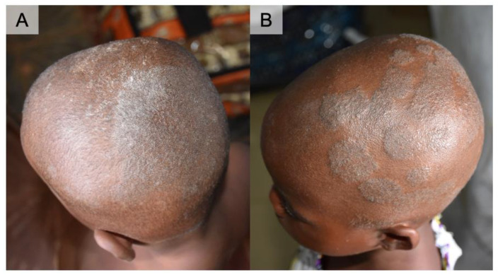
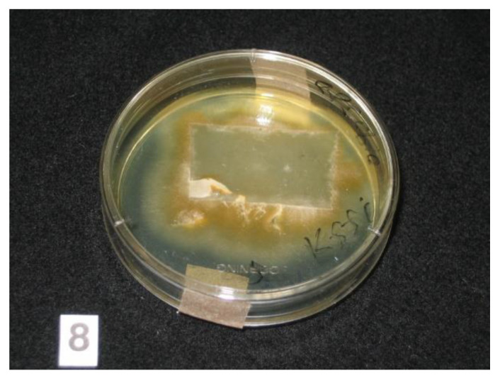
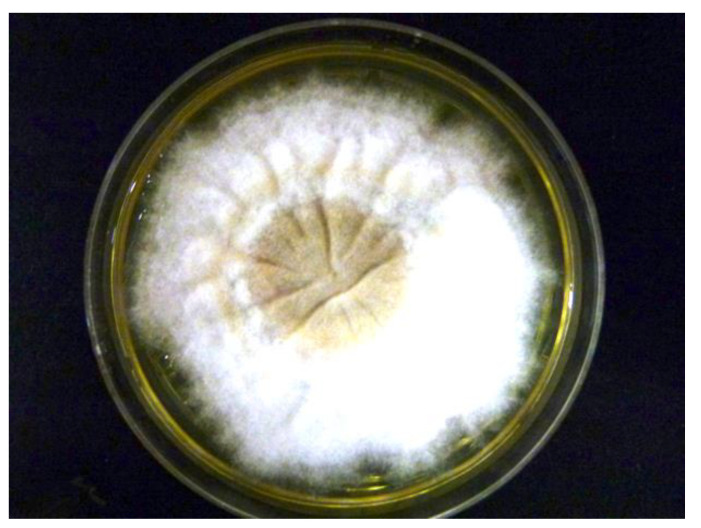
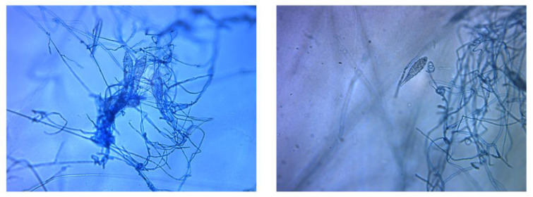
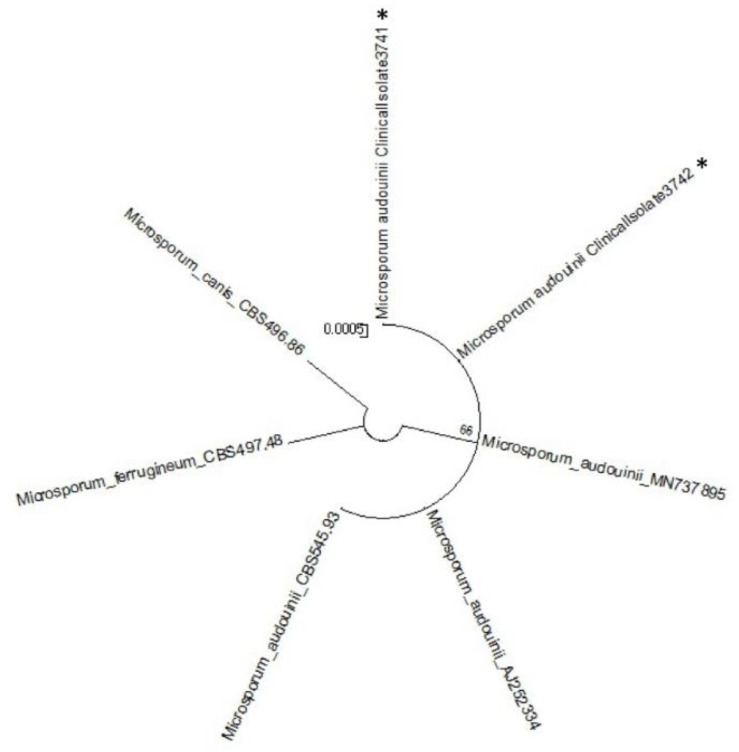
We report here two cases of tinea capitis caused by Microsporum (M.) audouinii in Côte d'Ivoire, West Africa. The patients were a three-year-old boy and a six-year-old girl who presented with scaly patches on the scalp. The causative fungus was isolated using an adhesive tape-sampling method and cultured on Sabouraud dextrose agar plates. It was identified as M. audouinii both by its macroscopic and microscopic features, confirmed by DNA sequencing. These are the first documented cases of M. audouinii infections confirmed with DNA sequencing to be reported from Côte d'Ivoire. The practicality of the tape-sampling method makes it possible to carry out epidemiological surveys evaluating the distribution of these dermatophytic infections in remote, resource-limited settings.
Keywords: Microsporum audouinii; dermatophyte; dermatophytosis; developing country; sub-Saharan Africa; tape sampling; tinea; tinea capitis.
Conflict of interest statement
The authors declare no conflict of interest.
Figures
References
-
- Hogewoning A.A., Adegnika A.A., Bouwes Bavinck J.N., Yazdanbakhsh M., Kremsner P.G., van der Raaij-Helmer E.M., Staats C.C.G., Willemze R., Lavrijsen A.P.M. Prevalence and causative fungal species of tinea capitis among schoolchildren in Gabon. Mycoses. 2011;54:e354–e359. doi: 10.1111/j.1439-0507.2010.01923.x. - DOI - PubMed
-
- World Health Organization . Epidemiology and Management of Common Skin Diseases in Children in Developing Countries. World Health Organization/Department of Child and Adolescent Health and Development; Geneva, Switzerland: 2005.
-
- Yotsu R.R., Comoé C.C., Ainyakou G.T., Konan N., Akpa A., Yao A., Aké J., Vagamon B., Abbet R.A., Bedimo R., et al. Impact of common skin diseases on children in rural Côte d’Ivoire with leprosy and Buruli ulcer co-endemicity: A mixed methods study. PLoS Negl. Trop. Dis. 2020;14:e0008291. doi: 10.1371/journal.pntd.0008291. - DOI - PMC - PubMed
Publication types
Grants and funding
LinkOut - more resources
Full Text Sources
Other Literature Sources
