. 2021 Jan 12;10(2):260.
doi: 10.3390/jcm10020260.

Uterine Fundus Remodeling after Hysteroscopic Metroplasty: A Prospective Pilot Study

Affiliations

Uterine Fundus Remodeling after Hysteroscopic Metroplasty: A Prospective Pilot Study

Paolo Casadio et al. J Clin Med. .

Abstract

The septate uterus is the most common congenital uterine malformation and is treated by hysteroscopic metroplasty. There are few studies on the fundal uterine changes that occur after surgery. We designed a pilot prospective observational study to evaluate by three-dimensional transvaginal ultrasound (3D-TVS) the changes not only of the internal fundal uterine profile, but also of the external one, after hysteroscopic metroplasty. Sixty women who underwent hysteroscopic metroplasty for partial or complete uterine septum (U2a and U2b subclasses of ESHRE/ESGE classification) were enrolled. We performed 3D-TVS after surgery confirming optimal removal of the septum. However, at ultrasound follow-up after three months, we observed a significant increase (p < 0.001) in the residual septum (Zr) (3.7 mm (95% CI: 3.1-4.4)), the myometrial wall thickness (Y) (2.5 mm (95% CI: 2.0-3.0)) and the total fundal wall thickness (Y + Zr) (6.2 mm (95% CI: 5.5-6.9)). Forty-three patients (72%) required a second step of hysteroscopic metroplasty. Moreover, the shape of uterine fundus changed in 58% of cases. We actually observed a remodeling of the uterine fundus with modifications of its external and internal profiles. Therefore, we propose to always perform a second ultrasound look at least three months after the metroplasty to identify cases that require a second- step metroplasty.

Keywords: hysteroscopy; remodeling; septate uterus; ultrasound; uterine fundus; uterine malformation.


Conflict of interest statement

The authors declare no conflict of interest.

Figures

Figure 1
Figure 1
Hysteroscopic view of a complete and partial uterine septum before and after hysteroscopic metroplasty. (A) Hysteroscopic view of a complete uterine septum at the beginning of surgery; (B) Hysteroscopic view of the previous uterine cavity at the end of the metroplasty; (C) Hysteroscopic view of a partial complete uterine septum at the beginning of surgery; (D) Hysteroscopic view of the previous uterine cavity at the end of the metroplasty.
Figure 2
Figure 2
Ultrasound images of a complete septate uterus (U2b according to ESHRE/ ESGE) before and after surgery. (A) Ultrasound midcoronal view of the complete septate uterus before hysteroscopic metroplasty with all the measurements of the septum (the uterine wall thickness (Y), the base of the septum (X) and the length of the septum (Z)); (B) Ultrasound midcoronal view of the same uterus three hours after surgery. The fundal uterine wall thickness is 1 cm, confirming the complete incision of the septum.
Figure 3
Figure 3
Ultrasound comparison between external and inner uterine profiles of septate uterus before surgery and three months after surgery. In each image, all the measurements of the septum are shown (the uterine wall thickness (Y), the base of the septum (X), the length of the septum before surgery (Z) and the length of the new inner uterine edge after surgery (Zr)). (A) Ultrasound midcoronal view of a partial septate uterus (U2a according to ESHRE/ ESGE) before surgery; (B) Ultrasound midocoronal image of the same uterus three months after surgery, characterized by the remodeling of the fundus; (C) Ultrasound midcoronal view of another partial septate uterus (U2a according to ESHRE/ ESGE) before surgery; (D) Ultrasound midcoronal image of the previous uterus three months after surgery, characterized by a residual partial septum. This patient was scheduled for a second- step metroplasty.
Figure 4
Figure 4
Distribution of absolute changes in Zr, Y and (Y + Zr) at three-month follow-up (n = 60).
Figure 5
Figure 5
The positive relationship between (Y + Zr) values, measured three months after hysteroscopic metroplasty, and Z (A) and (Y + Z) (B) values measured before surgery.
Figure 6
Figure 6
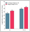
Mean Z and (Y + Z) values measured before surgery and their relationship with the change of the fundal uterine shape at three months after surgery.
Figure 7
Figure 7
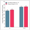
Mean Z and (Y + Z) values measured before surgery and their relationship with the change of the fundal uterine shape at three months after surgery in patients scheduled for second surgery (n = 43).

References

    1. Raga F., Bauset C., Remohi J., Bonilla-Musoles F., Simón C., Pellicer A. Reproductive impact of congenital Mullerian anomalies. Hum. Reprod. 1997;12:2277–2281. doi: 10.1093/humrep/12.10.2277. - DOI - PubMed
    1. Rikken J.F.W., Leeuwis-Fedorovich N.E., Letteboer S., Emanuel M.H., Limpens J., van der Veen F., Goddijn M., van Wely M. The pathophysiology of the septate uterus: A systematic review. BJOG Int. J. Obstet. Gynaecol. 2019;126:1192–1199. doi: 10.1111/1471-0528.15798. - DOI - PubMed
    1. Saravelos S.H., Cocksedge K.A., Li T.C. Prevalence and diagnosis of congenital uterine anomalies in women with reproductive failure: A critical appraisal. Hum. Reprod. Update. 2008;14:415–429. doi: 10.1093/humupd/dmn018. - DOI - PubMed
    1. Grimbizis G.F., Camus M., Tarlatzis B.C., Bontis J.N., Devroey P. Clinical implications of uterine malformations and hysteroscopic treatment results. Hum. Reprod. Update. 2001;7:161–174. doi: 10.1093/humupd/7.2.161. - DOI - PubMed
    1. Grimbizis G.F., Gordts S., Di Spiezio Sardo A., Brucker S., De Angelis C., Gergolet M., Li T.C., Tanos V., Brölmann H., Gianaroli L., et al. The ESHRE/ESGE consensus on the classification of female genital tract congenital anomalies. Hum. Reprod. 2013;28:2032–2044. doi: 10.1093/humrep/det098. - DOI - PMC - PubMed

LinkOut - more resources