Review
. 2021 Jan 12;22(2):690.
doi: 10.3390/ijms22020690.

Neurofibromatosis Type 2 (NF2) and the Implications for Vestibular Schwannoma and Meningioma Pathogenesis

Affiliations
Review

Neurofibromatosis Type 2 (NF2) and the Implications for Vestibular Schwannoma and Meningioma Pathogenesis

Suha Bachir et al. Int J Mol Sci. .

Abstract

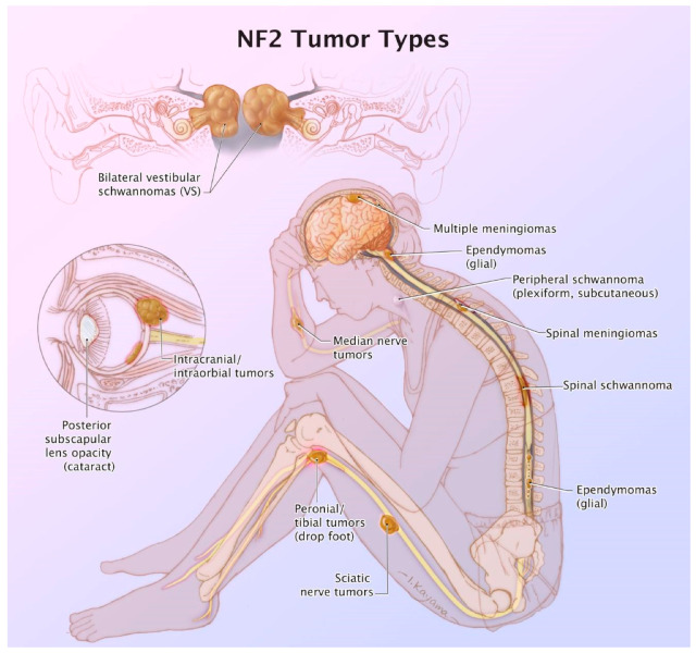
Patients diagnosed with neurofibromatosis type 2 (NF2) are extremely likely to develop meningiomas, in addition to vestibular schwannomas. Meningiomas are a common primary brain tumor; many NF2 patients suffer from multiple meningiomas. In NF2, patients have mutations in the NF2 gene, specifically with loss of function in a tumor-suppressor protein that has a number of synonymous names, including: Merlin, Neurofibromin 2, and schwannomin. Merlin is a 70 kDa protein that has 10 different isoforms. The Hippo Tumor Suppressor pathway is regulated upstream by Merlin. This pathway is critical in regulating cell proliferation and apoptosis, characteristics that are important for tumor progression. Mutations of the NF2 gene are strongly associated with NF2 diagnosis, leading to benign proliferative conditions such as vestibular schwannomas and meningiomas. Unfortunately, even though these tumors are benign, they are associated with significant morbidity and the potential for early mortality. In this review, we aim to encompass meningiomas and vestibular schwannomas as they pertain to NF2 by assessing molecular genetics, common tumor types, and tumor pathogenesis.

Keywords: meningiomas; neurofibromatosis type 2 (NF2); vestibular schwannomas.


Conflict of interest statement

Samy declares a conflict of interest due to research funding from Cochlear Corporation. The funders had no role in the design of the study; in the collection, analyses, or interpretation of data; in the writing of the manuscript, or in the decision to publish the results.

Figures

Figure 1
Figure 1
46-year-old woman with neurofibromatosis type 2 (NF2). Contrast-enhanced T1-weighted-Fat saturation MR images (A,F,I) show avidly enhancing schwannoma involving left cerebellopontine angle and internal auditory canal (black asterisk in (A)) and avidly enhancing meningiomas in the right anterior temporal convexity, sphenoid wing, and left lateral posterior fossa (white asterisks in (A)). Post-operative changes from right suboccipital craniotomy and mastoidectomy with residual meningioma (white arrows in (A)). Avidly enhancing dumbbell-shaped right C5 intradural schwannoma extending through right C5 foramen (B,C). Large avidly enhancing right paraspinal T10 schwannoma extending through right T10 foramen (D) with associated heterogenous T2 hyperintensity on axial T2-weighted image (G). Avidly enhancing intramedullary C6 ependymoma (E,F). Numerous multilevel tiny enhancing rounded nodules along the cauda equina (H,I) consistent with schwannomas which demonstrate T2 hypointensity on axial and sagittal T2-weighted images (white arrows in (J,K)).
Figure 2
Figure 2
Immunohistochemistry of an atypical meningioma (WHO Grade II) from an NF2 patient. Left panel: 40× magnification, MIB-1(Ki67) nuclear immunostain shows an elevated index (25%). Right panel: 40× H&E shows increased cellularity, prominent nucleoli, mitoses, and necrosis (as represented in the bottom right hand section and highlighted by the arrow in bold).
Figure 3
Figure 3
12-year-old boy with NF2. Contrast-enhanced T1-weighted-Fat saturation MR images (B,F) show avidly enhancing masses involving cerebellopontine angles and internal auditory canals (black asterisks in (B)) with associated T2 hypointensity on axial high resolution T2 FIESTA (white asterisks in (A)), consistent with bilateral vestibular schwannomas. Also noted are asymmetric enhancement and enlargement of right geniculate ganglion of right CN7 (white arrow in (B)) and right CN3 (black arrow in (C)) consistent with additional schwannomas. Large third ventricular meningioma with obstructing hydrocephalus (D). Additional left parafalcine and left sphenoid wing meningiomas are noted (arrowheads in (E,F)).
Figure 4
Figure 4
Summary figure of the various morbidities associated with NF2. An original illustration by Ikumi Kayama.

Similar articles

Cited by

References

    1. Evans D.G.R. Neurofibromatosis Type 2 (NF2): A Clinical and Molecular Review. Orphanet J. Rare Dis. 2009;4:16. doi: 10.1186/1750-1172-4-16. - DOI - PMC - PubMed
    1. Ruggieri M., Praticò A.D., Evans D.G. Diagnosis, Management, and New Therapeutic Options in Childhood Neurofibromatosis Type 2 and Related Forms. Semin. Pediatr. Neurol. 2015;22:240–258. doi: 10.1016/j.spen.2015.10.008. - DOI - PubMed
    1. Pećina-Šlaus N. Merlin, the NF2 gene product. Pathol. Oncol. Res. 2013;19:365–373. doi: 10.1007/s12253-013-9644-y. - DOI - PubMed
    1. Kresak J.L., Walsh M. Neurofibromatosis: A Review of NF1, NF2, and Schwannomatosis. J. Pediatr. Genet. 2016;5:98–104. doi: 10.1055/s-0036-1579766. - DOI - PMC - PubMed
    1. Tiwari R., Singh A.K. StatPearls. StatPearls Publishing; Treasure Island, FL, USA: 2020. Neurofibromatosis Type 2.