Review
. 2021 Jan 12;22(2):705.
doi: 10.3390/ijms22020705.

Bone Marrow Failure Syndromes, Overlapping Diseases with a Common Cytokine Signature

Affiliations
Review

Bone Marrow Failure Syndromes, Overlapping Diseases with a Common Cytokine Signature

Valentina Giudice et al. Int J Mol Sci. .

Abstract

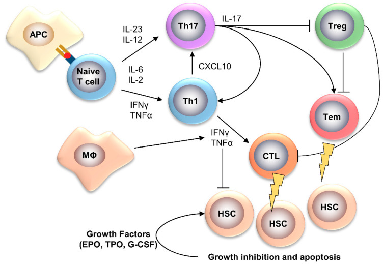
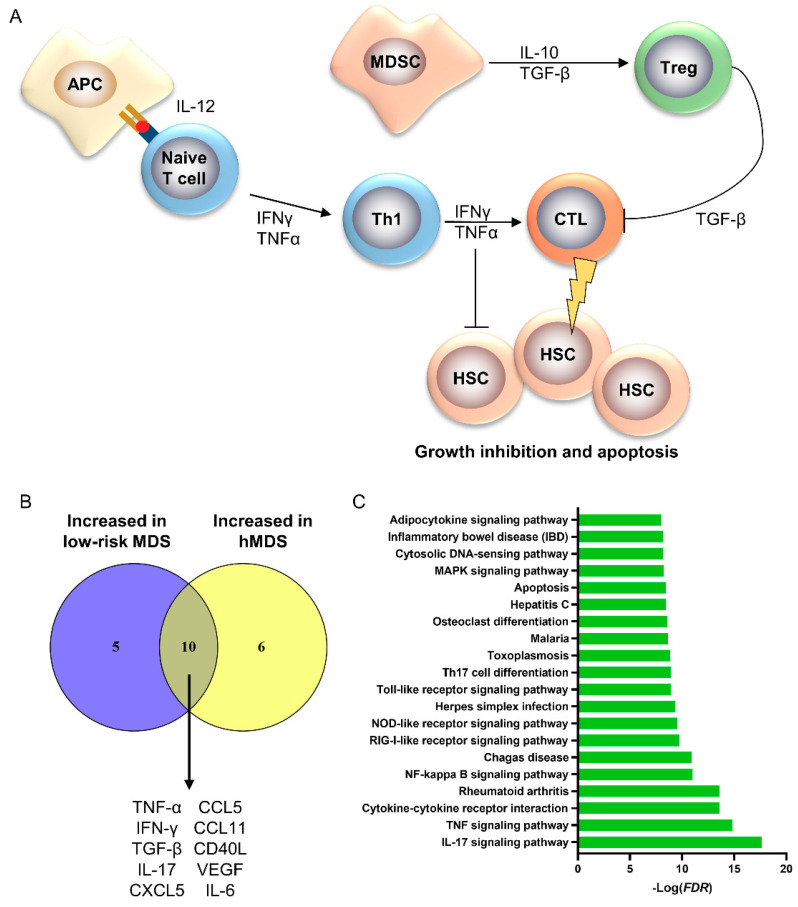
Bone marrow failure (BMF) syndromes are a heterogenous group of non-malignant hematologic diseases characterized by single- or multi-lineage cytopenia(s) with either inherited or acquired pathogenesis. Aberrant T or B cells or innate immune responses are variously involved in the pathophysiology of BMF, and hematological improvement after standard immunosuppressive or anti-complement therapies is the main indirect evidence of the central role of the immune system in BMF development. As part of this immune derangement, pro-inflammatory cytokines play an important role in shaping the immune responses and in sustaining inflammation during marrow failure. In this review, we summarize current knowledge of cytokine signatures in BMF syndromes.

Keywords: aplastic anemia; bone marrow failure syndromes; cytokines; myelodysplastic syndromes.


Conflict of interest statement

The authors declare no conflict of interest.

Figures

Figure 1
Figure 1
Bone marrow failure (BMF) syndromes’ pathophysiology. BMF syndromes are characterized by empty bone marrow and peripheral blood cytopenia(s), and can be divided in congenital disorders, iatrogenic aplastic anemia (AA), and immune-mediated BMF. In congenital disorders, such as Shwachman–Diamond syndrome (SDS) or Fanconi anemia, hematopoietic stem cells (HSCs) harbor mutations in genes important for normal hemopoiesis, which becomes insufficient over time in maintaining normal ranges of circulating cells. In iatrogenic AA, HSCs are directly damaged by external stressors, such as chemicals and radiation. In immune-mediated BMF, dysregulated immune responses can cause an autologous immune attack of cytotoxic T lymphocytes (CTLs) against HSCs or can suppress hemopoiesis through changes in BM microenvironment. Clinical presentation of BMF syndromes differs among diseases; however, immunosuppressive therapies (ISTs) can often restore bone marrow cellularity, which is one of the major pieces of evidence for the immune-mediated pathogenesis in BMF. Abbreviations. PNH, paroxysmal nocturnal hemoglobinuria; LGL, T-large granular lymphocyte leukemia; AA, acquired aplastic anemia; MDS, myelodysplastic syndromes; AML, acute myeloid leukemia.
Figure 2
Figure 2
Pathophysiology of AA. In AA, an unknown antigen might trigger a T helper (Th)1 immune response, causing the expansion of cytotoxic T lymphocytes (CTLs), which directly kill hematopoietic stem cells (HSCs) [2,6,7,8]. Chronic antigen exposure might also polarize naïve T cells toward the Th17 phenotype, leading to expansion of effector memory T cells (Tem) with cytotoxic activities and inhibition of T regulatory cells (Treg) [2]. Several interleukins (ILs) and cytokines, such as interferon (IFN)-gamma and tumor necrosis factor (TNF)-alpha, are involved in immune response polarization and direct growth inhibition of HSCs, which leads to increased levels of growth factors for maintaining normal hemopoiesis [8]. APC, antigen-presenting cells; MΦ, macrophage; CXCL, C-X-C motif chemokine; G-CSF, granulocyte colony-stimulating factor; TPO, thrombopoietin; EPO, erythropoietin.
Figure 3
Figure 3
Pathophysiology of hypoplastic myelodysplastic syndrome (hMDS). (A) In hMDS, naïve T cells differentiate into Th1 cells, causing CTL activation, which directly kills HSCs [61]. Myeloid derived suppressor cells (MDSC) can also induce expansion of Treg, especially in high-risk MDS. IFN-γ, TNF-α, and tumor growth factor-beta (TGF-β) drive in immune response polarization and direct growth inhibition of HSCs [66,70,71,72,76]. (B) Reported cytokines increased in low-risk MDS and hMDS were interpolated using Venn’s diagram, and shared cytokines (n = 10) were used for protein pathway analysis to identify common pathways in BMF disease pathophysiology. (C) The top 20 related pathways are reported.
Figure 4
Figure 4
Pathophysiology of T-LGL leukemia. In T-LGL leukemia, an unknown chronic antigen causes the clonal drift of a T cell clonotype [88,91]. Acquisition of somatic mutations in STAT3 and overexpression of IL-15 and/or PDGF drive the monoclonal expansion of the leukemic clone [94,97]. Alterations in apoptosis pathways, such as inhibition of Fas-mediated apoptosis through binding of soluble Fas-ligand (sFas-L), also favor survival of the T-LGL clone [88,98,99,100].

References

    1. Rodgers G.P., Young N.S. The Bethesda Handbook of Clinical Hematology (English Edition) 4th ed. Lippincott Williams & Wilkins; Philadelphia, PA, USA: 2013.
    1. Young N.S. Aplastic Anemia. N. Engl. J. Med. 2018;379:1643–1656. doi: 10.1056/NEJMra1413485. - DOI - PMC - PubMed
    1. Young N.S., Calado R.T., Scheinberg P. Current concepts in the pathophysiology and treatment of aplastic anemia. Blood. 2006;108:2509–2519. doi: 10.1182/blood-2006-03-010777. - DOI - PMC - PubMed
    1. Young N.S. Current concepts in the pathophysiology and treatment of aplastic anemia. Hematol. Am. Soc. Hematol. Educ. Program. 2013;2013:76–81. doi: 10.1182/asheducation-2013.1.76. - DOI - PMC - PubMed
    1. Park M., Park C.J., Cho Y.W., Jang S., Lee J.H., Lee J.H., Lee K.H., Lee Y.H. Alterations in the bone marrow microenvironment may elicit defective hematopoiesis: A comparison of aplastic anemia, chronic myeloid leukemia, and normal bone marrow. Exp. Hematol. 2017;45:56–63. doi: 10.1016/j.exphem.2016.09.009. - DOI - PubMed

MeSH terms

LinkOut - more resources