

Predictive Model of Nail Consistency Using Scanning Electron Microscopy with Energy-Dispersive X-Ray
- PMID: 33445794
- PMCID: PMC7828269
- DOI: 10.3390/biology10010053
Predictive Model of Nail Consistency Using Scanning Electron Microscopy with Energy-Dispersive X-Ray
Abstract
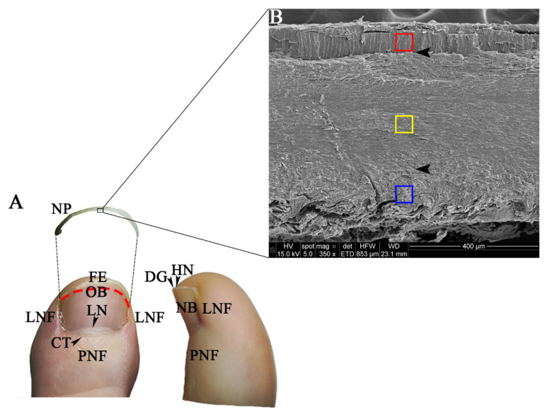
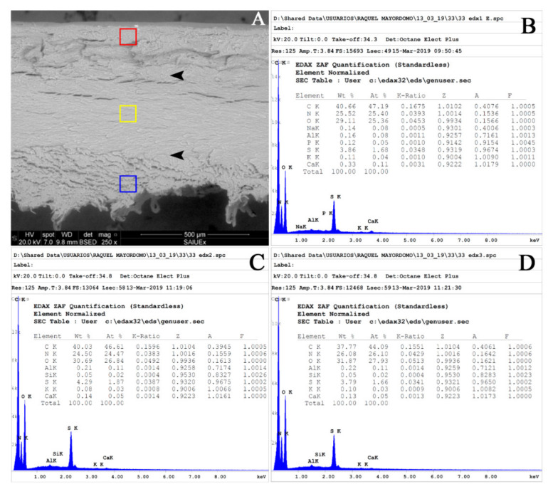
The nail plate is made up of tightly packed keratin-rich cells. Factors such as the special distribution of the intermediate filaments in each layer (dorsal, intermediate, and ventral), the relative thickness of the layers, and their chemical composition define the characteristics of each nail. The main objective of this study is to determine nail consistency by calculating a predictive model based on elemental composition analysis using scanning electron microscopy. Nail consistency was determined in 57 participants (29 women and 28 men) in two age groups (young people and adults). Elemental composition was analysed in each layer using scanning SEM-EDS, and nail plate thickness was measured by image analysis. A total of 12 elements were detected in nail plates, of which carbon, nitrogen, phosphorus, sulphur, and calcium showed significant differences between layers (p-values ≤ 0.01). The level of calcium in the dorsal layer was the main predictive variable in calculating the predictive model of consistency, with 75.4% correctly classified cases. Elemental analysis in each layer of the nail plate by SEM-EDS can be used to develop a predictive model of nail consistency that will help health professionals to objectively determine nail consistency.
Keywords: SEM-EDS; binary logistic regression; nail apparatus; nail consistency; nail plate; predictive model; thickness.
Conflict of interest statement
The authors declare no conflict of interest. The funders had no role in the design of the study; in the collection, analyses, or interpretation of data; in the writing of the manuscript, or in the decision to publish the results.
Figures
Similar articles
-
Drug permeation through the three layers of the human nail plate.J Pharm Pharmacol. 1999 Mar;51(3):271-8. doi: 10.1211/0022357991772448. J Pharm Pharmacol. 1999. PMID: 10344627
-
Application of scanning electron microscopy and X-ray microanalysis: FE-SEM, ESEM-EDS, and EDS mapping for studying the characteristics of topographical microstructure and elemental mapping of human cardiac calcified deposition.Anal Bioanal Chem. 2014 Jan;406(1):359-66. doi: 10.1007/s00216-013-7414-z. Epub 2013 Nov 8. Anal Bioanal Chem. 2014. PMID: 24202192
-
Histological structure of human nail as studied by synchrotron X-ray microdiffraction.Cell Mol Biol (Noisy-le-grand). 2000 Sep;46(6):1025-34. Cell Mol Biol (Noisy-le-grand). 2000. PMID: 10976860
-
Ultrastructure and chemical composition of the proboscis hooks of Acanthocephalus lucii (Müller, 1776) (Acanthocephala: Palaeacanthocephala) using X-ray elemental analysis.Folia Parasitol (Praha). 2014 Dec;61(6):549-57. Folia Parasitol (Praha). 2014. PMID: 25651697
-
Performing elemental microanalysis with high accuracy and high precision by scanning electron microscopy/silicon drift detector energy-dispersive X-ray spectrometry (SEM/SDD-EDS).J Mater Sci. 2015;50(2):493-518. doi: 10.1007/s10853-014-8685-2. Epub 2014 Nov 12. J Mater Sci. 2015. PMID: 26346887 Free PMC article. Review.
Cited by
-
Trace Element Distribution and Arsenic Speciation in Toenails as Affected by External Contamination and Evaluation of a Cleaning Protocol.Anal Chem. 2024 Mar 12;96(10):4039-4047. doi: 10.1021/acs.analchem.3c03962. Epub 2024 Feb 29. Anal Chem. 2024. PMID: 38422552 Free PMC article.
References
-
- Fleckman P., Allan C. Surgical anatomy of the nail unit. Dermatol. Surg. 2001;27:257–260. - PubMed
Grants and funding
LinkOut - more resources
Full Text Sources
Other Literature Sources
