. 2021 Jan 12;22(2):709.
doi: 10.3390/ijms22020709.

Delphinidin Increases the Sensitivity of Ovarian Cancer Cell Lines to 3-Bromopyruvate

Affiliations

Delphinidin Increases the Sensitivity of Ovarian Cancer Cell Lines to 3-Bromopyruvate

Natalia Pieńkowska et al. Int J Mol Sci. .

Abstract

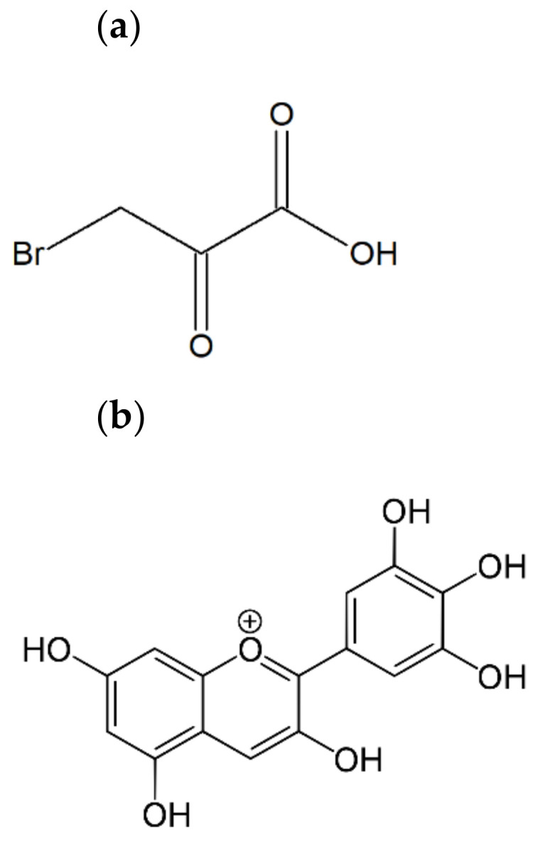
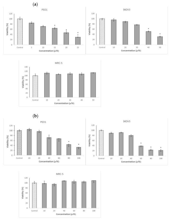
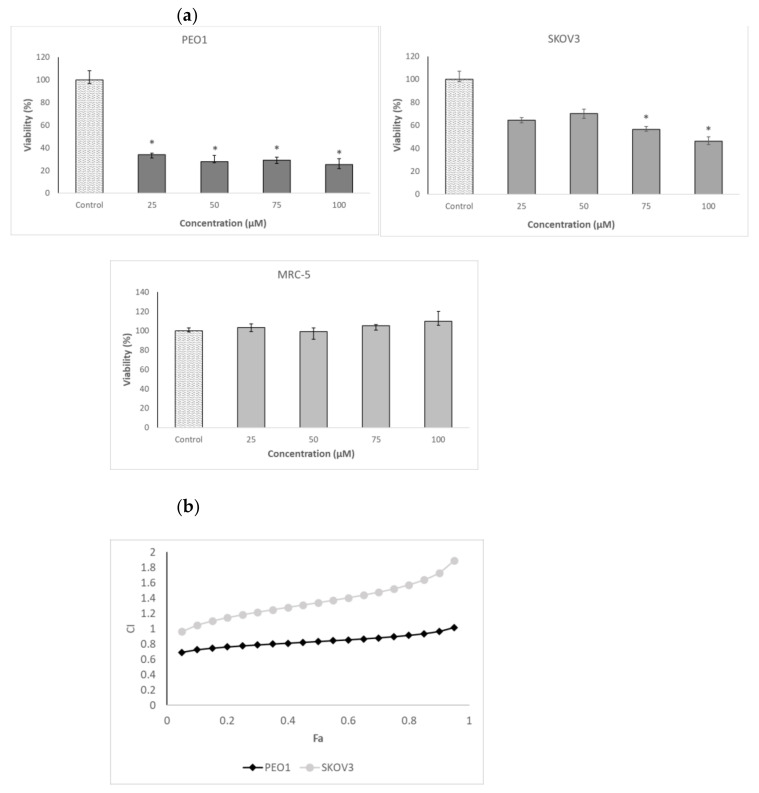
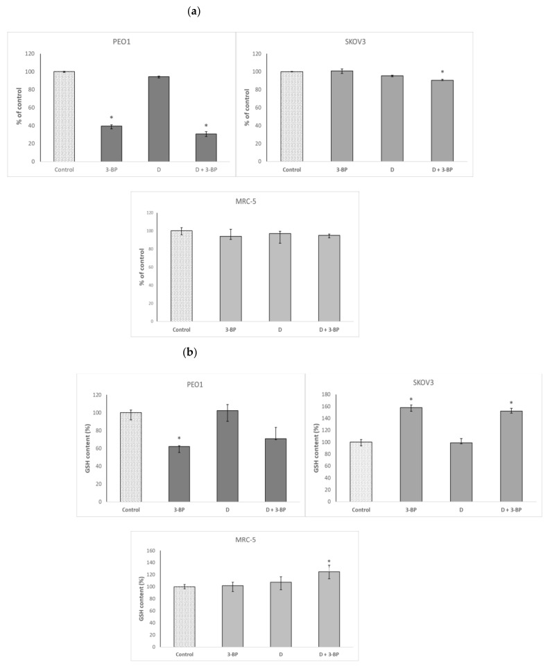
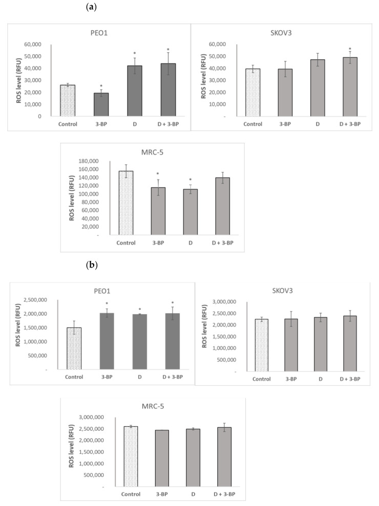
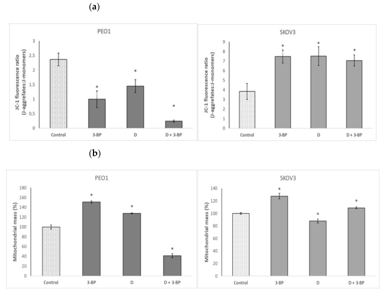
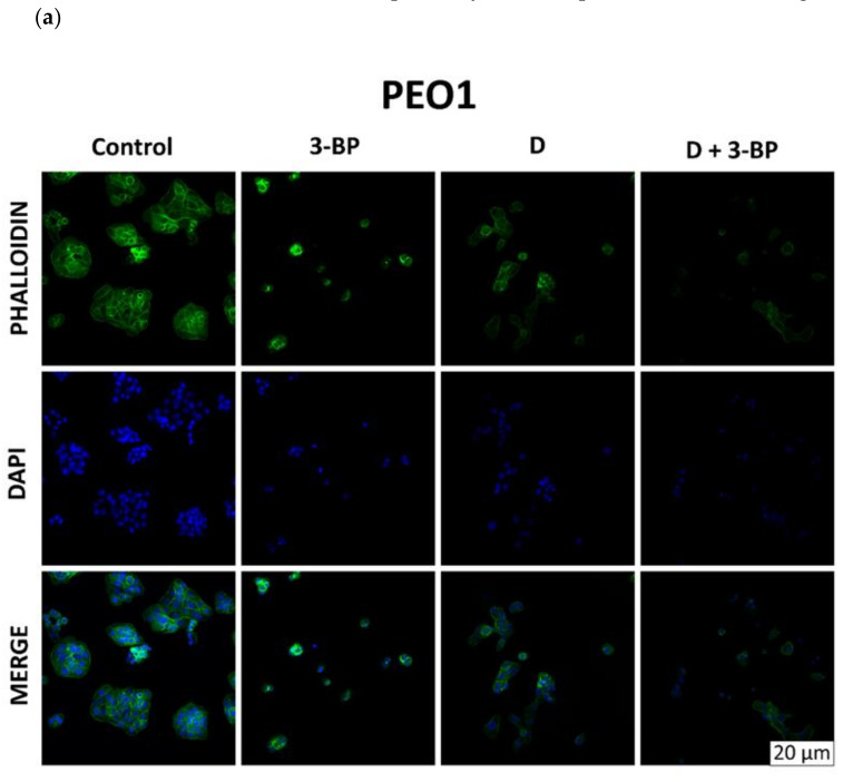
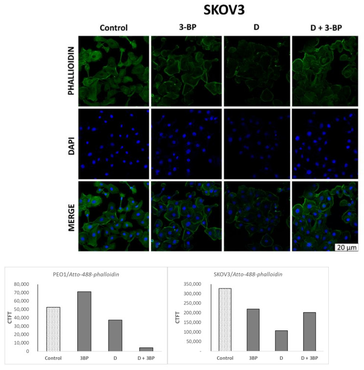
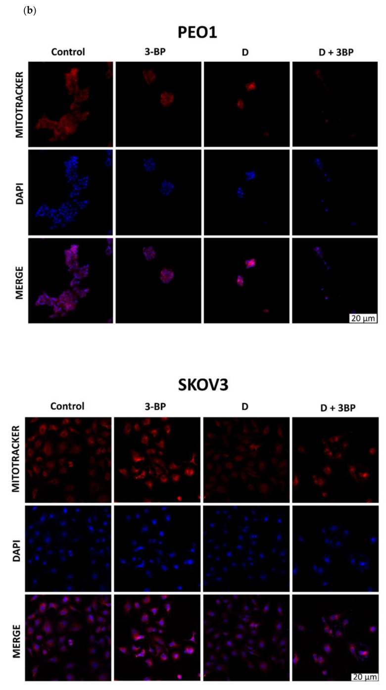
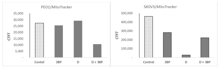
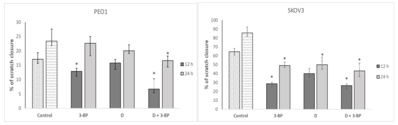
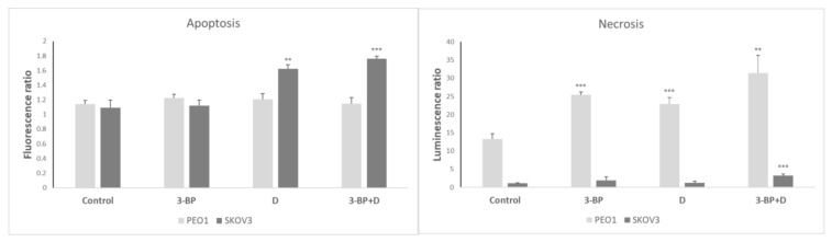
3-Bromopyruvic acid (3-BP) is a promising anticancer compound. Two ovary cancer (OC) cell lines, PEO1 and SKOV3, showed relatively high sensitivity to 3-BP (half maximal inhibitory concentration (IC50) of 18.7 and 40.5 µM, respectively). However, the further sensitization of OC cells to 3-BP would be desirable. Delphinidin (D) has been reported to be cytotoxic for cancer cell lines. We found that D was the most toxic for PEO1 and SKOV3 cells from among several flavonoids tested. The combined action of 3-BP and D was mostly synergistic in PEO1 cells and mostly weakly antagonistic in SKOV3 cells. The viability of MRC-5 fibroblasts was not affected by both compounds at concentrations of up to 100 µM. The combined action of 3-BP and D decreased the level of ATP and of dihydroethidium (DHE)-detectable reactive oxygen species (ROS), cellular mobility and cell staining with phalloidin and Mitotracker Red in both cell lines but increased the 2',7'-dichlorofluorescein (DCFDA)-detectable ROS level and decreased the mitochondrial membrane potential and mitochondrial mass only in PEO1 cells. The glutathione level was increased by 3-BP+D only in SKOV3 cells. These differences may contribute to the lower sensitivity of SKOV3 cells to 3-BP+D. Our results point to the possibility of sensitization of at least some OC cells to 3-BP by D.

Keywords: 3-bromopyruvate; anthocyanidins; delphinidin; glycolytic inhibitors; ovarian cancer.


Conflict of interest statement

The authors declare no conflict of interest.

Figures

Figure 1
Figure 1
The chemical structure of 3-bromopyruvic acid (a) and delphinidin (3,3′,4′,5,5′,7-hexahydroxyflavylium) (b).
Figure 2
Figure 2
Effect of 3-bromopyruvic acid (3-BP) (a) and delphinidin (D) (b) on the survival of PEO1, SKOV3 and MRC-5 cells estimated by the Neutral Red assay. The cells were treated with D in the concentration range of 10–100 μM and 3-BP at concentrations of 5–25 μM for PEO1 and 10–50 μM for MRC-5, as well as SKOV3. Nontreated cell were used as a control. * p < 0.05 with respect to the control (Kruskal-Wallis test; n ≥ 6).
Figure 3
Figure 3
Effect of the combined action of 3-BP and D on the viability of the PEO1, SKOV3 and MRC-5 cells. Survival of the cells treated with 3-BP at a concentration corresponding to ~half maximal inhibitory concentration (IC50) (SKOV3 and MRC-5: 40 μM and PEO1: 20 μM) and various concentrations of D indicated on the axis of the abscissae (a). Plot of the combination index (CI) as a function of the effect level (Fa) for the combined action of 3-BP and D. Concentrations of 3-BP and D: 20, 40, 60 and 80 μM (b). * p < 0.05 with respect to the control (Kruskal-Wallis test; n ≥ 6).
Figure 4
Figure 4
Effect of 3-BP and delphinidin (D) on the ATP (a) and GSH content (b) of PEO1, as well as SKOV3 and MRC-5 cells. *p < 0.05 with respect to the control (Kruskal-Wallis test; n ≥ 6).
Figure 5
Figure 5
Effect of 3-BP and delphinidin (D) on the level of reactive oxygen species (ROS) estimated with dihydroethidium (DHE, (a) and 2’,7’-dihydrodichlorofluorescein diacetate (DCFDA, (b). * p < 0.05 with respect to the control (one-way ANOVA and Least Significant Difference (LSD) post-hoc test; n ≥ 3).
Figure 6
Figure 6
Effect of 3-BP and delphinidin (D) on mitochondrial staining with the JC-1 probe (a) and mitochondrial mass (b) of PEO1 and SCOV3 cells. * p < 0.05 with respect to the control (one-way ANOVA and LSD post-hoc test; n ≥ 3).
Figure 7
Figure 7
Morphological changes in PEO1 and SKOV3 cells (confocal images, 65×). Effect of 3-BP, D and D plus 3-BP on the staining of PEO1 and SKOV3 cells with phalloidin (a) and Mitotracker Red (b).
Figure 7
Figure 7
Morphological changes in PEO1 and SKOV3 cells (confocal images, 65×). Effect of 3-BP, D and D plus 3-BP on the staining of PEO1 and SKOV3 cells with phalloidin (a) and Mitotracker Red (b).
Figure 7
Figure 7
Morphological changes in PEO1 and SKOV3 cells (confocal images, 65×). Effect of 3-BP, D and D plus 3-BP on the staining of PEO1 and SKOV3 cells with phalloidin (a) and Mitotracker Red (b).
Figure 7
Figure 7
Morphological changes in PEO1 and SKOV3 cells (confocal images, 65×). Effect of 3-BP, D and D plus 3-BP on the staining of PEO1 and SKOV3 cells with phalloidin (a) and Mitotracker Red (b).
Figure 8
Figure 8
Effect of 3-BP and delphinidin (D) on the migration of PEO1 and SCOV3 cells. * p < 0.05 with respect to the control (Kruskal-Wallis test; n = 12).
Figure 9
Figure 9
The induction of apoptosis and necrosis by 3-BP, D and 3-BP+D in PEO1 and SKOV3 cells (after 24 h). Fluorescence and luminescence, respectively, of control cells at 0 time assumed as 1. ** p < 0.01 and *** p < 0.001 with respect to the control (one-way ANOVA and LSD post-hoc test; n = 3).

References

    1. Boussios S., Karihtala P., Moschetta M., Abson C., Karathanasi A., Zakynthinakis-Kyriakou N., Ryan J.E., Sheriff M., Rassy E., Pavlidis N. Veliparib in ovarian cancer, a new synthetically lethal therapeutic approach. Investig. New Drugs. 2020;38:181–193. doi: 10.1007/s10637-019-00867-4. - DOI - PubMed
    1. Ren F., Shen J., Shi H., Hornicek F.J., Kan Q., Duan Z. Novel mechanisms and approaches to overcome multidrug resistance in the treatment of ovarian cancer. Biochim. Biophys. Acta. 2016;1866:266–275. doi: 10.1016/j.bbcan.2016.10.001. - DOI - PubMed
    1. Gibson S.J., Fleming G.F., Temkin S.M., Chase D.M. The Application and Outcome of Standard of Care Treatment in Elderly Women with Ovarian Cancer, A Literature Review over the Last 10 Years. Front. Oncol. 2016;6:63. doi: 10.3389/fonc.2016.00063. - DOI - PMC - PubMed
    1. Webber K., Friedlander M. Chemotherapy for epithelial ovarian; fallopian tube and primary peritoneal cancer. Best Pract. Res. Clin. Obstet. Gynaecol. 2017;41:126–138. doi: 10.1016/j.bpobgyn.2016.11.004. - DOI - PubMed
    1. Mirandola L., Cannon M.J., Cobos E., Bernardini G., Jenkins M.R., Kast W.M., Chiriva-Internati M. Cancer testis antigens, novel biomarkers and targetable proteins for ovarian cancer. Int. Rev. Immunol. 2011;30:127–137. doi: 10.3109/08830185.2011.572504. - DOI - PubMed

MeSH terms

LinkOut - more resources