. 2021 Jan 14;22(1):80.
doi: 10.1186/s12891-021-03956-9.

Short-term effectiveness of precise safety decompression via double percutaneous lumbar foraminoplasty and percutaneous endoscopic lumbar decompression for lateral lumbar spinal canal stenosis: a prospective cohort study

Affiliations

Short-term effectiveness of precise safety decompression via double percutaneous lumbar foraminoplasty and percutaneous endoscopic lumbar decompression for lateral lumbar spinal canal stenosis: a prospective cohort study

Yu Wang et al. BMC Musculoskelet Disord. .

Abstract

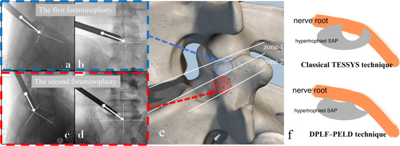
Purpose: This prospective cohort study reports on a modified technique, namely precise safety decompression via double percutaneous lumbar foraminoplasty (DPLF) and percutaneous endoscopic lumbar decompression (PELD) for lateral lumbar spinal canal (LLSC) stenosis, and its short-term clinical outcomes.

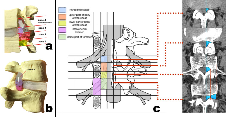
Methods: The study analyzed 69 patients with single-level LLSC stenosis simultaneously occurring in both zones 1 and 2 (defined as retrodiscal space and upper bony lateral recess respectively by new LLSC classification) who underwent DPLF-PELD from November 2018 to April 2019. Clinical outcomes were evaluated according to preoperative, 3 months postoperatively, and last follow-up, via leg pain/low back pain (LBP) visual analog scale (VAS) scores, Oswestry disability index (ODI) scores, and the Macnab criteria. The postoperative MRI and CT were used to confirm the complete decompression, and flexion-extension x-rays at the last follow-up were used to observe lumbar stability.

Results: All patients successfully underwent DPLF-PELD, and the stenosis was completely decompressed, confirmed by postoperative MRI and CT. The mean follow-up duration was 13 months (range: 8-17 months). The mean preoperative leg pain VAS score is 7.05 ± 1.04 (range 5-9), which decreased to 1.03 ± 0.79(range: 0-3) at 3 months postoperatively and to 0.75 ± 0.63 (range: 0-2) by the last follow-up visit (p < 0.05). The mean preoperative ODI was 69.8 ± 9.05 (range: 52-85), which decreased to 20.3 ± 5.52 (range: 10-35) at the third month postoperatively and to 19.6 ± 5.21 (range: 10-34) by the final follow-up visit (p < 0.05). The satisfactory (excellent or good) results were 94.2%. There was one patient with aggravated symptoms, which were relieved after an open surgery. Two patients had a dural tear, and two patients suffered postoperative LBP. No recurrence or segmental instability was observed at the final follow-up.

Conclusion: DPLF-PELD could be a good alternative for the treatment of LLSC stenosis patients whose stenosis occurred in both zones 1 and 2.

Trial registration: Chinese Clinical Trial Registry ( ChiCTR1800019551 ). Registered 18 November 2018.

Keywords: Double percutaneous lumbar foraminoplasty; Lumbar lateral spinal canal; Percutaneous endoscopic lumbar decompression.


Conflict of interest statement

All authors declare no conflict of interest.

Figures

Fig. 1
Fig. 1
Five zones of the lateral lumbar spinal canal (LLSC) divided by accurate boundaries. a,b Different zones shown in an artificial model in both medial and lateral views. c schematic diagram of the five zones. The boundaries of each zone is described in the text. The right four axial CT scanning image shows the different views of cross-sections (the red solid lines) and the labeled regions correspond to each zone
Fig. 2
Fig. 2
The specially designed depth-limited trephine
Fig. 3
Fig. 3
a, b Schematic diagram of the inclination of the trephine trajectory in lateral and AP (anterior-posterior) views in the first foraminoplasty. c, d Schematic diagram of the inclination of the trephine trajectory in lateral and AP views in the second foraminoplasty. The cross point of the white solid line is described in the text. e schematic diagram of the two-time percutaneous lumbar foraminoplasty procedure. f schematic diagram of the difference between classical TESSYS technique and double percutaneous lumbar foraminoplasty and PELD (DPLF–PELD) technique
Fig. 4
Fig. 4
The patient complained of severe left radicular pain for 12 months. He could not walk for 3 months due to severe left buttock and leg pain. Left L4/5 LLSC stenosis in both zones 1 and 2 was confirmed. We confirmed the totally decompression by postoperative CT and MRI. The leg pain was relieved immediately after the operation. No lumbar instability was indicated in the final follow-up. a, b Preoperative X-ray in AP and lateral view. c Preoperative sagittal CT scans indicated stenosis of the retrodiscal space (zone 1) on the left at L4/5 (red circle). d Preoperative axial CT scans indicated upper bony lateral recess (zone 2) stenosis on the left at L4/5 (red circle). e, f Preoperative sagittal and axial T2-weighted MRI scans showing the L4/5 left zone 1 stenosis caused by a lumbar disk bulge anteriorly, and hypertrophied, curled ligamentum flavum posteriorly (red circle). g, h Fluoroscopy during surgery demonstrates the trajectory of the trephine in the first foraminoplasty in the AP and lateral views. i, j Fluoroscopy during the operation shows the trajectory of the trephine in the second foraminoplasty in the AP and lateral views. k, l Three months postoperative sagittal and axial CT bony-window scans clearly demonstrate complete decompression of zones 1 and 2. m, n Three months postoperative sagittal and axial T2-weighted MRI scans indicate that the nerve root was decompressed without recurrence. o, p Postoperative flexion-extension x-rays at the final follow-up confirm that no lumbar instability occurred

Similar articles

Cited by

References

    1. Lurie J, Tomkins-Lane C. Management of lumbar spinal stenosis. BMJ. 2016;352:h6234. doi: 10.1136/bmj.h6234. - DOI - PMC - PubMed
    1. Inoue G, Miyagi M, Takaso M. Surgical and nonsurgical treatments for lumbar spinal stenosis. Eur J Orthop Surg Traumatol. 2016;26(7):695–704. doi: 10.1007/s00590-016-1818-3. - DOI - PubMed
    1. Ahn Y. Percutaneous endoscopic decompression for lumbar spinal stenosis. Expert Rev Med Devices. 2014;11(6):605–616. doi: 10.1586/17434440.2014.940314. - DOI - PubMed
    1. Issack PS, Cunningham ME, Pumberger M, Hughes AP, Cammisa FP., Jr Degenerative lumbar spinal stenosis: evaluation and management. J Am Acad Orthop Surg. 2012;20(8):527–535. doi: 10.5435/JAAOS-20-08-527. - DOI - PubMed
    1. Lee CK, Rauschning W, Glenn W. Lateral lumbar spinal canal stenosis: classification, pathologic anatomy and surgical decompression. Spine (Phila Pa 1976) 1988;13(3):313–320. doi: 10.1097/00007632-198803000-00015. - DOI - PubMed