. 2021 Jan 14;4(3):e202000809.
doi: 10.26508/lsa.202000809. Print 2021 Mar.

Reduced RNA turnover as a driver of cellular senescence

Affiliations

Reduced RNA turnover as a driver of cellular senescence

Nowsheen Mullani et al. Life Sci Alliance. .

Abstract

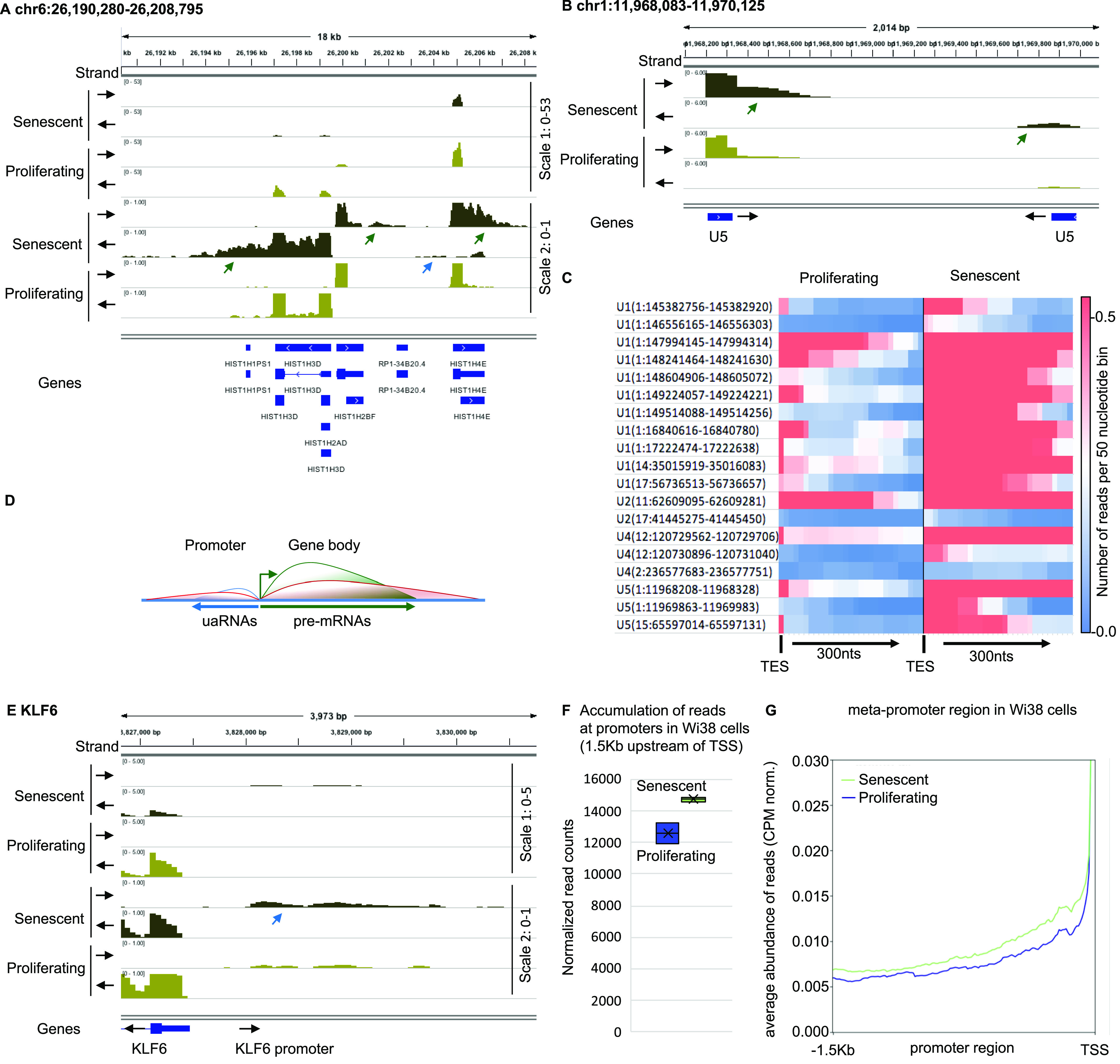
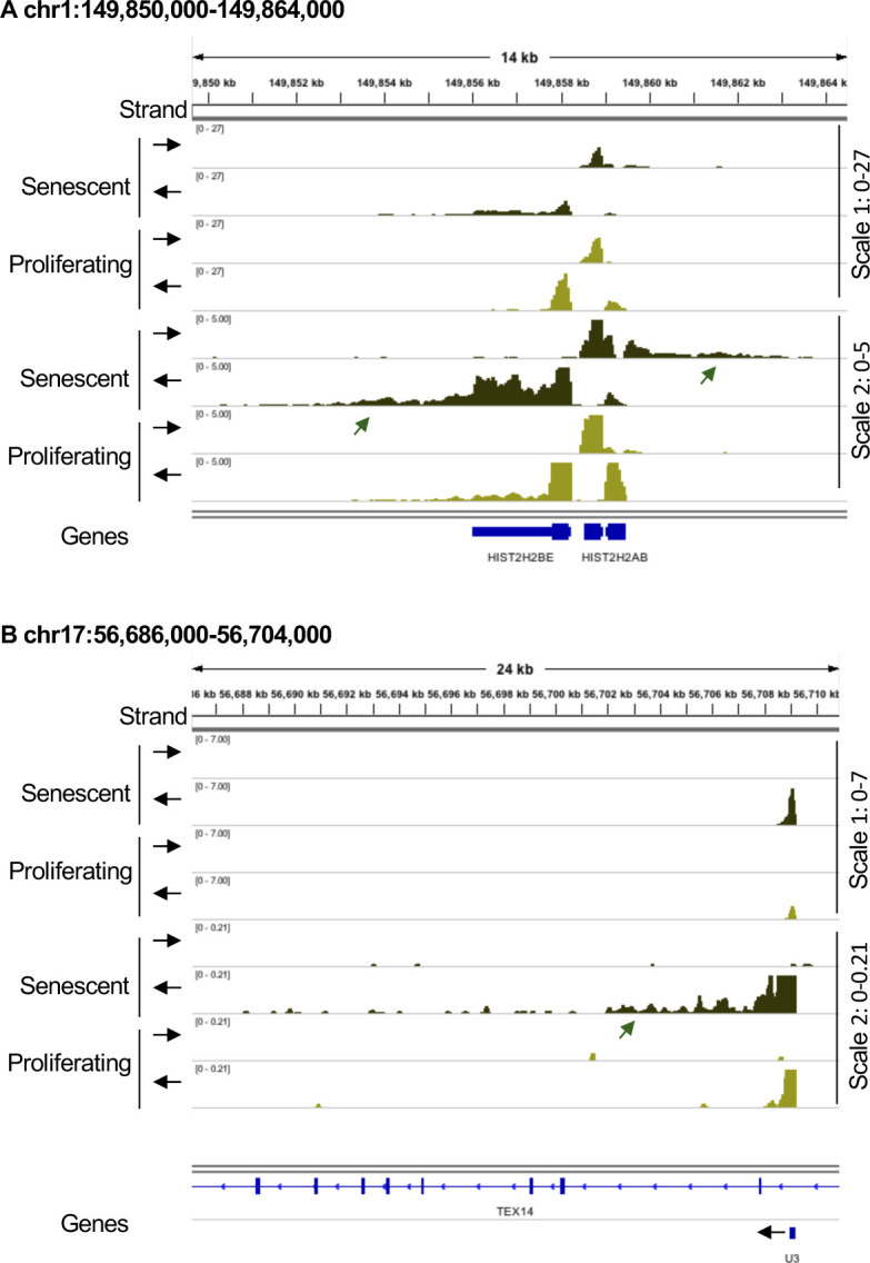
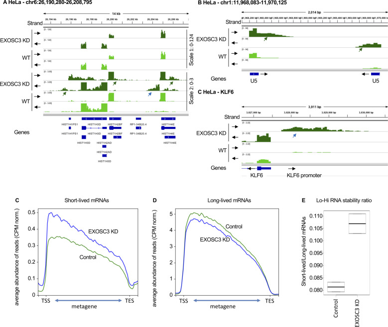
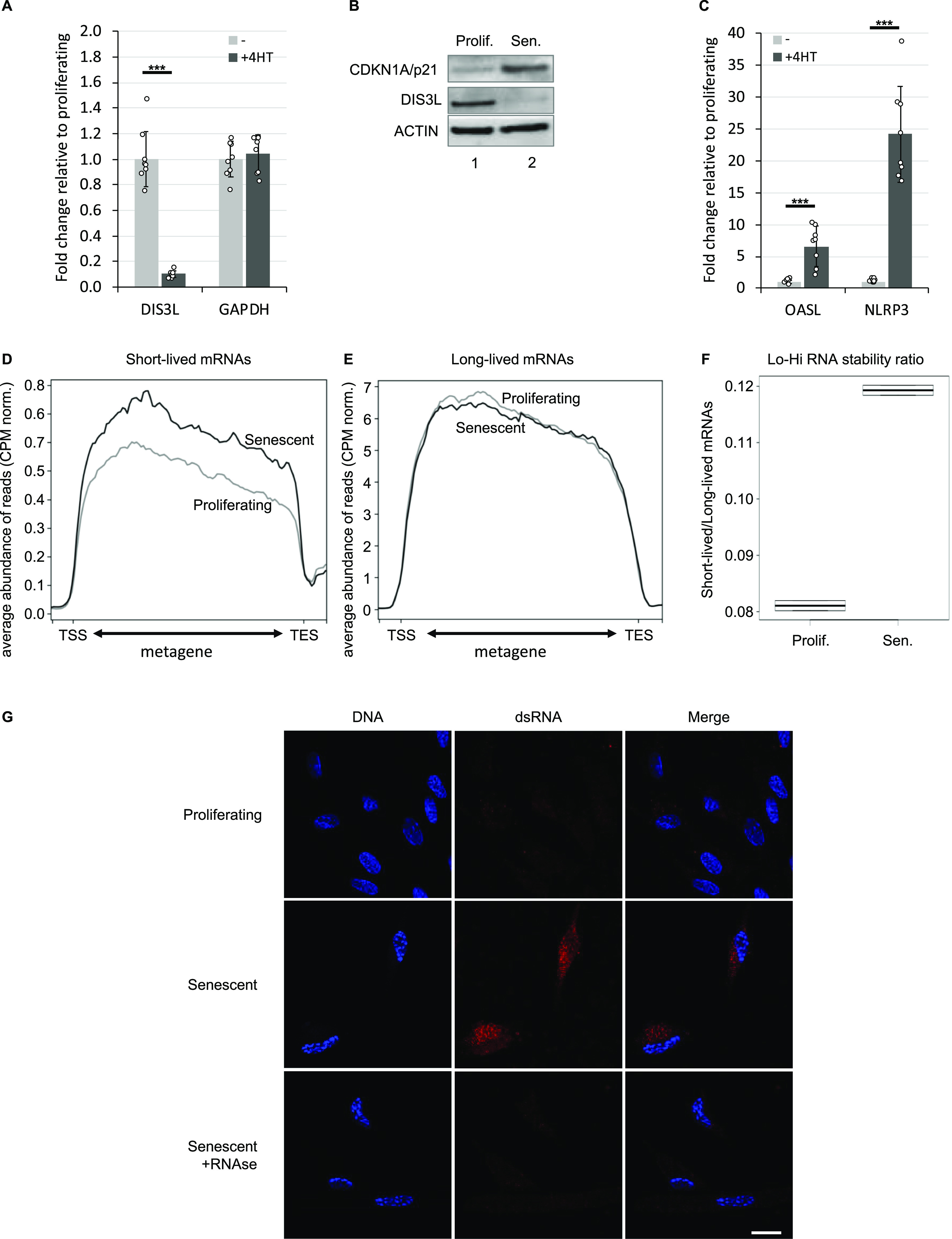
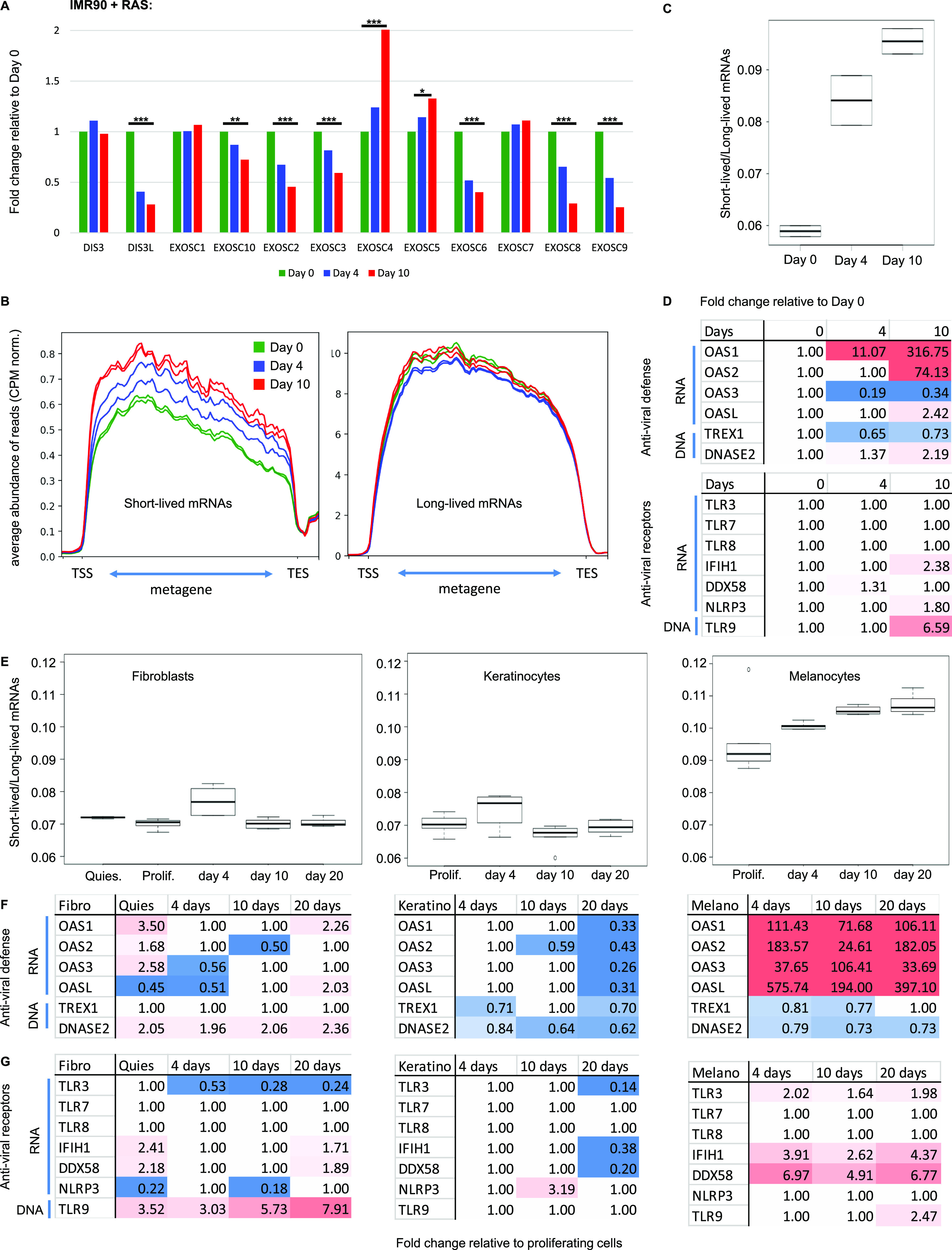
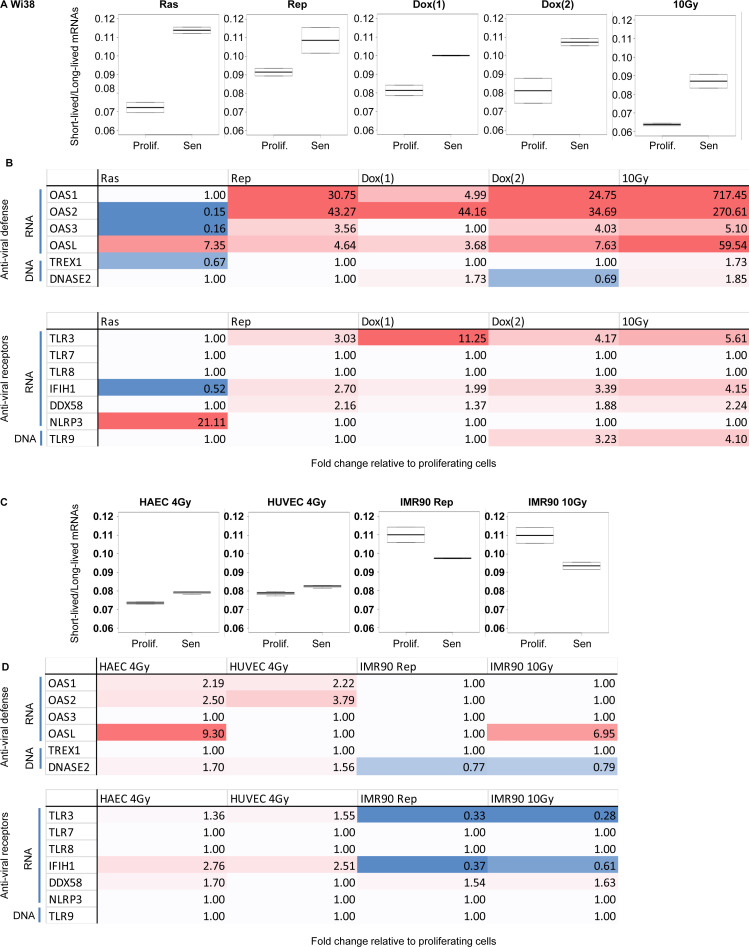
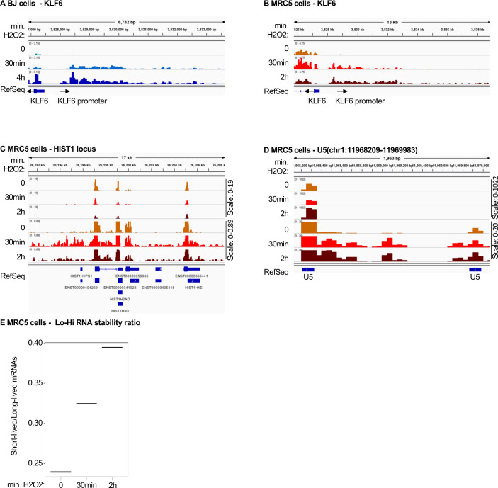
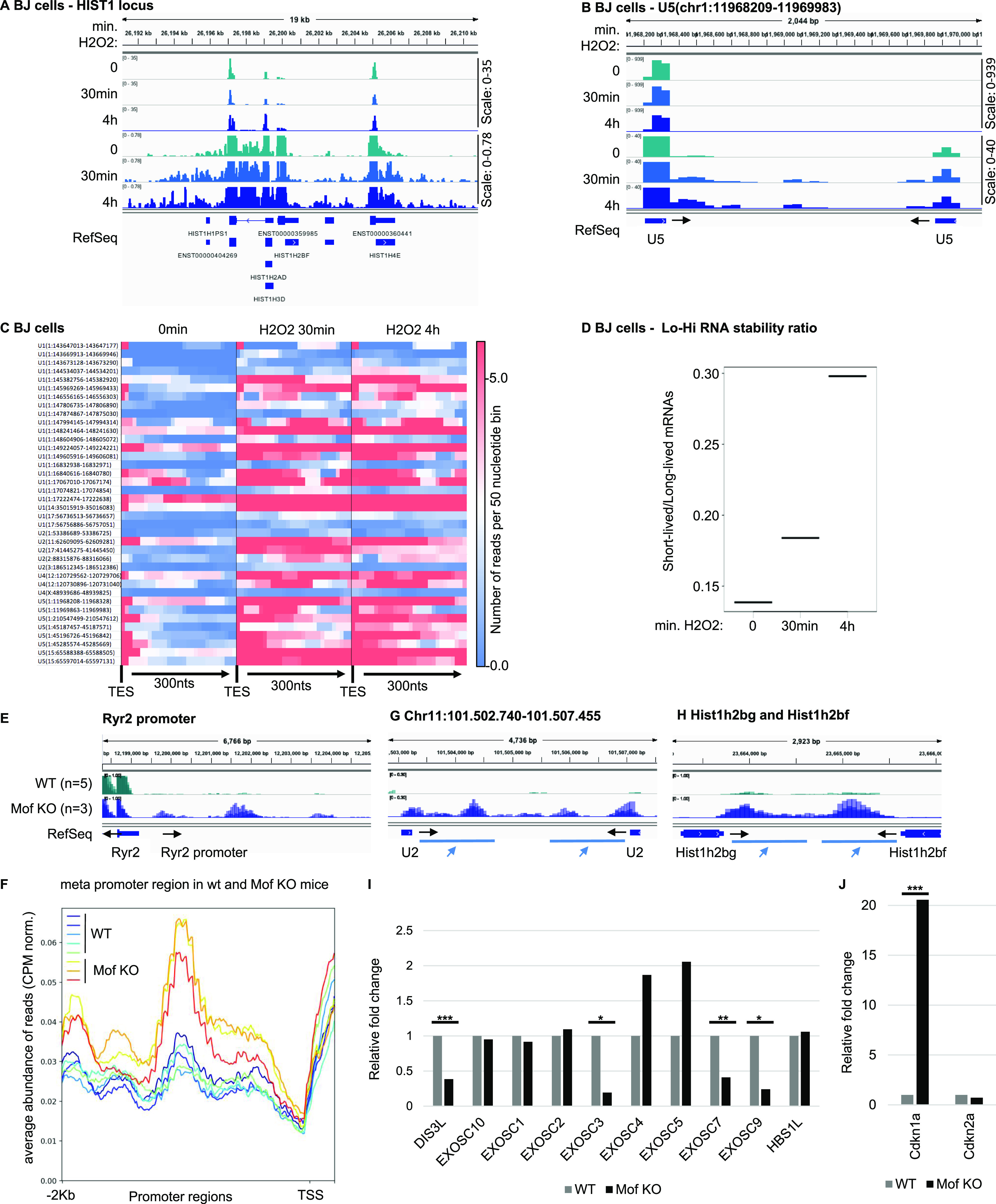
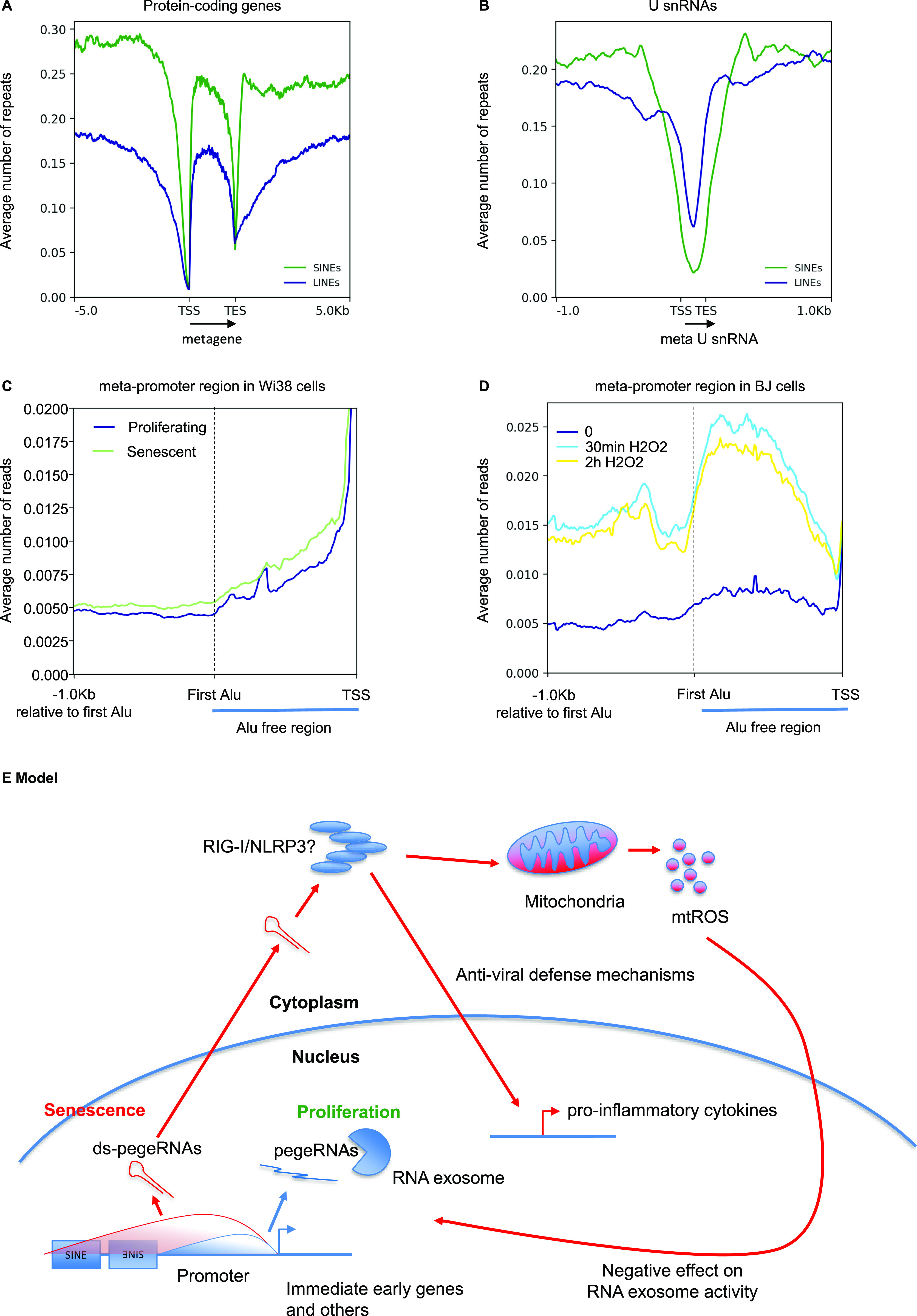
Accumulation of senescent cells is an important contributor to chronic inflammation upon aging. The inflammatory phenotype of senescent cells was previously shown to be driven by cytoplasmic DNA. Here, we propose that cytoplasmic double-stranded RNA has a similar effect. We find that several cell types driven into senescence by different routes share an accumulation of long promoter RNAs and 3' gene extensions rich in retrotransposon sequences. Accordingly, these cells display increased expression of genes involved in response to double stranded RNA of viral origin downstream of the interferon pathway. The RNA accumulation is associated with evidence of reduced RNA turnover, including in some cases, reduced expression of RNA exosome subunits. Reciprocally, depletion of RNA exosome subunit EXOSC3 accelerated expression of multiple senescence markers. A senescence-like RNA accumulation was also observed in cells exposed to oxidative stress, an important trigger of cellular senescence. Altogether, we propose that in a subset of senescent cells, repeat-containing transcripts stabilized by oxidative stress or reduced RNA exosome activity participate in driving and maintaining the permanent inflammatory state characterizing cellular senescence.


Conflict of interest statement

The authors declare that they have no conflict of interest.

Figures

Figure 1.
Figure 1.. Senescent WI38 cells accumulate pegeRNAs.
RNA-seq data from WI38 hTERT RAF1-ER human fibroblasts either proliferating or driven into senescence by induction of RAF1-ER (Muniz et al, 2017). (A, B, E) Indicated loci were visualized with Integrative Genomics Viewer. Black arrows indicate the orientation of the track. Green arrows indicate 3′ extensions, blue arrows, promoter RNAs. (C) Heat map illustrating increased accumulation of 3′ extensions of U snRNAs in senescent versus proliferating WI38 hTERT RAF1-ER cells. Transcription end site indicates the 3′ end of the U snRNA gene. (D) Schematic representation of divergent transcription at promoters. Green line represents pre-mRNA, blue line, normal accumulation of upstream antisense RNAs, red line, accumulation of upstream antisense RNAs in senescent cells. (F) At 5,260 promoters not overlapping with coding regions of any gene, reads were counted within a region of 1,500 nucleotides upstream of the transcription start site (TSS) in either proliferating or senescent WI38 hTERT RAF1-ER cells. (G) Average profile of read distribution along the 5,260 promoters in proliferating and senescent WI38 hTERT RAF1-ER cells.
Figure S1.
Figure S1.. Senescent WI38 cells accumulate pegeRNAs.
RNA-seq data from WI38 hTERT RAF1-ER human fibroblasts either proliferating or driven into senescence by induction of RAF1-ER (Lazorthes et al, 2015). (A, B) Indicated loci were visualized with Integrative Genomics Viewer. Black arrows indicate the orientation of the track. Green arrows indicate 3′ extensions.
Figure S2.
Figure S2.. Reduced expression of RNA exosome subunits in senescence.
RNA-seq data from HeLa cells transfected either with siLuc (control) or siEXOSC3 (Schlackow et al, 2017). (A, B, C) Indicated loci were visualized with Integrative Genomics Viewer. Black arrows indicate the orientation of the track. Green arrows indicate 3′ extensions, blue arrows, promoter RNAs. (C, D) Average profile of reads mapping to short-lived (less than 2 h) or long-lived (more than 10 h) mRNAs as listed in Tani et al (2012), either in proliferating or in senescent cells as indicated. (E) Lo-Hi RNA stability ratio: ratio of the number of reads mapping to short-lived (less than 2 h) over long-lived (more than 10 h).
Figure 2.
Figure 2.. Reduced expression of RNA exosome subunits in senescence.
(A, B, C) Total RNA or protein extracts were prepared from WI38 hTERT RAF1-ER human fibroblasts either proliferating or driven into senescence by induction of an activated form of C-RAF. (A, B, D) Expression of indicated genes were assessed by quantitative RT-PCR (A, D) or Western blots (B). Indicated values were averaged from eight PCR reactions. *** indicates P-values below 0.001. (D, E) Average profile of reads mapping to short-lived (less than 2 h) or long-lived (more than 10 h) mRNAs as listed in Tani et al (2012), either in proliferating or in senescent cells as indicated. (F) Ratio of the number of reads mapping to short-lived (less than 2 h) over long-lived (more than 10 h). (G) WI38 hTert pTripZ bRAFV600E cells, either proliferating or driven into senescence by inducing bRAFV600E expression with doxycycline for 7 d, were fixed, permeabilized, and analyzed for dsRNA using the mouse monoclonal antibody J2 (red). To visualize the cell nuclei, DNA was stained with DAPI (blue). Where indicated, fixed and permeabilized cells were treated with a cocktail of RNAseA and RNAseH before incubation with the J2 antibody. Scale bar: 5 μm. Source data are available for this figure.
Figure 3.
Figure 3.. Reduced RNA decay in senescent cells of various origin.
(A, B, C, D) RNA-seq data from IMR90 human fibroblasts induced into senescence via activation of the oncogene Ras (Lau et al, 2019). Samples (n = 2) had been collected at day 0 (growing phase), day 4 (beginning of SASP induction) and day 10 (senescent phase) of Ras induction. (A) Histograms show variations in expression levels of the indicated genes. *** and ** indicate P-values below 0.001 and 0.01, respectively. (B) Average profile of reads mapping to short-lived (less than 2 h) or long-lived (more than 10 h) mRNAs as in Fig 2 at the indicated time points. (C) Lo-Hi RNA stability ratio for the experiment at the indicated time points, calculated as in Fig 2. (D) Fold activation of the indicated genes at the indicated time point. Red gradient indicates up-regulation (max red at 10-fold), blue indicates down-regulation (max blue at 0.5). Variations with pVal > 0.05 were set to 1. (E, F, G) RNA-seq data from Hernandez-Segura et al (2017), n = 6 for each cell type and time point. HCA-2 (fibroblasts), keratinocytes, or melanocytes had been exposed to ionizing radiation. RNA had been harvested 4, 10, or 20 d later. (E) Lo-Hi RNA stability ratio for each experiment calculated as indicated in Fig 2. (D, F, G) Fold activation of the indicated genes represented as in (D).
Figure S3.
Figure S3.. Reduced RNA decay in senescent cells of various origin.
RNA-seq data from Casella et al (2019), n = 2 for all experiments. (A) Wi38 fibroblasts were driven into senescence either by forced expression of oncogenic RAS, continuous replication, or DNA damage with doxorubicin (two experiments) or γ irradiation (10 Gy). Lo-Hi RNA stability ratio for each experiment was calculated as in Fig 2. (A, B) Fold activation of the indicated genes in the experiments listed in (A). Red gradient indicates up-regulation (max red at 10-fold) and blue indicates down-regulation (max blue at 0.5). Variations with pVal > 0.05 were set to 1. (C, D) HAEC and HUVEC cell lines were driven into senescence by γ irradiation, and IMR90 cells were rendered senescent either by continuous replication or γ irradiation (same control for the two routes). (C) Lo-Hi RNA stability ratio for each experiment was calculated as in Fig 2. (B, D) Fold activation of the indicated genes represented as in (B).
Figure S4.
Figure S4.. Accumulation of pegeRNAs in cells exposed to oxidative stress.
BJ or MRC5 human fibroblasts having been exposed to 0.2 mM H2O2 for the indicated times (one sample per time point) were aligned on Hg19 (Giannakakis et al, 2015). (A, B, C, D) Indicated loci were visualized in the Integrative Genomics Viewer genome browser. (E) Lo-Hi RNA stability ratio for MRC5 cells at the indicated time points, calculated as indicated in Fig 2.
Figure 4.
Figure 4.. Accumulation of pegeRNAs in cells exposed to oxidative stress.
(A, B, C, D) BJ human fibroblasts exposed to 0.2 mM H2O2 for the indicated times (one sample per time point) (Giannakakis et al, 2015). Indicated loci were visualized in the Integrative Genomics Viewer genome browser. (C) Heat map illustrating increased accumulation of non-maturated U snRNAs in BJ cells exposed to H2O2 for the indicated times. Transcription end site indicates the 3′ end of the U snRNA gene. (D) Lo-Hi RNA stability ratio for BJ cells at the indicated time points, calculated as indicated in Fig 2. (E, F, G, H, I, J) Mouse cardiomyocytes either WT (n = 4) or inactivated for the Mof histone acetylase (n = 3) were analyzed by RNA-seq (Chatterjee et al, 2016). (E, G, H) Data were aligned on mouse genome mm9 and indicated loci were examined using the Integrative Genomics Viewer genome browser. (F) Average profile of read distribution along the promoters of 1,200 genes with similar expression levels in either WT or Mof KO cardiomyocytes. (I, J) Differential gene expression was estimated with DESeq2. Histograms show variations of the indicated genes. ***, **, and * indicate P-values below 0.001, 0.01, and 0.05 respectively.
Figure 5.
Figure 5.. Inactivation of the RNA exosome induce a senescent-like phenotype and deregulates mitochondrial genes.
(A) Schematic: WI38 hTert pTripZ bRAFV600E cell were transfected with either non-targetted siRNAs (siNT) or two different EXOSC3 siRNAs (red and white dots in histograms), then treated with 25 ng/ml of doxycycline at the time indicated in the schematic. All samples were harvested and the same time, and total RNA was extracted. (B, C, D, E, F), Expression of indicated genes were assessed by quantitative RT-PCR. Indicated values were averaged from eight PCR reactions. ***, and ** indicate P-values below 0.001, and 0.01 respectively. (G, H, I, J, K, L) RNA-seq data (n = 2) from mouse ES cells inactivated for Exosc3 and harboring an inducible Exosc3 expression construct human (Chiu et al, 2018). Exosc3 expression is initially induced (WT) then the induced is removed from the medium and cells are cultured for 3 d (Exosc3 KO). (G, H) Indicated loci were visualized with Integrative Genomics Viewer. Black arrows indicate the orientation of the gene. Blue arrows indicate regions of interest. (I, K, L) GO term analysis of genes differentially expressed upon Exosc3 KO was carried out with Enrichr. (J) Differential gene expression was estimated with DESeq2. Histograms show variations of the indicated genes. ***, and ** indicate P-values below 0.001, and 0.01 respectively.
Figure 6.
Figure 6.. pegeRNAs from senescent cells reach into SINE- and LINE-containing regions.
(A, B) Average number of either SINEs (green profiles) or LINEs (blue profiles) per 50 nucleotide bin in the neighborhood of either protein-coding genes (A) or snRNAs (B). TSS, transcription start site; TES, transcription end site. Gene bodies are all scaled to 2 Kb. U snRNA bodies are scaled to 200 nucleotides. Location of SINEs and LINEs was obtained from the Hg19 version of RepeatMasker. (C, D) At the 5,260 promoters not overlapping with coding regions of any gene described in Fig 1, the region from the TSS to the first Alu sequence was scaled to 1 Kb (indicated as Alu-free region). The profile then continues 1 Kb after the start of this first Alu. The average number of reads per 50 nucleotide bin was then calculated within and upstream of this region. (C, D) This was carried out for the RNA-seq data from (C) WI38 hTERT RAF1-ER human fibroblasts either proliferating or driven into senescence (Lazorthes et al, 2015) or (D) BJ cells exposed to H2O2 for the indicated times (Giannakakis et al, 2015). (E) Hypothetical model: an initial source of oxidative stress increases elongation and reduces degradation of pegeRNAs that will eventually contain sequences encoded by repeats originating from retrotransposons. Because of their abundance, a fraction of the pegeRNAs reaches the cytoplasm (Giannakakis et al, 2015). In the cytoplasm, dsRNAs generated by inverted repeats are detected by antiviral defense mechanisms. Activation of these RNA receptors results in mitochondrial dysfunction (Djafarzadeh et al, 2011). This leads to production of mitochondrial reactive oxygen species (mtROS) that hampers the RNA exosome activity and feeds the inflammatory phenotype of senescent cells.

References

    1. Allmang C, Kufel J, Chanfreau G, Mitchell P, Petfalski E, Tollervey D (1999) Functions of the exosome in rRNA, snoRNA and snRNA synthesis. EMBO J 18: 5399–5410. 10.1093/emboj/18.19.5399 - DOI - PMC - PubMed
    1. Ben-Porath I, Weinberg RA (2005) The signals and pathways activating cellular senescence. Int J Biochem Cell Biol 37: 961–976. 10.1016/j.biocel.2004.10.013 - DOI - PubMed
    1. Cao K, Blair CD, Faddah DA, Kieckhaefer JE, Olive M, Erdos MR, Nabel EG, Collins FS (2011) Progerin and telomere dysfunction collaborate to trigger cellular senescence in normal human fibroblasts. J Clin Invest 121: 2833–2844. 10.1172/jci43578 - DOI - PMC - PubMed
    1. Carvalho C, L’Hote V, Courbeyrette R, Kratassiouk G, Pinna G, Cintrat JC, Denby-Wilkes C, Derbois C, Olaso R, Deleuze JF, et al. (2019) Glucocorticoids delay RAF-induced senescence promoted by EGR1. J Cell Sci 132: jcs230748 10.1242/jcs.230748 - DOI - PubMed
    1. Casella G, Munk R, Kim KM, Piao Y, De S, Abdelmohsen K, Gorospe M (2019) Transcriptome signature of cellular senescence. Nucleic Acids Res 47: 7294–7305. 10.1093/nar/gkz555 - DOI - PMC - PubMed

Publication types

LinkOut - more resources