

An in vivo screen of noncoding loci reveals that Daedalus is a gatekeeper of an Ikaros-dependent checkpoint during haematopoiesis
- PMID: 33446502
- PMCID: PMC7826330
- DOI: 10.1073/pnas.1918062118
An in vivo screen of noncoding loci reveals that Daedalus is a gatekeeper of an Ikaros-dependent checkpoint during haematopoiesis
Abstract
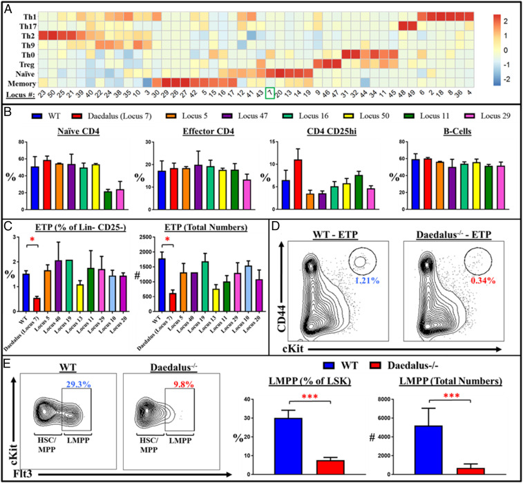
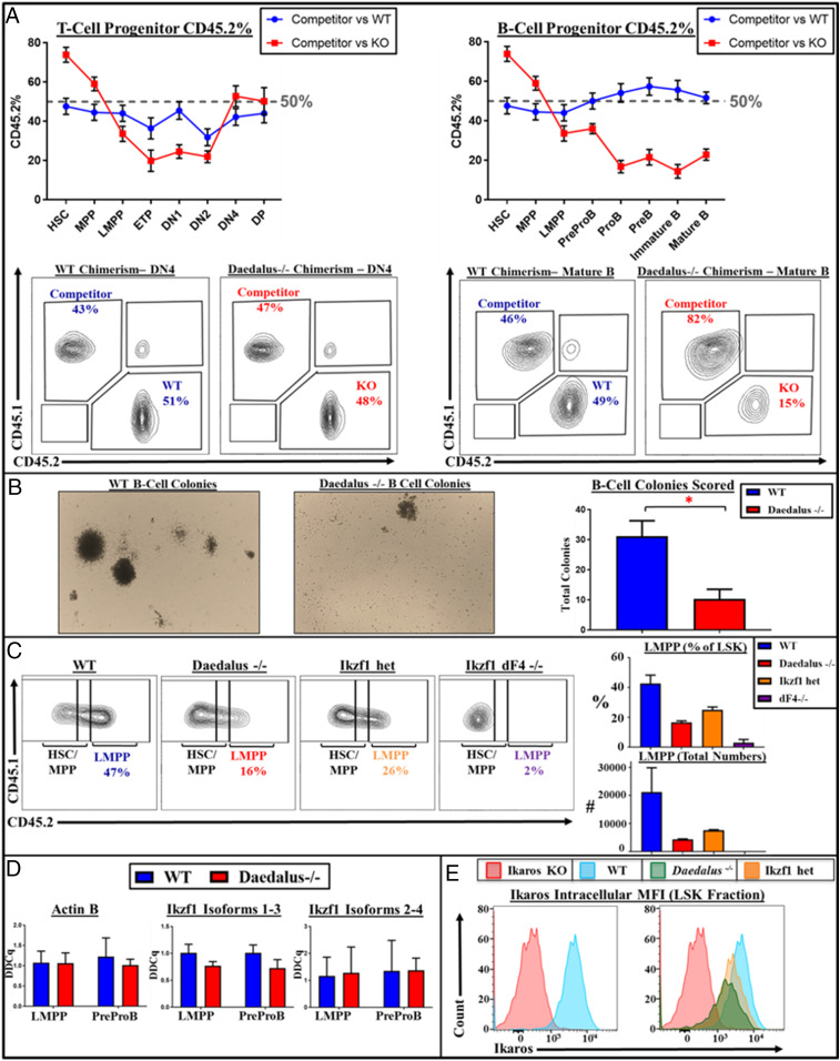
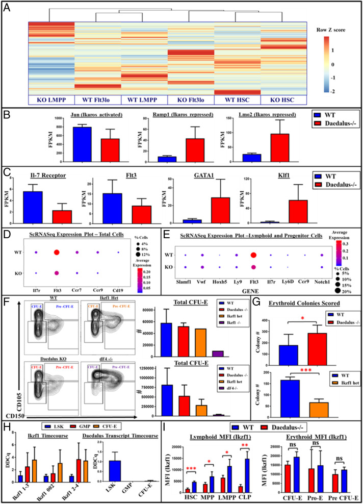
Haematopoiesis relies on tightly controlled gene expression patterns as development proceeds through a series of progenitors. While the regulation of hematopoietic development has been well studied, the role of noncoding elements in this critical process is a developing field. In particular, the discovery of new regulators of lymphopoiesis could have important implications for our understanding of the adaptive immune system and disease. Here we elucidate how a noncoding element is capable of regulating a broadly expressed transcription factor, Ikaros, in a lymphoid lineage-specific manner, such that it imbues Ikaros with the ability to specify the lymphoid lineage over alternate fates. Deletion of the Daedalus locus, which is proximal to Ikaros, led to a severe reduction in early lymphoid progenitors, exerting control over the earliest fate decisions during lymphoid lineage commitment. Daedalus locus deletion led to alterations in Ikaros isoform expression and a significant reduction in Ikaros protein. The Daedalus locus may function through direct DNA interaction as Hi-C analysis demonstrated an interaction between the two loci. Finally, we identify an Ikaros-regulated erythroid-lymphoid checkpoint that is governed by Daedalus in a lymphoid-lineage-specific manner. Daedalus appears to act as a gatekeeper of Ikaros's broad lineage-specifying functions, selectively stabilizing Ikaros activity in the lymphoid lineage and permitting diversion to the erythroid fate in its absence. These findings represent a key illustration of how a transcription factor with broad lineage expression must work in concert with noncoding elements to orchestrate hematopoietic lineage commitment.
Keywords: Ikaros; hematopoiesis; lymphocytes; noncoding.
Conflict of interest statement
Competing interest statement: R.A.F. is a consultant for GSK and Zai Lab Ltd.
Figures
Similar articles
-
Ikaros isoform x is selectively expressed in myeloid differentiation.J Immunol. 2003 Mar 15;170(6):3091-8. doi: 10.4049/jimmunol.170.6.3091. J Immunol. 2003. PMID: 12626565
-
Ikaros fingers on lymphocyte differentiation.Int J Hematol. 2014 Sep;100(3):220-9. doi: 10.1007/s12185-014-1644-5. Epub 2014 Aug 2. Int J Hematol. 2014. PMID: 25085254 Free PMC article. Review.
-
Awakening lineage potential by Ikaros-mediated transcriptional priming.Curr Opin Immunol. 2010 Apr;22(2):154-60. doi: 10.1016/j.coi.2010.02.011. Epub 2010 Mar 17. Curr Opin Immunol. 2010. PMID: 20299195 Free PMC article. Review.
-
Ikaros expression in human hematopoietic lineages.Exp Hematol. 2000 Nov;28(11):1232-8. doi: 10.1016/s0301-472x(00)00530-0. Exp Hematol. 2000. PMID: 11063871
-
Genome-wide identification of Ikaros targets elucidates its contribution to mouse B-cell lineage specification and pre-B-cell differentiation.Blood. 2013 Mar 7;121(10):1769-82. doi: 10.1182/blood-2012-08-450114. Epub 2013 Jan 9. Blood. 2013. PMID: 23303821
Cited by
-
A Treg-specific long noncoding RNA maintains immune-metabolic homeostasis in aging liver.Nat Aging. 2023 Jul;3(7):813-828. doi: 10.1038/s43587-023-00428-8. Epub 2023 Jun 5. Nat Aging. 2023. PMID: 37277640
-
Multi-omics integration analysis identifies INPP4B as a T-cell-specific activation suppressor.Clin Transl Med. 2025 Aug;15(8):e70430. doi: 10.1002/ctm2.70430. Clin Transl Med. 2025. PMID: 40754682 Free PMC article.
-
The ubiquitin ligase Cul5 regulates CD4+ T cell fate choice and allergic inflammation.Nat Commun. 2022 May 19;13(1):2786. doi: 10.1038/s41467-022-30437-x. Nat Commun. 2022. PMID: 35589717 Free PMC article.
References
-
- Boehm T., Design principles of adaptive immune systems. Nat. Rev. Immunol. 11, 307–317 (2011). - PubMed
-
- Greenberg P. D., Riddell S. R., Deficient cellular immunity: Finding and fixing the defects. Science 285, 546–551 (1999). - PubMed
-
- Buckley R. H., Primary cellular immunodeficiencies. J. Allergy Clin. Immunol. 109, 747–757 (2002). - PubMed
-
- Buckley R. H., Primary immunodeficiency diseases due to defects in lymphocytes. N. Engl. J. Med. 343, 1313–1324 (2000). - PubMed
-
- Cobaleda C., Schebesta A., Delogu A., Busslinger M., Pax5: The guardian of B cell identity and function. Nat. Immunol. 8, 463–470 (2007). - PubMed
Publication types
MeSH terms
Substances
Grants and funding
LinkOut - more resources
Full Text Sources
Other Literature Sources
Molecular Biology Databases
