. 2021 Feb 19;7(1):a005454.
doi: 10.1101/mcs.a005454. Print 2021 Feb.

Identification and characterization of novel ACD variants: modulation of TPP1 protein level offsets the impact of germline loss-of-function variants on telomere length

Affiliations

Identification and characterization of novel ACD variants: modulation of TPP1 protein level offsets the impact of germline loss-of-function variants on telomere length

Gabrielle Henslee et al. Cold Spring Harb Mol Case Stud. .

Abstract

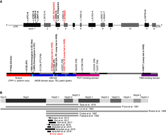
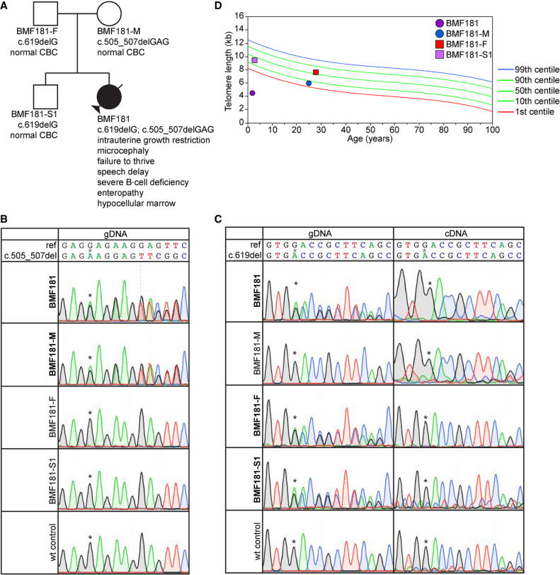
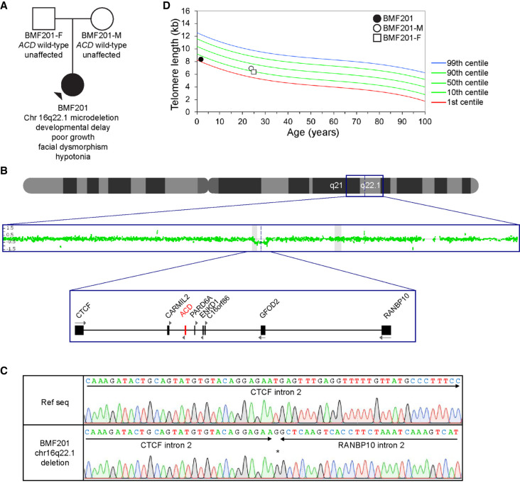
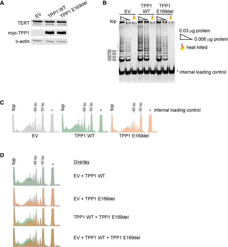
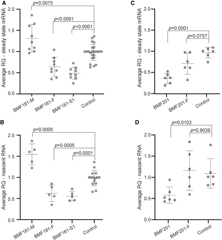
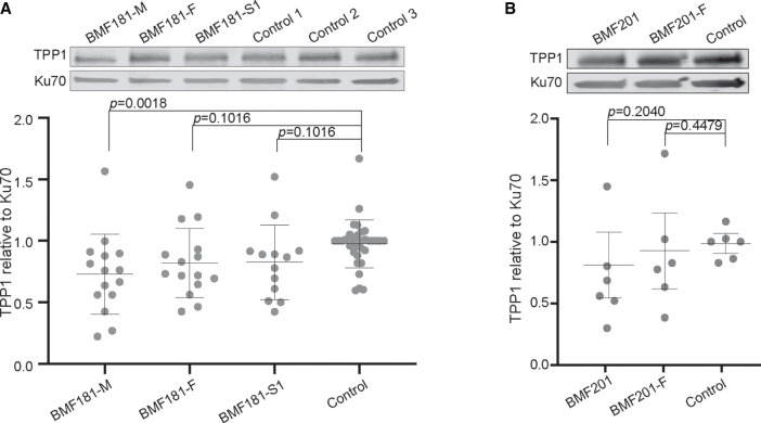
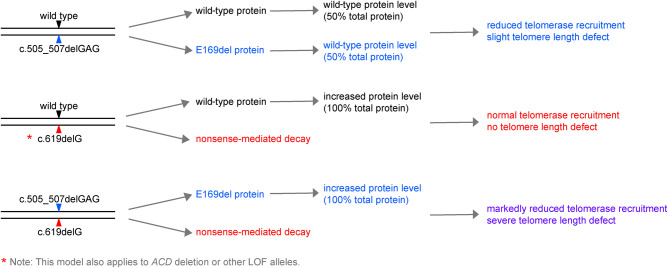
Telomere biology disorders, largely characterized by telomere lengths below the first centile for age, are caused by variants in genes associated with telomere replication, structure, or function. One of these genes, ACD, which encodes the shelterin protein TPP1, is associated with both autosomal dominantly and autosomal recessively inherited telomere biology disorders. TPP1 recruits telomerase to telomeres and stimulates telomerase processivity. Several studies probing the effect of various synthetic or patient-derived variants have mapped specific residues and regions of TPP1 that are important for interaction with TERT, the catalytic component of telomerase. However, these studies have come to differing conclusions regarding ACD haploinsufficiency. Here, we report a proband with compound heterozygous novel variants in ACD (NM_001082486.1)-c.505_507delGAG, p.(Glu169del); and c.619delG, p.(Asp207Thrfs*22)-and a second proband with a heterozygous chromosomal deletion encompassing ACD: arr[hg19] 16q22.1(67,628,846-67,813,408)x1. Clinical data, including symptoms and telomere length within the pedigrees, suggested that loss of one ACD allele was insufficient to induce telomere shortening or confer clinical features. Further analyses of lymphoblastoid cell lines showed decreased nascent ACD RNA and steady-state mRNA, but normal TPP1 protein levels, in cells containing heterozygous ACD c.619delG, p.(Asp207Thrfs*22), or the ACD-encompassing chromosomal deletion compared to controls. Based on our results, we conclude that cells are able to compensate for loss of one ACD allele by activating a mechanism to maintain TPP1 protein levels, thus maintaining normal telomere length.

Keywords: abnormality of B cell number; bone marrow hypocellularity; fingernail dysplasia; microcephaly; oral leukoplakia; reticulated skin pigmentation; toenail dysplasia.


Figures

Figure 1.
Figure 1.
Previously reported ACD variants and Chr 16q22 deletions. (A) Reported ACD variants. Nucleotide positions are depicted in the upper graphic, whereas amino acid positions are depicted in the lower graphic. Red text indicates variants described in this report. Bold text indicates variants associated with short telomeres. Non-bold text indicates variants found in cancer-prone families and associated with long telomeres. (DC) Dyskeratosis congenita, (AA) aplastic anemia, (PF) pulmonary fibrosis, (PTC) papillary thyroid carcinoma, (FM) familial melanoma, (HHS) Hoyeraal–Hreidarsson syndrome, (comp het) compound heterozygous variant found in the context of another disease-causing variant. (B) Reported Chromosome 16q22.1 deletions. Gray lines indicate deletions for which specific nucleotide information is not available. Asterisks indicate deletions that do not encompass ACD.
Figure 2.
Figure 2.
The ACD c.619delG variant is subject to nonsense-mediated decay but is not associated with telomere shortening. (A) Pedigree for proband BMF181. (B) Genomic DNA (gDNA) Sanger sequence chromatograms surrounding nucleotides c.505_507. Asterisks denote the location of the first deleted G in BMF181 and BMF181-M. (C) gDNA and complementary DNA (cDNA) Sanger sequence chromatograms surrounding nucleotide c.619. Asterisks denote the location of the deleted G in BMF181, BMF181-F, and BMF181-S1. (D) Lymphocyte telomere lengths as measured by telomere flow FISH for BMF181 and family members.
Figure 3.
Figure 3.
Deletion of one ACD allele is not associated with telomere shortening. (A) Pedigree for proband BMF201. (B) Chromosomal microarray (CMA) indicating a deletion encompassing ACD and neighboring genes. Scanning from the top down, the graphics depict a schematic of Chromosome 16, the CMA data indicating deletion of 185 kb in Chr 16q22.1, and a schematic of the genes deleted or disrupted in BMF201. (C) Sanger sequence chromatograms confirming the heterozygous deletion of Chr 16q22.1(67,628,846-67,813,408) in BMF201. The asterisk denotes a single inserted G. (D) Lymphocyte telomere lengths as measured by telomere flow FISH for BMF201 and parents.
Figure 4.
Figure 4.
TPP1 p.Glu169del is defective for telomerase stimulation. Shown are representative images for five independent experiments. (A) Western blot of lysates of 293T cells transiently transfected with plasmids expressing TERT, hTR, and either empty vector (EV), wild-type TPP1 (TPP1 WT), or TPP1 p.Glu169del (TPP1 E169del) used for the telomerase repeated amplification protocol (TRAP) assay shown in B. β-actin was used as a loading control. (B) TRAP assays performed with 0.03 µg protein, 0.006 µg protein, or 0.03 µg of heat-killed protein. Internal control is denoted by *. (C) Densitometry traces of the 0.03 µg TRAP reactions shown in B. The top of each lane aligns with the left of the densitometry trace. Internal control is denoted by *. Traces were generated using ImageJ software. (D) Overlay images of the densitometry traces shown in C.
Figure 5.
Figure 5.
ACD steady-state mRNA and nascent transcript levels are reduced in the presence of the c.619delG variant. (A) Reverse transcriptase quantitative PCR (RT-qPCR) analysis of steady-state ACD mRNA expression relative to GAPDH control in BMF181 family LCLs. Three independent control LCL lines were used and grouped together for analysis. Control 2 was used to normalize values across experiments. Mean and standard deviation are shown. Results were log-transformed and then analyzed using one-way ANOVA followed by the Holm–Sidak test to adjust for multiple comparisons. N = 19 for control, N = 9 for all other samples. (RQ) Relative quantification, fold change compared to calibrator sample. (B) RT-qPCR analysis of nascent ACD RNA relative to GAPDH control in BMF181 family LCLs. Three independent control LCL lines were used and grouped together for analysis. Nascent transcripts were specifically amplified using primers spanning the exon 1-intron 1 junction. Results analyzed as in A. N = 15 for control, N = 5 for all other samples. (C) RT-qPCR analysis of steady-state ACD mRNA expression relative to GAPDH control in BMF201 family LCLs. A single control LCL line was used for comparison. Results analyzed as in A. N = 6. (D) RT-qPCR analysis of nascent ACD RNA relative to GAPDH control in BMF201 family LCLs. A single control LCL line was used for comparison. Results analyzed as in A. N = 6.
Figure 6.
Figure 6.
TPP1 protein levels are maintained in the presence of heterozygous ACD c.619delG. (A) Representative western blot and quantification of TPP1 in BMF181 family LCLs. Ku70 was used as a loading control. Three independent control LCL lines were used and grouped together for analysis. Control 3 was used to normalize values across experiments. Mean and standard deviation are shown. Results were log-transformed and then analyzed using one-way ANOVA followed by the Holm–Sidak test to adjust for multiple comparisons. N = 15 for BMF181-M, BMF181-F, and BMF181-S1. N = 4 for controls 1 and 2. N = 24 for control 3. See Supplemental Figure 2C for compilation of western blots used for analysis. (B) Representative western blot and quantification of TPP1 in BMF201 family LCLs. A single control LCL line was used for comparison. Results analyzed as in A. No statistically significant difference was detected between the cell lines. N = 6. See Supplemental Figure 2D for compilation of western blots used for analysis.
Figure 7.
Figure 7.
Working model for the impact of the BMF181 family and BMF201 ACD variants on TPP1 protein expression and telomere length. Presented is a schematic for our working model to explain the results described. According to our model, BMF181-M (top schematic) would express TPP1 WT and p.Glu169del in equal amounts, leading to reduced telomerase recruitment due to the interaction defect of TPP1 p.Glu169del with TERT, thus leading to telomere shortening. When combined with a second variant in trans, as in BMF181 (bottom schematic), the absence of WT TPP1 would result in rapid and dramatic telomere shortening. However, our model predicts that variants resulting in NMD, as in BMF181-F and BMF181-S1, or whole-gene deletions, as in BMF201, would result in increased WT protein to compensate for loss of a second functional allele, thus allowing normal telomerase recruitment (middle schematic).

Similar articles

Cited by

References

    1. Abdullah S, Helal M, Dupuis L, Stavropoulos DJ, Louro P, Ramos L, Mendoza-Londono R. 2019. 16q22.1 microdeletion and anticipatory guidance. Am J Med Genet A 179: 1287–1292. 10.1002/ajmg.a.61155 - DOI - PubMed
    1. Allsopp RC, Harley CB. 1995. Evidence for a critical telomere length in senescent human fibroblasts. Exp Cell Res 219: 130–136. 10.1006/excr.1995.1213 - DOI - PubMed
    1. Alter BP, Baerlocher GM, Savage SA, Chanock SJ, Weksler BB, Willner JP, Peters JA, Giri N, Lansdorp PM. 2007. Very short telomere length by flow fluorescence in situ hybridization identifies patients with dyskeratosis congenita. Blood 110: 1439–1447. 10.1182/blood-2007-02-075598 - DOI - PMC - PubMed
    1. Alter BP, Giri N, Savage SA, Rosenberg PS. 2009. Cancer in dyskeratosis congenita. Blood 113: 6549–6557. 10.1182/blood-2008-12-192880 - DOI - PMC - PubMed
    1. Alter BP, Giri N, Savage SA, Peters JA, Loud JT, Leathwood L, Carr AG, Greene MH, Rosenberg PS. 2010. Malignancies and survival patterns in the National Cancer Institute inherited bone marrow failure syndromes cohort study. Br J Haematol 150: 179–188. 10.1111/j.1365-2141.2010.08212.x - DOI - PMC - PubMed

Publication types

MeSH terms

LinkOut - more resources