. 2021 Feb 1;35(3-4):234-249.
doi: 10.1101/gad.342378.120. Epub 2021 Jan 14.

SIX2 and SIX3 coordinately regulate functional maturity and fate of human pancreatic β cells

Affiliations

SIX2 and SIX3 coordinately regulate functional maturity and fate of human pancreatic β cells

Romina J Bevacqua et al. Genes Dev. .

Abstract

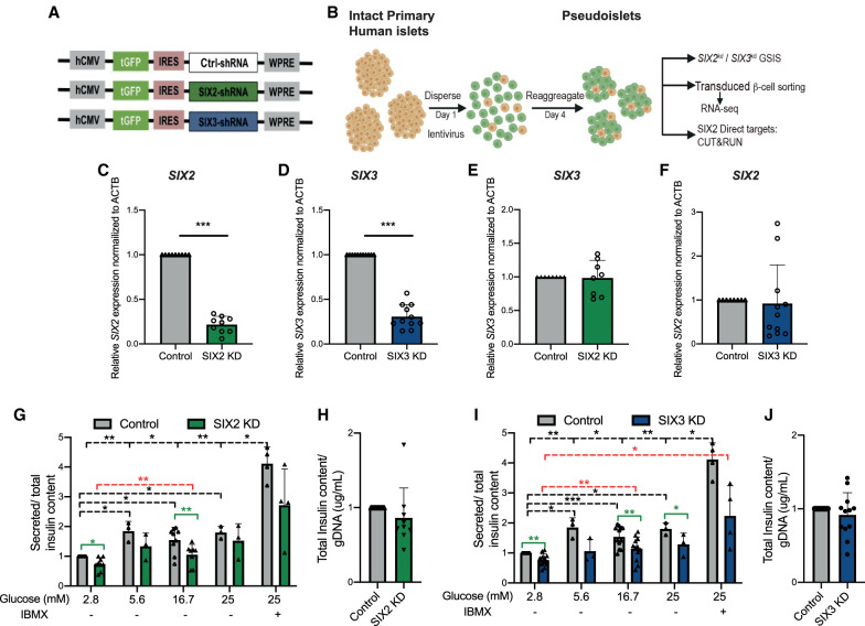
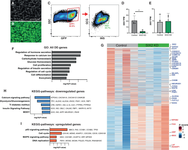
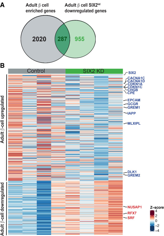
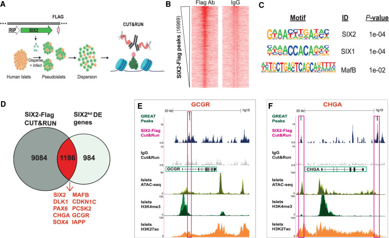
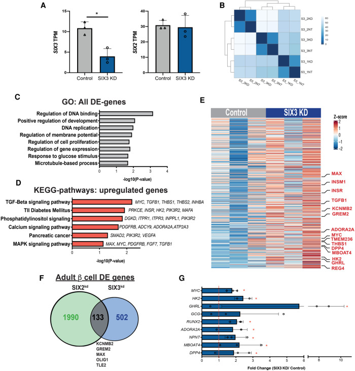
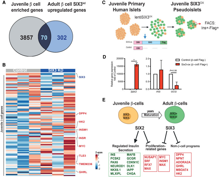
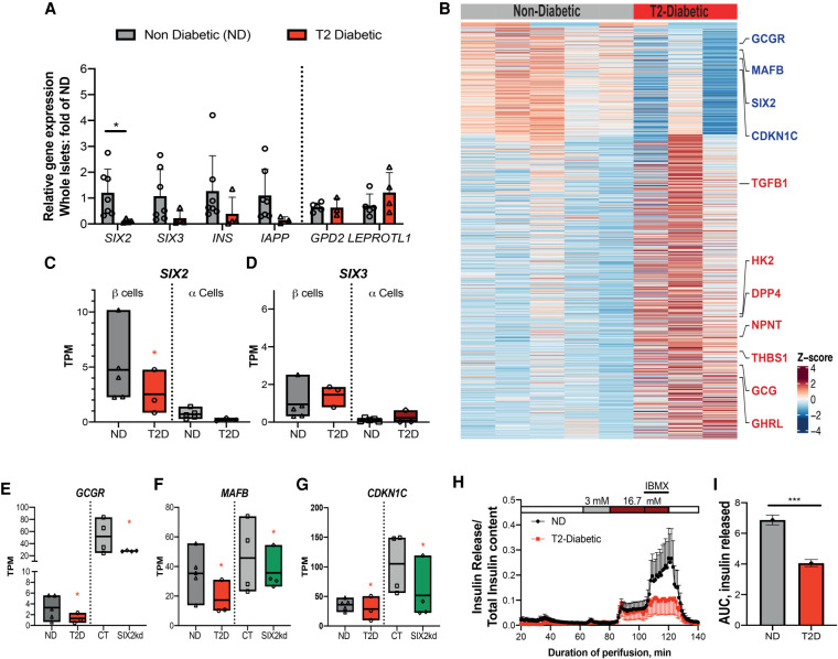
The physiological functions of many vital tissues and organs continue to mature after birth, but the genetic mechanisms governing this postnatal maturation remain an unsolved mystery. Human pancreatic β cells produce and secrete insulin in response to physiological cues like glucose, and these hallmark functions improve in the years after birth. This coincides with expression of the transcription factors SIX2 and SIX3, whose functions in native human β cells remain unknown. Here, we show that shRNA-mediated SIX2 or SIX3 suppression in human pancreatic adult islets impairs insulin secretion. However, transcriptome studies revealed that SIX2 and SIX3 regulate distinct targets. Loss of SIX2 markedly impaired expression of genes governing β-cell insulin processing and output, glucose sensing, and electrophysiology, while SIX3 loss led to inappropriate expression of genes normally expressed in fetal β cells, adult α cells, and other non-β cells. Chromatin accessibility studies identified genes directly regulated by SIX2. Moreover, β cells from diabetic humans with impaired insulin secretion also had reduced SIX2 transcript levels. Revealing how SIX2 and SIX3 govern functional maturation and maintain developmental fate in native human β cells should advance β-cell replacement and other therapeutic strategies for diabetes.

Keywords: diabetes mellitus; islet; pancreas; transcription factor; β cells.


Figures

Figure 1.
Figure 1.
shRNA-mediated suppression of SIX2 and SIX3 in primary human islets results in impaired glucose-stimulated insulin secretion. (A) Schematics of the lentiviral constructs coding for a short hairpin RNA (shRNA) and GFP. (B) Schematic detailing the pseudoislet technique. (C) SIX2 mRNA expression in primary human islets. (Gray bar) Control, (green bar) SIX2kd. n = 9 independent donor repetitions. (D) SIX3 mRNA expression in primary human islets. (Gray bar) Control, (blue bar) SIX3kd. n = 11 independent donor repetitions. SIX3 mRNA were not altered by SIX2kd (n = 8 independent donor repetitions) (E), and SIX2 mRNA levels were not affected by SIX3kd (n = 11 independent donor repetitions) (F). (G,I) In vitro glucose-stimulated insulin secretion from human pseudoislets control, SIX2kd (n = 9 independent donor repetitions) (G), or SIX3kd (n = 12 independent donor repetitions) (I). Secreted insulin normalized to insulin content. (Black lines) Significant differences within the control, (red lines) significant differences within the KD groups, (green lines) significant differences between control and KD conditions. (H,J) Total insulin from human pseudoislets after transduction with SIX2kd (n = 9 independent donor repetitions) (H) or SIX3kd (n = 12 independent donor repetitions) (J). Data presented as mean; error bars represent the standard error. Two-tailed t-tests used to generate P-values: (*) P < 0.05, (**) P < 0.01, (***) P < 0.0001.
Figure 2.
Figure 2.
RNA-seq of SIX2kd β cells reveals genes regulated by SIX2 in primary human islets. (A,B) SIX2kd human pseudoislets. (A) Bright field. (B) Blue light (488 nm). Scale bars, 500 µm. (C) FACS scheme used to sort GFP+ β cells. (D,E) Normalized transcript levels of SIX2 (D) and SIX3 (E) in GFP+ β−cells. (Gray bar) Control, (green bar) SIX2kd. n = 4. (F) GO term enrichment in genes deregulated in β cells post-SIX2kd. (G,H) KEGG pathway enrichment in genes up-regulated (G) or down-regulated (H) in β cells post-SIX2kd (n = 4). (I) Heat map showing all differentially expressed (DE) genes in β cells post-SIX2kd. The data are presented as mean; error bars represent the standard error. (*) P < 0.05
Figure 3.
Figure 3.
SIX2kd in primary β cells results in down-regulation of genes enriched in adult β-cell. (A) Venn diagram showing adult β-cell genes down-regulated post-SIX2kd. (B) Heat map of adult down-regulated genes and juvenile up-regulated genes in adult β cells post-SIX2kd.
Figure 4.
Figure 4.
Identification of presumptive SIX2 genetic targets in primary human β cells using CUT&RUN. (A) Schematic of the CUT&RUN approach: Pseudoislets overexpressing SIX2-FLAG under the RIP promoter were used for CUT&RUN with anti-FLAG antibody (n = 3 independent donors). (B) Heat map showing enrichment of peak read densities at the center of the peak for the CUT&RUN libraries generated with FLAG antibody, but not for IgG. Peaks were called using HOMER. (C) Enriched motifs in the differential peaks were identified by HOMER. (D) Overlap of the SIX2-associated genomic regions and the SIX2kd DE genes. (E,F) Tracks showing SIX2-FLAG genomic regions associated with GCGR (E) or CHGA (F). Accessible chromatin regions in human islets are shown by ATAC-seq, H3K4me3, and H3K27ac ChiP-seq tracks. SIX2-FLAG CUT&RUN peaks are shown in pink boxes (note: for GCGR, two peaks are shown), and regulated genes are highlighted in green boxes.
Figure 5.
Figure 5.
RNA-seq of SIX3kd β cells reveals a distinct gene set regulated by SIX3. (A) Normalized transcript levels of SIX3 and SIX2 in GFP-expressing β cells. (Gray bar) Control, (blue bar) SIX3kd . n = 3. (B) Heat map of the sample-to-sample distances for all the samples used in this experiment. (C) GO term enrichment in genes deregulated in β cells post-SIX3kd. (D) KEGG pathways enriched in genes up-regulated in β cells post-SIX3kd (n = 3). (E) Heat map showing all up-regulated genes upon SIX3kd in β cells. (F) Overlapped DE genes in adult β cells post SIX2kd and SIX3kd. (G) Fold transcript levels of non-β-cell genes significantly altered in β cells post-SIX3kd (n = 3). The data are presented as mean; error bars represent the standard error. (*) P < 0.05.
Figure 6.
Figure 6.
SIX3 represses non-β-cell programs in the adult β-cell. (A) Venn diagram showing juvenile β-cell genes up-regulated post-SIX3kd in adult β cells. (B) Heat map of adult down-regulated and juvenile up-regulated genes in adult β cells post-SIX3kd. (C) Schematic detailing the juvenile pseudoislet technique used to overexpress SIX3-FLAG (SIX3-ox) in juvenile pseudoislets (n = 2) and of the constructs used to overexpress SIX3 in juvenile pseudoislets: FACS was used to sort FLAG+ β cells. (D) SIX3, INS, and GCG mRNA expression in FLAG+-expressing β cells post FACS. (White bar) Control , (red bar) SIX3-ox. n = 2. (E) Schematics showing proposed coordinated regulation of maturation by SIX2 and SIX3 in the β-cell. The data are presented as mean; error bars represent the standard error. (*) P < 0.05.
Figure 7.
Figure 7.
SIX2 expression is reduced in β cells from T2D donors. (A) Gene expression levels in whole islets from nondiabetic (ND; gray bars) (n = 5) or type 2 diabetic (T2D; red bar) (n = 3). (B) Heat map showing DE genes in nondiabetic versus T2 diabetic β cells. (C,D) Box plots displaying TPM counts of SIX2 (C) and SIX3 (D) in nondiabetic (ND) (n = 5) β cells (dark-gray bars) and α cells (light-gray bars) or type 2 diabetic (T2D) β cells (red bars) and α cells (dark-red bars) (n = 3). (EG) Box plots displaying TPM GCGR (E), MAFB (F), and CDKN1C (G) in β-cells of ND (n = 5) and T2D (n = 3), the expression of which is DE in adult β cells post-SIX2kd. (Gray bars) Çontrol, (green bars) SIX2kd. (H) Insulin secretion of ND (n = 5) versus T2D (n = 3; see the Materials and Methods). (I) Plot of the total area under the curve of the released insulin. The data in A are presented as mean; error bars represent the standard error. Box plots show the mean. (Red *) P ≤ 0.01, (black *) P < 0.05.

Similar articles

Cited by

  • Engineering islets from stem cells for advanced therapies of diabetes.
    Siehler J, Blöchinger AK, Meier M, Lickert H. Siehler J, et al. Nat Rev Drug Discov. 2021 Dec;20(12):920-940. doi: 10.1038/s41573-021-00262-w. Epub 2021 Aug 10. Nat Rev Drug Discov. 2021. PMID: 34376833 Review.
  • HNF1α maintains pancreatic α and β cell functions in primary human islets.
    Qian MF, Bevacqua RJ, Coykendall VM, Liu X, Zhao W, Chang CA, Gu X, Dai XQ, MacDonald PE, Kim SK. Qian MF, et al. JCI Insight. 2023 Dec 22;8(24):e170884. doi: 10.1172/jci.insight.170884. JCI Insight. 2023. PMID: 37943614 Free PMC article.
  • Loci for insulin processing and secretion provide insight into type 2 diabetes risk.
    Broadaway KA, Yin X, Williamson A, Parsons VA, Wilson EP, Moxley AH, Vadlamudi S, Varshney A, Jackson AU, Ahuja V, Bornstein SR, Corbin LJ, Delgado GE, Dwivedi OP, Fernandes Silva L, Frayling TM, Grallert H, Gustafsson S, Hakaste L, Hammar U, Herder C, Herrmann S, Højlund K, Hughes DA, Kleber ME, Lindgren CM, Liu CT, Luan J, Malmberg A, Moissl AP, Morris AP, Perakakis N, Peters A, Petrie JR, Roden M, Schwarz PEH, Sharma S, Silveira A, Strawbridge RJ, Tuomi T, Wood AR, Wu P, Zethelius B, Baldassarre D, Eriksson JG, Fall T, Florez JC, Fritsche A, Gigante B, Hamsten A, Kajantie E, Laakso M, Lahti J, Lawlor DA, Lind L, März W, Meigs JB, Sundström J, Timpson NJ, Wagner R, Walker M, Wareham NJ, Watkins H, Barroso I, O'Rahilly S, Grarup N, Parker SC, Boehnke M, Langenberg C, Wheeler E, Mohlke KL. Broadaway KA, et al. Am J Hum Genet. 2023 Feb 2;110(2):284-299. doi: 10.1016/j.ajhg.2023.01.002. Epub 2023 Jan 23. Am J Hum Genet. 2023. PMID: 36693378 Free PMC article.
  • Sine oculis homeobox homolog family function in gastrointestinal cancer: Progression and comprehensive analysis.
    Lan YZ, Wu Z, Chen WJ, Yu XN, Wu HT, Liu J. Lan YZ, et al. World J Clin Oncol. 2025 Jan 24;16(1):97163. doi: 10.5306/wjco.v16.i1.97163. World J Clin Oncol. 2025. PMID: 39867730 Free PMC article. Review.
  • Transcriptional mechanisms of pancreatic β-cell maturation and functional adaptation.
    Wortham M, Sander M. Wortham M, et al. Trends Endocrinol Metab. 2021 Jul;32(7):474-487. doi: 10.1016/j.tem.2021.04.011. Epub 2021 May 21. Trends Endocrinol Metab. 2021. PMID: 34030925 Free PMC article. Review.

References

    1. Aguayo-Mazzucato C, Koh A, El Khattabi I, Li W-C, Toschi E, Jermendy A, Juhl K, Mao K, Weir GC, Sharma A, et al. 2011. Mafa expression enhances glucose-responsive insulin secretion in neonatal rat β cells. Diabetologia 54: 583–593. 10.1007/s00125-010-2026-z - DOI - PMC - PubMed
    1. Allen HL, Flanagan SE, Shaw-Smith C, De Franco E, Akerman I, Caswell R, Ferrer J, Hattersley AT, Ellard S, The International Pancreatic Agenesis Consortium. 2012. GATA6 haploinsufficiency causes pancreatic agenesis in humans. Nat Genet 44: 20–22. 10.1038/ng.1035 - DOI - PMC - PubMed
    1. Arda HE, Li L, Tsai J, Torre EA, Rosli Y, Peiris H, Spitale RC, Dai C, Gu X, Qu K, et al. 2016. Age-dependent pancreatic gene regulation reveals mechanisms governing human β cell function. Cell Metab 23: 909–920. 10.1016/j.cmet.2016.04.002 - DOI - PMC - PubMed
    1. Arda HE, Tsai J, Rosli YR, Giresi P, Bottino R, Greenleaf WJ, Chang HY, Kim SK. 2018. A chromatin basis for cell lineage and disease risk in the human pancreas. Cell Syst 7: 310–322.e4. 10.1016/j.cels.2018.07.007 - DOI - PMC - PubMed
    1. Artner I, Blanchi B, Raum JC, Guo M, Kaneko T, Cordes S, Sieweke M, Stein R. 2007. Mafb is required for islet β cell maturation. Proc Natl Acad Sci 104: 3853–3858. 10.1073/pnas.0700013104 - DOI - PMC - PubMed

Publication types

MeSH terms