. 2021 Jan 14;11(1):1474.
doi: 10.1038/s41598-020-80072-z.

Dermal fibroblasts cultured from donors with type 2 diabetes mellitus retain an epigenetic memory associated with poor wound healing responses

Affiliations

Dermal fibroblasts cultured from donors with type 2 diabetes mellitus retain an epigenetic memory associated with poor wound healing responses

Aaiad H A Al-Rikabi et al. Sci Rep. .

Abstract

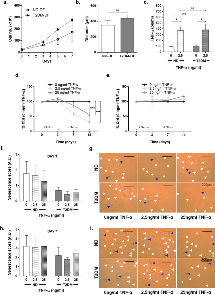
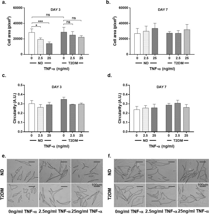
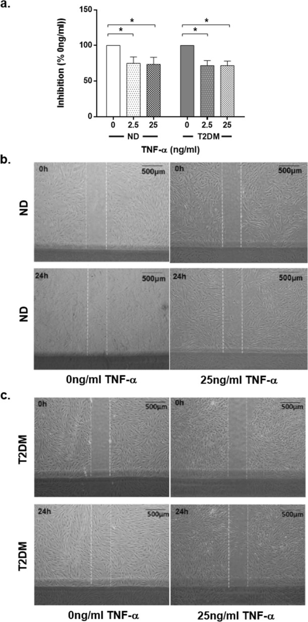
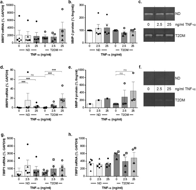
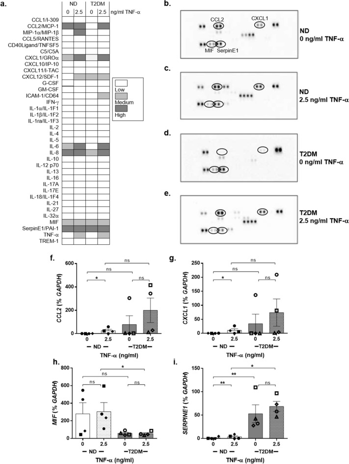
The prevalence of Type 2 diabetes mellitus (T2DM) is escalating globally. Patients suffer from multiple complications including the development of chronic wounds that can lead to amputation. These wounds are characterised by an inflammatory environment including elevated tumour necrosis factor alpha (TNF-α). Dermal fibroblasts (DF) are critical for effective wound healing, so we sought to establish whether there were any differences in DF cultured from T2DM donors or those without diabetes (ND-DF). ND- and T2DM-DF when cultured similarly in vitro secreted comparable concentrations of TNF-α. Functionally, pre-treatment with TNF-α reduced the proliferation of ND-DF and transiently altered ND-DF morphology; however, T2DM-DF were resistant to these TNF-α induced changes. In contrast, TNF-α inhibited ND- and T2DM-DF migration and matrix metalloprotease expression to the same degree, although T2DM-DF expressed significantly higher levels of tissue inhibitor of metalloproteases (TIMP)-2. Finally, TNF-α significantly increased the secretion of pro-inflammatory cytokines (including CCL2, CXCL1 and SERPINE1) in ND-DF, whilst this effect in T2DM-DF was blunted, presumably due to the tendency to higher baseline pro-inflammatory cytokine expression observed in this cell type. Collectively, these data demonstrate that T2DM-DF exhibit a selective loss of responsiveness to TNF-α, particularly regarding proliferative and secretory functions. This highlights important phenotypic changes in T2DM-DF that may explain the susceptibility to chronic wounds in these patients.


Conflict of interest statement

The authors declare no competing interests.

Figures

Figure 1
Figure 1
TNF-α has a differential effect on ND-DF and T2DM-DF proliferation. DF were quiesced for 24 h and then treated with DMEM + 10% FBS. (a) Basal proliferation rates were quantified by cell counting at days 2, 3 and 7 (n = 3 donors ND-DF, n = 4 donors T2DM-DF). (b) Basal migration rates were quantified using scratch wound assay (both n = 4 donors). (c) Confluent cells were quiesced for 24 h before being stimulated ± 2.5 ng/ml TNF-α for 24 h. Supernatants were collected and the concentration of TNF-α quantified (both n = 4 donors). (d) Cells were quiesced for 24 h and treated with 2.5 and 25 ng/ml TNF-α for 72 h before being transferred into full growth medium in the absence of TNF-α. Fluorescence was measured on days 0, 3, 7 and 14 and the percentage inhibition from control calculated for ND-DF and (e) T2DM-DF (both n = 4 donors). (fg) Parallel cultures were fixed and stained for senescence-associated β-galactosidase in ND-DF and T2DM-DF at day 3 and (hi) day 7 (both n = 4 donors). Blue arrowheads indicate senescent cells and white arrowheads indicate unstained cells. Two-way ANOVA with Sidak post-hoc test *P < 0.05, ***P < 0.001, ns = non-significant.
Figure 2
Figure 2
TNF-α has a differential effect on ND-DF and T2DM-DF morphology. The morphology of DF in images taken from the senescence assay were analysed using Image J. (a) Mean spread cell area and (b) circularity of ND- and T2DM-DF at day 3 post-TNF-α withdrawal (both n = 4 donors). (c) Representative images. (d) Mean spread cell area and (e) circularity of ND-DF and T2DM-DF at day 7 post-TNF-α withdrawal (both n = 4 donors). (f) Representative images. Two-way ANOVA with Sidak post-hoc test *P  < 0.05, ***P < 0.001, ns = non-significant.
Figure 3
Figure 3
Migratory capacity of ND-DF and T2DM-DF. (a) Confluent cells were quiesced for 24 h before being scratched, stimulated ± 0–25 ng/ml TNF-α and imaged. Repeat images were taken of the same wounds 24 h later. The percentage inhibition of TNF-α on migration was calculated by dividing the distance migrated in control cells by the distance migrated in treated cells (both n = 4 donors). (b) ND-DF representative images, (c) T2DM-DF representative images. Two-way ANOVA with Sidak post-hoc test, *P < 0.05, ns = non-significant.
Figure 4
Figure 4
Matrix remodelling in ND-DF and T2DM-DF. (a) Cells used in the migration assay were collected and total RNA extracted. MMP2 gene expression was quantified and expressed relative to GAPDH (both n = 4 donors). (b) Conditioned media from the migration assays was analysed for MMP-2 secretion using gelatin zymography (both n = 4 donors), (c) representative images. (d) MMP9 gene expression and (ef) protease activity were analysed using the same method (both n = 4 donors). (g) TIMP1 and (h) TIMP2 gene activity in the same cells (both n = 4 donors). Two-way ANOVA with Sidak post-hoc test ***P < 0.001, ns = non-significant.
Figure 5
Figure 5
Cytokine secretion in ND-DF and T2DM-DF. The secretion of multiple cytokines was measured in conditioned media from the migration assays using R&D Systems Human Cytokine Array Panel A. (a) Heatmap of the cytokines contained within the array and expression in ND and T2DM-DF, both basally and in response to 2.5 ng/ml TNF-α. (b) Representative dot blot used in the heatmap analysis for ND-DF under control (basal conditions), (c) ND-DF in response to TNF-α, (d) T2DM-DF under control (basal conditions) and (e) T2DM-DF in response to TNF-α. (f) Gene expression of CCL2, (g) CXCL1, (h) MIF and (i) SERPINE1 was measured in the cells from the migration assays (both n = 4 donors). Two-way ANOVA with Sidak post-hoc test, **P < 0.01, *P < 0.05, ns = non-significant.

Similar articles

Cited by

References

    1. Dinh T, et al. Mechanisms involved in the development and healing of diabetic foot ulceration. Diabetes. 2012;61:2937–2947. doi: 10.2337/db12-0227. - DOI - PMC - PubMed
    1. Martin P, Nunan R. Cellular and molecular mechanisms of repair in acute and chronic wound healing. Br. J. Dermatol. 2015;173:370–378. doi: 10.1111/bjd.13954. - DOI - PMC - PubMed
    1. Singer AJ, Clark RA. Cutaneous wound healing. N. Engl. J. Med. 1999;341:738–746. doi: 10.1056/NEJM199909023411006. - DOI - PubMed
    1. Thangapazhum RL, Darling TN, Meyerle J. Alternation of skin properties with autologous dermal fibroblasts. Int. J. Mol. Sci. 2014;15:8407–8427. doi: 10.3390/ijms15058407. - DOI - PMC - PubMed
    1. Smith RS, Smith TJ, Blieden TM, Phipps RP. Fibroblasts as sentinel cells: Synthesis of chemokines and regulation of inflammation. Am. J. Pathol. 1997;151:317–322. - PMC - PubMed

Publication types

MeSH terms

Substances