. 2021 Jan 14;11(1):1279.
doi: 10.1038/s41598-020-80270-9.

Deuterium-labeled Raman tracking of glucose accumulation and protein metabolic dynamics in Aspergillus nidulans hyphal tips

Affiliations

Deuterium-labeled Raman tracking of glucose accumulation and protein metabolic dynamics in Aspergillus nidulans hyphal tips

Mitsuru Yasuda et al. Sci Rep. .

Abstract

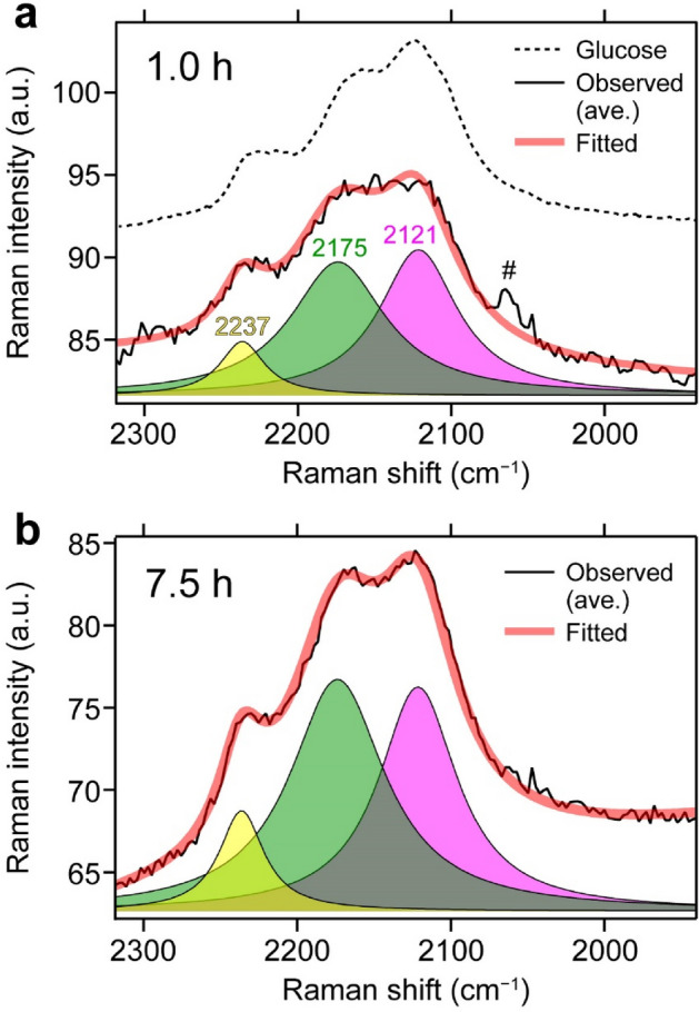
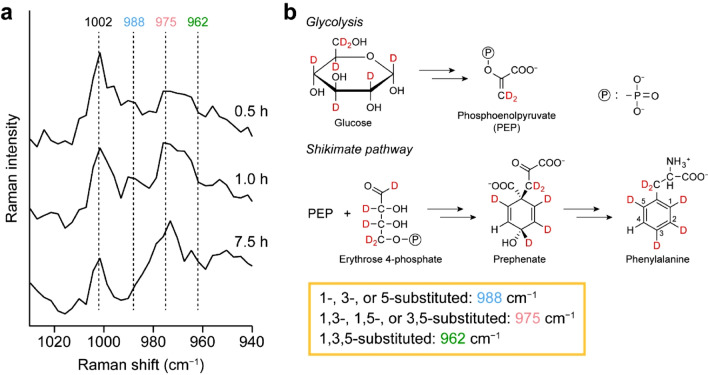
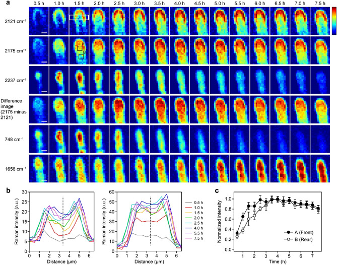
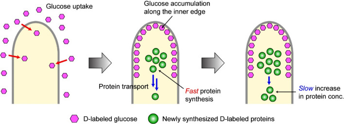
Filamentous fungi grow exclusively at their tips, where many growth-related fungal processes, such as enzyme secretion and invasion into host cells, take place. Hyphal tips are also a site of active metabolism. Understanding metabolic dynamics within the tip region is therefore important for biotechnology and medicine as well as for microbiology and ecology. However, methods that can track metabolic dynamics with sufficient spatial resolution and in a nondestructive manner are highly limited. Here we present time-lapse Raman imaging using a deuterium (D) tracer to study spatiotemporally varying metabolic activity within the hyphal tip of Aspergillus nidulans. By analyzing the carbon-deuterium (C-D) stretching Raman band with spectral deconvolution, we visualize glucose accumulation along the inner edge of the hyphal tip and synthesis of new proteins from the taken-up D-labeled glucose specifically at the central part of the apical region. Our results show that deuterium-labeled Raman imaging offers a broadly applicable platform for the study of metabolic dynamics in filamentous fungi and other relevant microorganisms in vivo.


Conflict of interest statement

The authors declare no competing interests.

Figures

Figure 1
Figure 1
Averaged Raman spectra of an A. nidulans hyphal tip at different culture times in D medium. (a) Optical images at 0.5, 3.0, and 7.5 h of the A. nidulans hypha studied in the present work. The formation of a septum is seen after 3.0 h. White dashed rectangular in the top image indicates the scanned area. Scale bar = 5 μm. (b) Average of Raman spectra measured within the hypha at 0.5, 1.0, 2.0, 4.0, 6.0, and 7.5 h after replacing H medium with D medium, together with a typical Raman spectrum of A. nidulans hyphal tips in H medium. The spectra are vertically offset for clarity of display. Asterisk denotes a broad background due to fluorescence emission from reduced cytochrome c. str. = stretching.
Figure 2
Figure 2
Least-squares fitting analysis of the C–D stretching band assuming three Lorentzian functions and a linear baseline. Observed averaged (thin black line) and fitted (thick red line) spectra at 1.0 (a) and 7.5 (b) h, together with the deconvolved subbands at 2121 (magenta), 2175 (green), and 2237 (yellow) cm−1. Also displayed in a is the Raman spectrum of D-labeled glucose dissolved in water (dashed line; not to scale). Hash sign (#) in a denotes a combination band of cytochromes.
Figure 3
Figure 3
Biosynthetic incorporation of deuterium from D-labeled glucose into proteins. (a) Raman spectra in the 940–1030 cm−1 region of the A. nidulans hypha at 0.5, 1.0, and 7.5 h. Each spectrum is an average of the Raman spectra recorded at all positions inside the hypha and vertically offset for clarity of display. (b) Simplified scheme for the biosynthetic pathways of the aromatic amino acid phenylalanine from glucose precursor via glycolysis and the shikimate pathway. This scheme was drawn by using ChemDraw 15.1.0.144 software, https://www.perkinelmer.com/category/chemdraw.
Figure 4
Figure 4
Time-lapse deuterium-labeled Raman imaging of the A. nidulans hyphal tip. (a) Time-lapse Raman images of the three deconvolved bands in the C–D stretching region at 2121, 2175, and 2237 cm−1, together with the difference images between the images at 2175 and 2121 cm−1 and Raman images of the 748 and 1656 cm−1 bands. In these images, Raman intensities are encoded in the “jet” color scale. The same color scale applies to Raman images in each row. White rectangular in the 2121 cm−1 image at 1.5 h indicates the region from which the line profiles shown in b were calculated; black squares (A and B) in the 2175 cm−1 image at 1.5 h indicate the regions from which the temporal profiles shown in c were obtained. Scale bar = 2 μm. (b) Line profiles of the 2121 and 2175 cm−1 images at 0.5, 1.0, 1.5, 2.0, 2.5, 4.0, 5.5, and 7.5 h. Dashed lines indicate the middle of the hypha. (c) Temporal profiles of the 2175 cm−1 band intensity in the front (A, filled circle) and rear (B, open circle) regions of the middle part of the hyphal tip. The intensity at each time point was calculated by averaging those at nine pixels within regions A and B and then normalized to the maximum value. Error bars represent standard deviation.
Figure 5
Figure 5
Cartoon picture showing the glucose uptake/accumulation and protein synthesis/transport processes in an A. nidulans hypha, as revealed by in vivo deuterium-labeled Raman imaging. conc. = concentration. This figure was created by using Adobe Illustrator CC 22.0.1 software, https://www.adobe.com/products/illustrator.html.

Similar articles

Cited by

References

    1. Garcia-Vidal C, Viasus D, Carratalà J. Pathogenesis of invasive fungal infections. Curr. Opin. Infect. Dis. 2013;26:270–276. doi: 10.1097/QCO.0b013e32835fb920. - DOI - PubMed
    1. Douglas AP, Chen SCA, Slavin MA. Emerging infections caused by non-Aspergillus filamentous fungi. Clin. Microbiol. Infect. 2016;22:670–680. doi: 10.1016/j.cmi.2016.01.011. - DOI - PubMed
    1. Punt PJ, et al. Filamentous fungi as cell factories for heterologous protein production. Trends Biotechnol. 2002;20:200–206. doi: 10.1016/S0167-7799(02)01933-9. - DOI - PubMed
    1. Kobayashi T, et al. Genomics of Aspergillus oryzae. Biosci. Biotechnol. Biochem. 2007;71:646–670. doi: 10.1271/bbb.60550. - DOI - PubMed
    1. Takeshita N, Manck R, Grün N, de Vega SH, Fischer R. Interdependence of the actin and the microtubule cytoskeleton during fungal growth. Curr. Opin. Microbiol. 2014;20:34–41. doi: 10.1016/j.mib.2014.04.005. - DOI - PubMed

Publication types

MeSH terms

LinkOut - more resources