. 2021 Jan 14;11(1):1425.
doi: 10.1038/s41598-020-80894-x.

Dulaglutide exerts beneficial anti atherosclerotic effects in ApoE knockout mice with diabetes: the earlier, the better

Affiliations

Dulaglutide exerts beneficial anti atherosclerotic effects in ApoE knockout mice with diabetes: the earlier, the better

Junpei Sanada et al. Sci Rep. .

Abstract

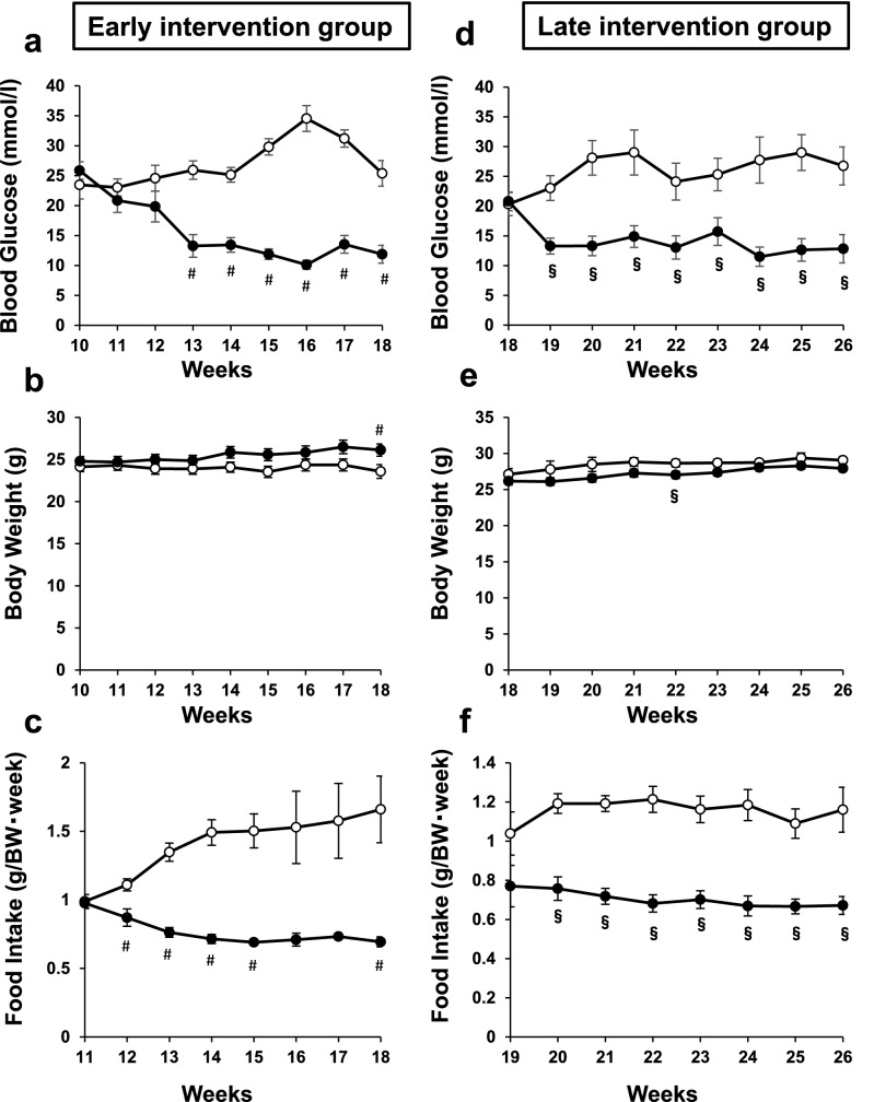
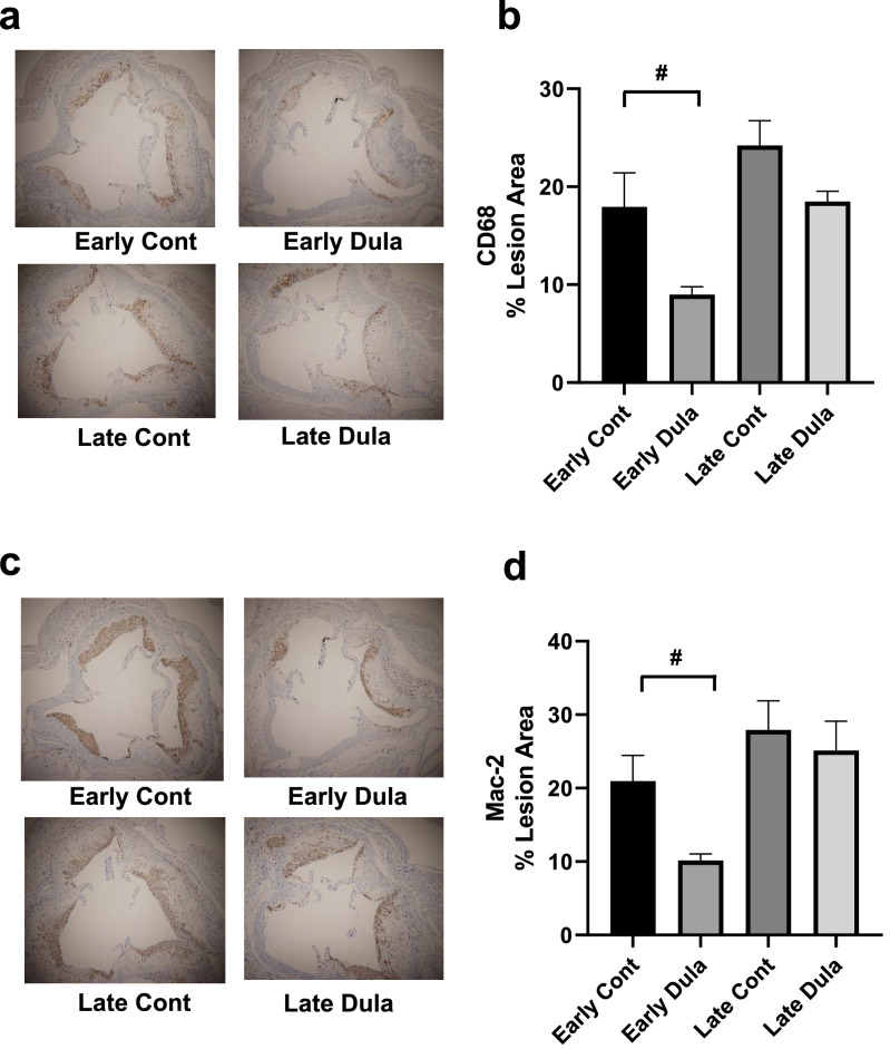
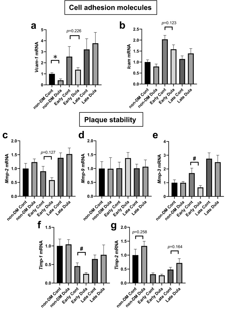
There has been no report about the mechanism for anti-atherosclerotic effects of dulaglutide (Dula) and/or about the difference of its effectiveness between in an early and a late phase of diabetes. To address such questions, streptozotocin (STZ) was intraperitoneally injected to ApoE knockout mice at 8 weeks of age. Either Dula or vehicle was administered to STZ-induced diabetic ApoE knockout mice from 10 to 18 weeks of age as an early intervention group and from 18 to 26 weeks as a late intervention group. Next, non-diabetic ApoE knockout mice without STZ injection were subcutaneously injected with either Dula or vehicle. In an early intervention group, atherosclerotic lesion in aortic arch and Mac-2 and CD68-positive areas in aortic root were significantly smaller in Dula group. In abdominal aorta, expression levels of some villain factors were lower in Dula group. In a late intervention group, there were no immunohistological differences in aortic root and expression levels of various factors between two groups. Furthermore, even in non-diabetic ApoE knockout mice, expression levels of inflammatory and macrophage markers were reduced by treatment with Dula. Taken together, Dula exerts more beneficial anti-atherosclerotic effects in an early phase of diabetes rather than in a late phase.


Conflict of interest statement

K.Ka. has been an advisor to, received honoraria for lectures from, and received scholarship grants from Novo Nordisk Pharma, Sanwa Kagaku Kenkyusho, Takeda, Taisho Pharma, MSD, Kowa, Sumitomo Dainippon Pharma, Novartis, Mitsubishi Tanabe Pharma, AstraZeneca, Boehringer Ingelheim, Chugai, Daiichi Sankyo, Sanofi. H.K. has received honoraria for lectures, received scholarship grants, and received research grant from Novo Nordisk Pharma, Sanofi, Eli Lilly, Boehringer Ingelheim, Taisho Pharma, Sumitomo Dainippon Pharma, Takeda Pharma, Ono Pharma, Daiichi Sankyo, Mitsubishi Tanabe Pharma, Kissei Pharma, MSD, AstraZeneca, Astellas, Novartis, Kowa.

Figures

Figure 1
Figure 1
Blood glucose levels, body weights and food intake in an early and a late intervention group in STZ-induced ApoE knockout mice. STZ-induced diabetic ApoE knockout mice were subcutaneously injected either Dula (0.6 mg/kg × 2/week) or vehicle in an early and a late intervention group. (a) Blood glucose levels, (b) body weights and (c) food intake in an early intervention group. Dula (black circle) (n = 9), control (white circle) (n = 9). (d) Blood glucose levels, (e) body weights and (f) food intake in a late intervention group. Dula (n = 8), control (n = 10). Values are the mean ± SEM of data obtained from each group. #p < 0.05 (Early Cont vs Early Dula), §p < 0.05 (Late Cont vs Late Dula).
Figure 2
Figure 2
Atherosclerotic lesion in aortic arch and Masson trichrome staining of aortic root in ApoE knockout mice. (a) Representative Sudan-IV staining pictures in aortic arch. (b) Sudan-IV positive area (% lesion area) (c) Representative Masson trichrome staining pictures in aortic root. (d) Masson trichrome staining (% lesion area). (e) Necrotic core in aortic root (% lesion area). Non-DM Cont (n = 7), non-DM Dula (n = 6), Early Cont (n = 5–6), Early Dula (n = 5 ~ 6), Late Cont (n = 6), Late Dula (n = 6). Values are the mean ± SEM of data obtained from each group. #p < 0.05 (Early Cont vs Early Dula), §p < 0.05 (Late Cont vs Late Dula).
Figure 3
Figure 3
CD68 and Mac2 staining of aortic root in early and late intervention groups in STZ-induced ApoE knockout mice. (a) Representative CD68 staining pictures, (b) CD68 positive area (% lesion area), (c) Representative Mac2 staining pictures, (d) Mac2 positive area (% lesion area). Early Cont (n = 5–6), Early Dula (n = 5–6), Late Cont (n = 6), Late Dula (n = 6). #p < 0.05 (Early Cont vs Early Dula).
Figure 4
Figure 4
mRNA expression levels of Glp-1r and inflammatory and coagulation markers in abdominal aorta. (a) Glp-1r mRNA expression levels, (bg) mRNA expression levels of inflammatory and coagulation markers expressed as fold change relative to non-diabetic control group. Non-diabetic Cont (n = 8), Non-diabetic Dula (n = 8), Early diabetic Cont (n = 8), Early diabetic Dula (n = 6), Late diabetic Cont (n = 8), Late diabetic Dula (n = 7). Values are the mean ± SEM of data obtained from each group. #p < 0.05 (Early Cont vs Early Dula).
Figure 5
Figure 5
mRNA expression levels related to cell adhesion molecules and plaque stability in abdominal aorta. (a, b) mRNA expression levels related to cell adhesion molecules such as Vcam-1 and Icam. (cg) mRNA expression levels related to plaque stability such as Mmp-2, Mmp-9, Mmp-3, Timp-1 and Timp2 expressed as fold change relative to non-diabetic control group. Non-diabetic Cont (n = 8), Non-diabetic Dula (n = 8), Early diabetic Cont (n = 8), Early diabetic Dula (n = 6), Late diabetic Cont (n = 8), Late diabetic Dula (n = 7). Values are the mean ± SEM of data obtained from each group. *p < 0.05 (non-DM Cont vs non-DM Dula), #p < 0.05 (Early Cont vs Early Dula).
Figure 6
Figure 6
mRNA expression levels of macrophage markers and M2-like macrophage markers in abdominal aorta. (a, b) mRNA expression levels of macrophage markers such as F4/80 and Cd68. (cg) mRNA expression levels M2-like macrophage markers such as Il-10, Cd206, Fizz1, Arg-1 and Ym-1 expressed as fold change relative to control group. Dula (n = 7), Control (n = 8). *p < 0.05 (non-DM cont vs non-DM Dula), #p < 0.05 (Early Cont vs Early Dula), §p < 0.05 (Late Cont vs Late Dula).

Similar articles

Cited by

References

    1. Drucker DJ. The cardiovascular biology of glucagon-like peptide-1. Cell Metab. 2016;24:15–30. doi: 10.1016/j.cmet.2016.06.009. - DOI - PubMed
    1. Koska J. Incretins and preservation of endothelial function. Cardiovasc. Hematol. Agents Med. Chem. 2012;10:295–308. doi: 10.2174/187152512803530289. - DOI - PubMed
    1. Marso SP, et al. LEADER Steering Committee; LEADER Trial Investigators. Liraglutide and cardiovascular outcomes in type 2 diabetes. N. Engl. J. Med. 2016;375:311–322. doi: 10.1056/NEJMoa1603827. - DOI - PMC - PubMed
    1. Marso SP, et al. SUSTAIN-6 Investigators. Semaglutide and cardiovascular outcomes in patients with type 2 diabetes. N. Engl. J. Med. 2016;375:1834–1844. doi: 10.1056/NEJMoa1607141. - DOI - PubMed
    1. Husain M, et al. PIONEER 6 Investigators. Oral semaglutide and cardiovascular outcomes in patients with type 2 diabetes. N. Engl. J. Med. 2019;381:841–851. doi: 10.1056/NEJMoa1901118. - DOI - PubMed

Publication types

MeSH terms