Role of prenatal imaging in the diagnosis and management of fetal facio-cervical masses
- PMID: 33446872
- PMCID: PMC7809128
- DOI: 10.1038/s41598-021-80976-4
Role of prenatal imaging in the diagnosis and management of fetal facio-cervical masses
Abstract
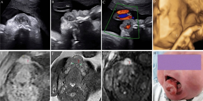
Congenital facio-cervical masses can be a developmental anomaly of cystic, solid, or vascular origin, and have an inseparable relationship with adverse prognosis. This retrospective cross-sectional study aimed at determining on the prenatal diagnosis of congenital facio-cervical masses, its management and outcome in a large tertiary referral center. We collected information on prenatal clinical data, pregnancy outcomes, survival information, and final diagnosis. Out of 130 cases of facio-cervical masses, a total of 119 cases of lymphatic malformations (LMs), 2 cases of teratoma, 2 cases of thyroglossal duct cyst, 4 cases of hemangioma, 1 case of congenital epulis, and 2 cases of dermoid cyst were reviewed. The accuracy of prenatal ultrasound was 93.85% (122/130). Observations of diameters using prenatal ultrasound revealed that the bigger the initial diameter is, the bigger the relative change during pregnancy. Magnetic resonance imaging (MRI) revealed that 2 cases of masses were associated with airway compression. In conclusion, ultrasound has a high overall diagnostic accuracy of fetal face and neck deformities. Prenatal US can enhance the management of ambulatory monitoring and classification. Furthermore, MRI provided a detailed assessment of fetal congenital malformations, as well as visualization of the trachea, presenting a multi-dimensional anatomical relationship.
Conflict of interest statement
The authors declare no competing interests.
Figures


Similar articles
-
[Iconographic imaging (ultrasound/MRI) in prenatal evaluation of cervical cystic lymphatic malformations].Gynecol Obstet Fertil. 2016 May;44(5):269-73. doi: 10.1016/j.gyobfe.2016.03.011. Epub 2016 Apr 22. Gynecol Obstet Fertil. 2016. PMID: 27118341 French.
-
Prenatal diagnosis of cervical masses by magnetic resonance imaging and 3D virtual models: perinatal and long-term follow-up outcomes.J Matern Fetal Neonatal Med. 2020 Jul;33(13):2181-2189. doi: 10.1080/14767058.2018.1543393. Epub 2018 Nov 20. J Matern Fetal Neonatal Med. 2020. PMID: 30458651
-
Prenatal magnetic resonance imaging enhances fetal diagnosis.J Pediatr Surg. 1998 Apr;33(4):553-8. doi: 10.1016/s0022-3468(98)90315-3. J Pediatr Surg. 1998. PMID: 9574750
-
Prenatal imaging - role of fetal MRI.Rofo. 2025 Apr;197(4):385-396. doi: 10.1055/a-2357-6997. Epub 2024 Dec 6. Rofo. 2025. PMID: 39642925 Review. English, German.
-
[Chances in current prenatal diagnosis].Ginekol Pol. 2003 Jul;74(7):565-72. Ginekol Pol. 2003. PMID: 14531331 Review. Polish.
Cited by
-
An operation on placental support in a fetus with a nasopharyngeal teratoma.Anaesth Rep. 2023 Mar 16;11(1):e12219. doi: 10.1002/anr3.12219. eCollection 2023 Jan-Jun. Anaesth Rep. 2023. PMID: 36936736 Free PMC article.
-
Masses of the fetal neck and oral cavity.Pediatr Radiol. 2025 Jun 25. doi: 10.1007/s00247-025-06307-1. Online ahead of print. Pediatr Radiol. 2025. PMID: 40560385 Review.
-
Large congenital facial teratoma.BMJ Case Rep. 2023 Apr 19;16(4):e253318. doi: 10.1136/bcr-2022-253318. BMJ Case Rep. 2023. PMID: 37076196 Free PMC article.
-
Cesarean section with operation on placental support as an adapted EXIT procedure for fetal thyroid goiter: A case report and literature review.Int J Gynaecol Obstet. 2025 May;169(2):474-484. doi: 10.1002/ijgo.16060. Epub 2024 Dec 5. Int J Gynaecol Obstet. 2025. PMID: 39636186 Free PMC article. Review.
References
Publication types
MeSH terms
LinkOut - more resources
Full Text Sources
Other Literature Sources
Medical