Review
. 2020 Nov 30;2020(2):e202029.
doi: 10.21542/gcsp.2020.29.

Microvascular dysfunction in septic and dengue shock: Pathophysiology and implications for clinical management

Affiliations
Review

Microvascular dysfunction in septic and dengue shock: Pathophysiology and implications for clinical management

Angela McBride et al. Glob Cardiol Sci Pract. .

Abstract

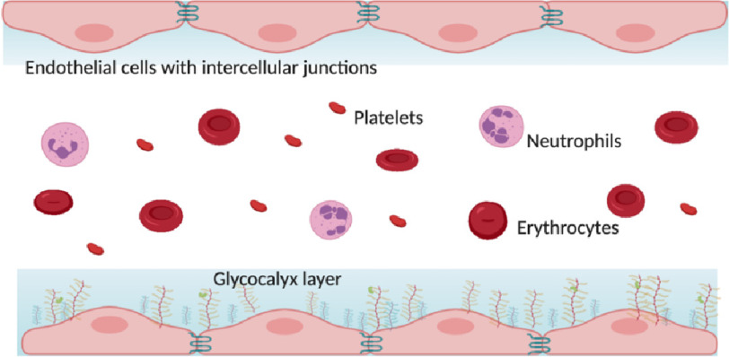
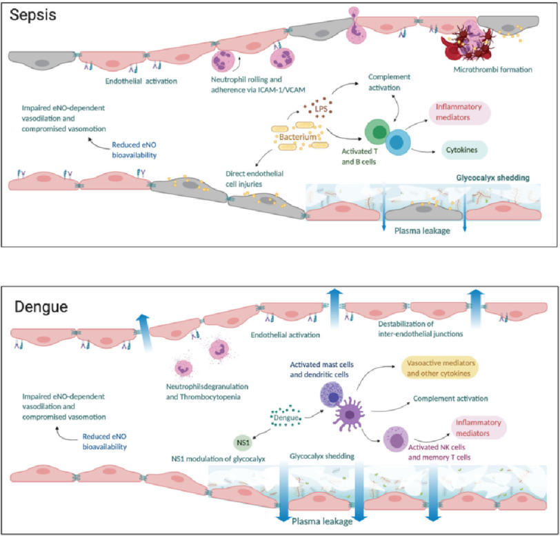
The microcirculation comprising of arterioles, capillaries and post-capillary venules is the terminal vascular network of the systemic circulation. Microvascular homeostasis, comprising of a balance between vasoconstriction, vasodilation and endothelial permeability in healthy states, regulates tissue perfusion. In severe infections, systemic inflammation occurs irrespective of the infecting microorganism(s), resulting in microcirculatory dysregulation and dysfunction, which impairs tissue perfusion and often precedes end-organ failure. The common hallmarks of microvascular dysfunction in both septic shock and dengue shock, are endothelial cell activation, glycocalyx degradation and plasma leak through a disrupted endothelial barrier. Microvascular tone is also impaired by a reduced bioavailability of nitric oxide. In vitro and in vivo studies have however demonstrated that the nature and extent of microvascular dysfunction as well as responses to volume expansion resuscitation differ in these two clinical syndromes. This review compares and contrasts the pathophysiology of microcirculatory dysfunction in septic versus dengue shock and the attendant effects of fluid administration during resuscitation.


Conflict of interest statement

SY receives consultancy fees from Janssen pharmaceuticals for dengue antiviral development and as a member of the ROCHE Advisory Board on Severe Dengue. All other authors declare no conflicts.

Figures

Figure 1.
Figure 1.. Appearance of the microcirculation under normal conditions with intact endothelial cell junctions and glycocalyx lining.
Figure 2.
Figure 2.. The microcirculation appearance in (a) sepsis and (b) dengue.
In sepsis, there is inflammation and direct injury of endothelial cells by bacterial proteins and toxins. Endothelial activation leads to neutrophil chemotaxis and adherence to intercellular adhesion molecule- 1 (ICAM-1) and vascular adhesion molecule (VCAM) expressed on endothelial cells. Concurrently, there is activation of complement, T and B immune cells leading to release of inflammatory mediators and cytokines which cause further inflammation and glycocalyx damage. Concurrently, the coagulation cascade is activated, and anticoagulant/fibrinolysis pathways are impaired leading to formation of microthrombi and compromised microvascular flow. In dengue, viral non-structural 1 (NS1) protein triggers hyperpermeability and directly alters the endothelial layer function through the activation of several enzymes responsible for glycocalyx degradation. Activation of immune cells leads to complement activation and release of inflammatory mediators. There is impaired endothelial nitric oxide (eNO) dependent vasodilation due to reduced eNO bioavailability leading to compromised microvascular flow. Plasma leakage in both sepsis and dengue occur as a result of glycocalyx shedding and disruption of tight and adherens junctions between endothelial cells.
Figure 3.
Figure 3.. Sublingual microcirculation in (a) normal (b) in septic shock.
The micro-vessels in septic shock have a comparatively larger diameter and do not run the entire course resulting in a dotted-line appearance characteristic of impaired microvascular flow in sepsis shown by the arrows in (b). Figures courtesy Critical Care Research Group (CCRG) laboratory, QLD, Australia.
Figure 4.
Figure 4.. Bleeding manifestations in dengue shock syndrome showing the appearance of linear petechiae after blood pressure cuff inflation.

References

    1. Singer M, Deutschman CS, Seymour CW, Shankar-Hari M, Annane D, Bauer M, Bellomo R, Bernard GR, Chiche JD, Coopersmith CM, et al. The third international consensus definitions for sepsis and septic shock (Sepsis-3) JAMA. 2016;315(8):801–810. - PMC - PubMed
    1. Wallez Y, Huber P. Endothelial adherens and tight junctions in vascular homeostasis, inflammation and angiogenesis. Biochim Biophys Acta. 2008;1778(3):794–809. - PubMed
    1. Yuan SY, Rigor RR. Regulation of endothelial barrier function. edn. San Rafael (CA); 2010. - PubMed
    1. Uchimido R, Schmidt EP, Shapiro NI. The glycocalyx: a novel diagnostic and therapeutic target in sepsis. Critical care. 2019;23(1):16. - PMC - PubMed
    1. Tarbell JM, Cancel LM. The glycocalyx and its significance in human medicine. J Intern Med. 2016;280(1):97–113. - PubMed

LinkOut - more resources