. 2021 Mar;47(3):04855.
doi: 10.3892/ijmm.2021.4855. Epub 2021 Jan 15.

Dihydroartemisinin attenuates osteoarthritis by inhibiting abnormal bone remodeling and angiogenesis in subchondral bone

Affiliations

Dihydroartemisinin attenuates osteoarthritis by inhibiting abnormal bone remodeling and angiogenesis in subchondral bone

Long Ma et al. Int J Mol Med. 2021 Mar.

Abstract

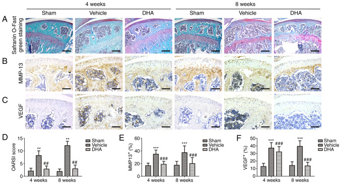
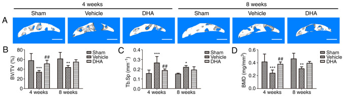
The present study aimed to investigate whether dihydroartemisinin (DHA) alleviates osteoarthritis (OA) in a mouse model of OA. Ten‑week‑old female C57BL/6j mice were used to establish OA models by anterior cruciate ligament transection (ACLT) and ovariectomized (OVX). DHA was then used to treat the OA in the ACLT and OVX mice. Safranin O‑fast green staining and Osteoarthritis Research Society International (OARSI)‑modified Mankin scores were used to grade articular cartilage degeneration. Expression of metalloproteinase‑13 (MMP‑13) and vascular endothelial growth factor (VEGF) in the articular cartilage and leukemia inhibitory factor (LIF), sclerostin, and β‑catenin in the subchondral bone were analyzed by immunohistochemistry. Expression of RANKL and CD31 were detected by immunofluorescence. Micro‑computed tomography was used to ascertain alterations in the microarchitecture of the subchondral bone. The results demonstrated that DHA decreased MMP‑13 and VEGF expression in the articular cartilage. DHA decreased OARSI scores and reduced articular cartilage degeneration. In addition, DHA reduced abnormal subchondral bone remodeling, as demonstrated by a reduction in trabecular separation (Tb.Sp), increased bone volume fractions (BV/TV), as well as bone mineral densities (BMD) compared with the ACLT+vehicle group and the OVX+vehicle group. Furthermore, DHA decreased the inhibition of sclerostin through reduction of LIF secretion by osteoclasts and, hence, attenuated aberrant bone remodeling and inhibited angiogenesis in subchondral bone, further reducing the progression of OA. The present study demonstrated that DHA attenuated OA by inhibiting abnormal bone remodeling and angiogenesis in subchondral bone, which may be a potential therapeutic target for this disease.

Keywords: osteoarthritis; subchondral bone; articular cartilage; angiogenesis; dihydroartemisinin; leukemia inhibitory factor.


Figures

Figure 1
Figure 1
Schematic of the experimental protocol. (A) Schematic of the experimental protocol of ACLT mice. (B) Schematic of the experimental protocol of OVX mice. ACLT, anterior cruciate ligament transection; OVX, ovariectomized.
Figure 2
Figure 2
DHA preserves the articular cartilage in ACLT mice. (A) Histological analysis of articular cartilage using safranin O-fast green staining of sagittal sections of the medial compartment of the tibia. Scale bar, 200 µm. (B, C, E and F) Measurement of matrix (B and E) MMP-13 and (C and F) VEGF expression by immunohistochemical staining and quantitative analysis. Scale bar, 100 µm. (D) OARSI-modified Mankin score of articular cartilage at various time-points. Sham, sham-operated group; Vehicle, ACLT+vehicle group; DHA, ACLT+DHA group. n=10 per group. **P<0.01 and ***P<0.001, compared with the the sham-operated group. ##P<0.01 and ###P<0.001, compared with the ACLT + vehicle group. DHA, dihydroartemisinin; ACLT, anterior cruciate ligament transection; MMP-13, metalloproteinase-13; VEGF, vascular endothelial growth factor; OARSI, Osteoarthritis Research Society International.
Figure 3
Figure 3
DHA restores the microarchitecture of the subchondral bone of ACLT mice. (A) 3D micro-CT reconstruction of sagittal views of the medial compartment of tibial subchondral bone at different time-points after sham or ACLT surgery. Scale bar, 500 µm. (B and D) Quantitative micro-CT analyses of the microarchitecture of tibial subchondral bone: (B) BV/TV (%), (C) Tb.Sp and (D) BMD. Sham, sham-operated group; Vehicle, ACLT+vehicle group; DHA, ACLT+DHA group. n=6 per group. *P<0.05, **P<0.01 and ***P<0.001, compared with the sham-operated group. ##P<0.01, compared with the vehicle group. DHA, dihydroartemisinin; ACLT, anterior cruciate ligament transection; micro-CT, micro-computed tomography; BV/TV, bone volume/tissue volume; Tb.Sp, trabecular separation; BMD, bone mineral density.
Figure 4
Figure 4
DHA inhibits abnormal bone remodeling in the subchondral bone of ACLT mice. (A and E) Immunofluorescence staining and quantification of the expression of RANKL (green) in tibial subchondral bone. Cell nuclei were stained blue using DAPI. Scale bar, 50 µm. (B-D and F-H) Immunohistochemical staining and quantification of the expression of (B and F) sclerostin, (C and G) β-catenin and (D and H) LIF in tibial subchondral bone. Scale bar, 50 µm. Sham, sham-operated group; Vehicle, ACLT+vehicle group; DHA, ACLT+DHA group. n=10 per group. **P<0.01 and ***P<0.001, compared with the sham-operated group. #P<0.05 and ##P<0.01, compared with the vehicle group. DHA, dihydroartemisinin; ACLT, anterior cruciate ligament transection; RANKL, receptor activator of nuclear factor κB ligand; LIF, leukemia inhibitory factor.
Figure 5
Figure 5
DHA inhibits abnormal angiogenesis in the subchondral bone of ACLT mice. (A and B) Immunofluorescence staining and quantification of the expression of CD31 (green) in tibial subchondral bone. Cell nuclei were stained blue using DAPI. Scale bar, 50 µm. Sham, sham-operated group; Vehicle, ACLT+vehicle group; DHA, ACLT+DHA group. n=10 per group. **P<0.01, compared with the sham-operated group. #P<0.05, compared with the vehicle group. DHA, dihydroartemisinin; ACLT, anterior cruciate ligament transection.
Figure 6
Figure 6
DHA preserves articular cartilage in OVX mice. (A) Histological analysis of articular cartilage using safranin O-fast green staining of sagittal sections of the medial compartment of the tibia. Scale bar, 200 µm. (B, C, E and F) Measurement of matrix (B and E) MMP-13 and (C and F) VEGF expression by immunohistochemical staining and quantitative analysis. Scale bar, 100 µm. (D) OARSI-modified Mankin score of articular cartilage at various time-points. Sham, sham-operated group; Vehicle, OVX+vehicle group; DHA, OVX+DHA group. n=10 per group. **P<0.01 and ***P<0.001, compared with the sham-operated group. ##P<0.01 and ###P<0.001, compared with the OVX+vehicle group. DHA, dihydroartemisinin; OVX, ovariectomized; MMP-13, metalloproteinase-13; VEGF, vascular endothelial growth factor; OARSI, Osteoarthritis Research Society International.
Figure 7
Figure 7
DHA restores the microarchitecture of the subchondral bone in OVX mice. (A) 3D micro-CT reconstruction of sagittal views of the medial compartment of tibial subchondral bone at different time-points after sham or OVX surgery. Scale bar, 500 µm. (B-D) Quantitative micro-CT analyses of the microarchitecture of tibial subchondral bone: (B) BV/TV (%), (C) Tb.Sp and (D) BMD. Sham, sham-operated group; Vehicle, OVX+vehicle group; DHA, OVX+DHA group. n=6 per group. **P<0.01 and ***P<0.001, compared with the sham-operated group. #P<0.05 and ##P<0.01, compared with the vehicle group. DHA, dihydroartemisinin; OVX, ovariectomized; micro-CT, micro-computed tomography; BV/TV, bone volume/tissue volume; Tb.Sp, trabecular separation; BMD, bone mineral density.
Figure 8
Figure 8
DHA inhibits abnormal bone remodeling in the subchondral bone of OVX mice. (A and E) Immunofluorescence staining and quantification of the expression of RANKL (green) in tibial subchondral bone. Cell nuclei were stained blue using DAPI. Scale bar, 50 µm. (B-D and F-H) Immunohistochemical staining and quantification of the expression of (B and F) sclerostin, (C and G) β-catenin and (D and H) LIF in tibial subchondral bone. Scale bar, 50 µm. Sham, sham-operated group. Vehicle, OVX+vehicle group. DHA, OVX+DHA group. n=10 per group. **P<0.01 and ***P<0.001, compared with the sham-operated group. #P<0.05 and ##P<0.01 and ###P<0.001 compared with the vehicle group. DHA, dihydroartemisinin; OVX, ovariectomized; RANKL, receptor activator of nuclear factor κB ligand; LIF, leukemia inhibitory factor.
Figure 9
Figure 9
DHA inhibits abnormal angiogenesis in the subchondral bone of OVX mice. (A and B) Immunofluorescence staining and quantification of the expression of CD31 (green) in tibial subchondral bone. Cell nuclei were stained blue using DAPI. Scale bar, 50 µm. Sham, sham-operated group; Vehicle, OVX+vehicle group; DHA, OVX+DHA group. n=10 per group. ***P<0.001, compared with the sham-operated group. ##P<0.01, compared with the vehicle group.

References

    1. Martel-Pelletier J, Barr AJ, Cicuttini FM, Conaghan PG, Cooper C, Goldring MB, Goldring SR, Jones G, Teichtahl AJ, Pelletier JP. Osteoarthritis. Nat Rev Dis Primers. 2016;2:16072. doi: 10.1038/nrdp.2016.72. - DOI - PubMed
    1. Goldring SR, Goldring MB. Changes in the osteochondral unit during osteoarthritis: Structure, function and cartilage-bone crosstalk. Nat Rev Rheumatol. 2016;12:632–644. doi: 10.1038/nrrheum.2016.148. - DOI - PubMed
    1. Hugle T, Geurts J. What drives osteoarthritis?-synovial versus subchondral bone pathology. Rheumatology (Oxford) 2017;56:1461–1471. - PubMed
    1. Huebner JL, Hanes MA, Beekman B, TeKoppele JM, Kraus VB. A comparative analysis of bone and cartilage metabolism in two strains of guinea-pig with varying degrees of naturally occurring osteoarthritis. Osteoarthritis Cartilage. 2002;10:758–767. doi: 10.1053/joca.2002.0821. - DOI - PubMed
    1. Eriksen EF. Cellular mechanisms of bone remodeling. Rev Endocr Metab Disord. 2010;11:219–227. doi: 10.1007/s11154-010-9153-1. - DOI - PMC - PubMed