Review
. 2021 Feb 26;49(1):107-118.
doi: 10.1042/BST20200043.

Aneuploidy in human eggs: contributions of the meiotic spindle

Affiliations
Review

Aneuploidy in human eggs: contributions of the meiotic spindle

Christopher Thomas et al. Biochem Soc Trans. .

Abstract

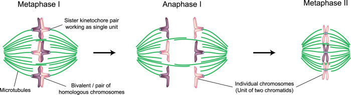
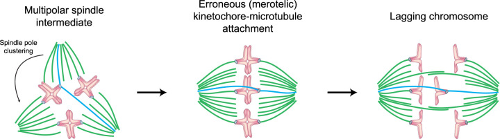
Human eggs frequently contain an incorrect number of chromosomes, a condition termed aneuploidy. Aneuploidy affects ∼10-25% of eggs in women in their early 30s, and more than 50% of eggs from women over 40. Most aneuploid eggs cannot develop to term upon fertilization, making aneuploidy in eggs a leading cause of miscarriages and infertility. The cellular origins of aneuploidy in human eggs are incompletely understood. Aneuploidy arises from chromosome segregation errors during the two meiotic divisions of the oocyte, the progenitor cell of the egg. Chromosome segregation is driven by a microtubule spindle, which captures and separates the paired chromosomes during meiosis I, and sister chromatids during meiosis II. Recent studies reveal that defects in the organization of the acentrosomal meiotic spindle contribute to human egg aneuploidy. The microtubules of the human oocyte spindle are very frequently incorrectly attached to meiotic kinetochores, the multi-protein complexes on chromosomes to which microtubules bind. Multiple features of human oocyte spindles favour incorrect attachments. These include spindle instability and many age-related changes in chromosome and kinetochore architecture. Here, we review how the unusual spindle assembly mechanism in human oocytes contributes to the remarkably high levels of aneuploidy in young human eggs, and how age-related changes in chromosome and kinetochore architecture cause aneuploidy levels to rise even higher as women approach their forties.

Keywords: aneuploidy; human oocyte; kinetochores; meiosis; spindle.


Conflict of interest statement

The authors declare that there are no competing interests associated with the manuscript.

Figures

Figure 1.
Figure 1.. Schematic representation of chromosome organization in human meiosis I and II.
(Left) Schematic of the meiosis I metaphase spindle. Green, microtubules. Light blue, kinetochores. Purple and magenta, parental chromosomes. The corresponding maternal and paternal chromosomes are joined by meiotic recombination to form a bivalent. Each bivalent contains two homologous chromosomes, one maternal and one paternal. In meiosis I, the sister kinetochores of each homologous chromosome act as a single unit and are attached to opposite spindle poles. (Middle) Schematic of the meiosis I anaphase spindle. Upon anaphase onset, the bivalent separates into two individual chromosomes, which then move to opposite spindle poles. One set of chromosomes will be eliminated into the first polar body (not represented), while the second set will remain in the egg and become aligned on the metaphase II spindle. (Right) Schematic of the metaphase II spindle. In meiosis II, the sister kinetochores do not act as a single unit anymore, but orient towards opposite spindle poles as in mitosis. Fertilization will trigger anaphase II (not shown), when sister chromatids will be separated. Half of the sister chromatids will be eliminated into the second polar body, while the second half will be enclosed in the maternal pronucleus.
Figure 2.
Figure 2.. Meiosis in human oocytes.
(Left) Schematic of chromatin organization and spindle assembly during human oocyte meiosis. Green, microtubules. Magenta, DNA. (Middle and right). Stills from a representative time-lapse movie of a human oocyte undergoing meiosis. Green, microtubules (EGFP-MAP4). Magenta, DNA (H2B-mRFP1). Merge with differential interference contrast in gray (right). Outlined regions magnified on the side (middle). Time, hours: minutes, 00:00 is nuclear envelope breakdown. Z-projections, 4 sections every 5 μm. Scale bar, 20 μm. Figure adapted with permission from figure 1 in [20].
Figure 3.
Figure 3.. Spindle instability during meiosis I leads to lagging chromosomes in anaphase I.
Schematic of a meiosis I spindle progressing through a transient multipolar spindle stage and then undergoing anaphase with lagging chromosomes. Green, microtubules forming correct attachments. Blue, microtubules forming erroneous attachments. Light blue, kinetochores. Magenta, DNA. The meiosis I spindle is often unstable and temporarily multipolar (left). Microtubules become incorrectly attached to kinetochores during the multipolar stages (see microtubules in blue). Error correction is incomplete, resulting in a high number of merotelically attached kinetochores prior to anaphase (middle). Such merotelic attachments are a common cause of lagging chromosomes in anaphase (right). The degree of spindle instability correlates with the degree of lagging chromosomes in anaphase.
Figure 4.
Figure 4.. Age-dependent changes in chromosome architecture lead to abnormal attachments with spindle microtubules and segregation errors.
(A) Schematic showing the ways in which chromosomes abnormally attach to spindle microtubules in human oocytes. Green, microtubules forming correct attachments. Blue, microtubules forming erroneous attachments. Light blue, kinetochores. Purple and magenta, chromosomes. Each bivalent contains two homologous chromosomes, one maternal and one paternal. (i) Sister kinetochores of the same chromosome should act as a single functional unit to allow for segregation of whole chromosomes in anaphase I. (ii) Sister kinetochores frequently separate in oocytes from older women and attach to spindle microtubules independently. (iii) Separation of sister kinetochores increases the probability of merotelic attachments. (iv) Separation of sister kinetochores allows bivalents to rotate on the spindle. Rotated bivalents can become inverted when their sister kinetochores attach to microtubules emanating from opposite spindle poles. (v) Bivalents may prematurely separate into univalents prior to anaphase I. These univalents often align on the metaphase spindle, with their two kinetochores facing in opposite directions and can give rise to both the reverse segregation and PSSC pattern of chromosome segregation in anaphase I. (vi) Kinetochores fragment into multiple lobes in oocytes from older females. The fragmented lobes of these kinetochores can attach independently to spindle microtubules. (B). Schematic showing the patterns of chromosome missegregation in human oocytes. Light blue, kinetochores. Purple and magenta, chromosomes. (i) Normal segregation — bivalents are separated in anaphase I into two individual chromosomes. Only one of these is retained within the oocyte and the other is excluded in the polar body. (ii) Nondisjunction — when chromosome segregation fails in anaphase I, leaving the entire bivalent in either the oocyte or polar body. (iii) Reverse segregation - sister chromatids, but not homologous chromosomes, segregate in meiosis I. Consequently, while the oocyte will contain the correct number of chromosomes in meiosis II, the chromatids of the missegregated chromosome will have different parental origins and are not linked. These mismatched chromatids will hence act independently of each other in meiosis II and are, therefore, likely to missegregate during anaphase II. (iv) Premature separation of sister chromatids (PSSC) — where an individual chromatid missegregates during anaphase I. This is a primary cause of aneuploidy in human oocytes. (C). Schematic showing cohesin dissociation from chromosomes in oocytes from older women. Green, microtubules forming correct attachments. Blue, microtubules forming erroneous attachments. Light blue, kinetochores. Pink, chromosomes. Purple, cohesin. Cohesin is a ring-like structure that is loaded onto chromosomes in the embryo. Cohesin tethers both homologous chromosomes and sister chromatids together prior to anaphase. In mouse oocytes, cohesin complexes have been shown to gradually dissociate from chromosomes with age. This can result in gaps between homologous chromosomes in oocytes from aged females and an increase in the frequency of abnormal attachments with spindle microtubules.

References

    1. Centers for Disease Control and Prevention. Assisted Reproductive Technology Fertility Clinic Success Rates Report - 2017. 2017;17:105–16. https://www.cdc.gov/art/reports/2017/fertility-clinic.html.
    1. Wong, C.C., Loewke, K.E., Bossert, N.L., Behr, B., De Jonge, C.J., Baer, T.M.et al. (2010) Non-invasive imaging of human embryos before embryonic genome activation predicts development to the blastocyst stage. Nat. Biotechnol. 28, 1115–1121 10.1038/nbt.1686 - DOI - PubMed
    1. Benkhalifa, M., Kasakyan, S., Clement, P., Baldi, M., Tachdjian, G., Demirol, A.et al. (2005) Array comparative genomic hybridization profiling of first-trimester spontaneous abortions that fail to grow in vitro. Prenat. Diagn. 25, 894–900 10.1002/pd.1230 - DOI - PubMed
    1. McCoy, R.C. (2017) Mosaicism in preimplantation human embryos: when chromosomal abnormalities are the norm. Trends Genet. 33, 448–463 10.1016/j.tig.2017.04.001 - DOI - PMC - PubMed
    1. Shahbazi, M.N., Wang, T., Tao, X., Weatherbee, B.A.T., Sun, L., Zhan, Y.et al. (2020) Developmental potential of aneuploid human embryos cultured beyond implantation. Nat. Commun. 11, 3987 10.1038/s41467-020-17764-7 - DOI - PMC - PubMed

Publication types

LinkOut - more resources