Review
. 2021 Apr;78(7):3205-3218.
doi: 10.1007/s00018-020-03735-0. Epub 2021 Jan 15.

Mechanisms of meiotic drive in symmetric and asymmetric meiosis

Affiliations
Review

Mechanisms of meiotic drive in symmetric and asymmetric meiosis

Alyssa N Kruger et al. Cell Mol Life Sci. 2021 Apr.

Abstract

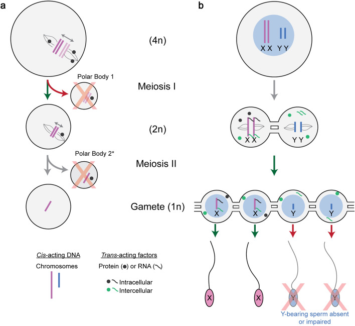
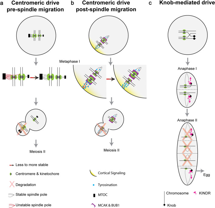
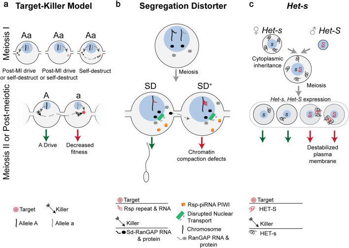
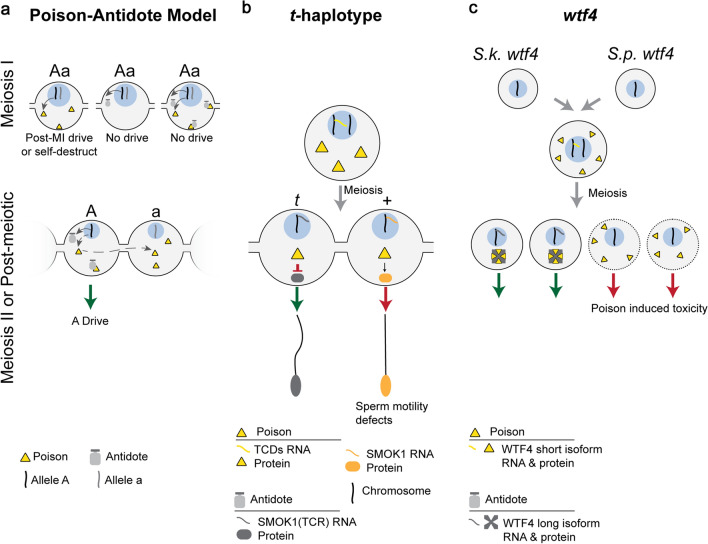
Meiotic drive, the non-Mendelian transmission of chromosomes to the next generation, functions in asymmetric or symmetric meiosis across unicellular and multicellular organisms. In asymmetric meiosis, meiotic drivers act to alter a chromosome's spatial position in a single egg. In symmetric meiosis, meiotic drivers cause phenotypic differences between gametes with and without the driver. Here we discuss existing models of meiotic drive, highlighting the underlying mechanisms and regulation governing systems for which the most is known. We focus on outstanding questions surrounding these examples and speculate on how new meiotic drive systems evolve and how to detect them.

Keywords: Germ cells; Meiosis; Meiotic drive; Selfish genetic elements.


Conflict of interest statement

The authors declare no competing interests.

Figures

Fig. 1
Fig. 1
The defining features of meiotic drive in asymmetric and symmetric meiosis. a Females undergo asymmetric meiosis—a single gamete is produced from a single round of meiosis. The driving cis-acting chromosomes (dark pink) biases its retention to the egg by interacting with the inward-facing egg pole (green arrow). The non-driving chromosome (light pink) binds the outward facing cortical pole and is extruded to a polar body (red arrow) which is degraded (red X). * The second polar body is extruded upon fertilization. b Males undergo symmetric meiosis—four gametes are produced from a single round of meiosis. Male meiotic drive systems bias fertilization by increasing the relative abundance of sperm carrying the driving chromosome (green arrow), or by decreasing the fitness of sperm with the non-driving chromosome (red arrow). Gray mRNA and protein represent X-linked trans-acting factors which remain in X-bearing cells (e.g., intracellular). Green mRNA and protein represent X-linked trans-acting factors which are shared and present in Y-bearing cells (e.g., intercellular) via cytoplasmic bridges. Cytoplasmic bridges are established prior to meiosis, in spermatogonia, and connect cells throughout meiosis and after meiosis. For simplicity, a single meiotic cell and the products of meiosis are shown.
Fig. 2
Fig. 2
Models of Asymmetric Meiotic Drive. This figure depicts the mechanistic understanding of two examples of centromeric drive (a and b) and one example of non-centromeric drive (c). a An example of female centromeric drive in hybrid mice where cis-acting chromosome reorientation occurs before trans-acting spindle migration. In this system, larger MTOCs (black box) give rise to denser spindle poles (gray line) which preferentially interact with larger kinetochores (green oval). If this favorable interaction is not initially established (red line), then proteins involved in fixing erroneous microtubule attachments are recruited and chromosomes reorient to the more favorable interaction (red–black arrow). Spindles migrate to the periphery, and the outward-facing larger kinetochore is extruded to the polar body and degraded (red X). b Another example of female centromeric drive in hybrid mice where chromosomes reorientation occurs after spindle migration. Cortical signaling (yellow gradient) leads to an enrichment of tyrosination (blue circle) on cortical spindle poles. Spindle pole tyrosination is less stable (red lines) on larger kinetochores (green) with more BUB1 kinase and MCAK (purple). If not in the more stable orientation, then proteins involved in fixing erroneous microtubule attachments cause chromosomes to reorient to the stable orientation (red–black arrow), causing the smaller kinetochore to be extruded to the polar body (red X). c Maize knob-mediated drive requires the kinesin KINDR (pink) which binds knob repeats (black circles) and migrates along the spindle microtubules toward the outward spindle poles (pink arrow), including the distal cell which becomes the egg
Fig. 3
Fig. 3
Symmetric Meiotic Drive—Target–Killer. This figure depicts the theoretical importance of intracellular versus intercellular regulation on target–killer meiotic drive systems (a), and the mechanisms underlying two target–killer meiotic drivers (b and c). a The killer protein (black arrow, encoded on the “A” haplotype) targets, directly or indirectly, the target (red bullseye, encoded on the “a” haplotype). During meiosis I (MI), expression of only the killer before or during MI can result in post-MI drive, if the target is a trans-acting factor expressed during or after meiosis II (MII). If the target is a cis-acting sequence, expression of the killer before or during MI could cause the driver to self-destruct. If only a trans-acting target is expressed during MI, post-MI drive occurs if the killer is expressed during or after MII, assuming the target acts intracellularly. However, if the target is partitioned into all MII cells and is therefore intercellular, then expression of the killer will result in the system self-destructing. If a trans-acting killer is expressed during MI and a cis-acting or trans-acting target are accessible, then the system will self-destruction. The target and killer are present in different cells and on different haplotypes in order to not self-destruct, as would be the case if both were expressed in meiotic cells. Expression of the target or killer following homologous chromosome separation in MII prevents self-destruction and results in meiotic drive (green and red arrows). Post-meiosis II expression of the killer necessitates sharing (dashed gray arrow) through cytoplasmic bridges, the target should not be shared in order to prevent self-destruction. b Drosophila melanogaster Segregation Distorter (SD) encodes the killer Sd-RanGAP. One model proposes that Sd-RanGAP mislocalization disrupts piRNA-based silencing (orange) of the target, the high copy number Rsp (red lines) resulting in chromatin compaction defects and SD drive. c The female strain of P. anserina encodes the killer allele, Het-s, and HET-s protein (black curly line) is therefore present upon gamete fusion as the cytoplasm (gray surrounding blue nucleus) is maternally inherited. The male strain’s Het-S allele and female Het-s are expressed following meiosis where they complex in Het-S spores and integrate and destabilize the plasma membrane (red arrow), resulting in Het-s drive (green arrow). Cells are contained within an ascus (large gray oval)
Fig. 4
Fig. 4
Symmetric Meiotic Drive—Poison-Antidote. This figure depicts the theoretical importance of intracellular versus intercellular regulation on poison–antidote meiotic drive systems (a), and the mechanisms underlying two poison-antidote meiotic drivers (b and c). a The poison protein (yellow triangle) and antidote (gray shape) are encoded by the same haplotype (“A”) in close genetic proximity. The antidote acts intracellularly (“A” haplotype) to spare driving chromosomes from the effects of the intercellular poison. Expression of the poison before or during MI can result in post-MI drive, if the poison acts specifically in MII or post-meiosis. However, if the poison acts on a biological pathway present before or during MI, expression before or during MI leads to self-destruction. Expression of the antidote before or during MI would result in it being partitioned into and consequently sparing all daughter cells, preventing drive. Expression of both the poison and antidote before homologous chromosomes separate would result in the antidote neutralizing the poison and preventing drive. Meiosis II or later expression of the poison requires that the poison is shared through cytoplasmic bridges (dashed gray arrow) to poison the other cell containing the “a” haplotype, resulting in A drive (green arrow). b In the mouse t-haplotype, the poisons, TCDs (yellow triangle), are expressed before meiosis and disrupt the post-meiotically expressed SMOK1 (orange oval) resulting in flagellar dysregulation (red arrow). The antidote, SMOK1(TCR) (gray oval), is a dominant negative variant of SMOK1 and is insensitive to TCD induced dysregulation, resulting in t-haplotype drive (green arrow). c In Schizosaccharomyces, the wtf4 gene encoded both the pre-meiotically expressed poison (yellow triangle) and the post-meiotically expressed antidote (gray shape). These represent distinct isoforms of wtf4. The antidote sequesters poison into distinct subcellular regions preventing its toxicity and results in drive (green arrow)

References

    1. Hurst GD, Werren JH. The role of selfish genetic elements in eukaryotic evolution. Nat Rev Genet. 2001;2(8):597–606. doi: 10.1038/35084545. - DOI - PubMed
    1. Werren JH. Selfish genetic elements, genetic conflict, and evolutionary innovation. Proc Natl Acad Sci USA. 2011;108(Suppl 2):10863–10870. doi: 10.1073/pnas.1102343108. - DOI - PMC - PubMed
    1. Agren JA, Clark AG. Selfish genetic elements. PLoS Genet. 2018;14(11):e1007700. doi: 10.1371/journal.pgen.1007700. - DOI - PMC - PubMed
    1. Hurst LD. A century of bias in genetics and evolution. Heredity (Edinb) 2019;123(1):33–43. doi: 10.1038/s41437-019-0194-2. - DOI - PMC - PubMed
    1. Helleu Q, Gerard PR, Montchamp-Moreau C. Sex chromosome drive. Cold Spring Harb Perspect Biol. 2014;7(2):a017616. doi: 10.1101/cshperspect.a017616. - DOI - PMC - PubMed

LinkOut - more resources