. 2021 Apr;78(7):3565-3576.
doi: 10.1007/s00018-020-03745-y. Epub 2021 Jan 15.

Ultrastructural modifications induced by SARS-CoV-2 in Vero cells: a kinetic analysis of viral factory formation, viral particle morphogenesis and virion release

Affiliations

Ultrastructural modifications induced by SARS-CoV-2 in Vero cells: a kinetic analysis of viral factory formation, viral particle morphogenesis and virion release

Sébastien Eymieux et al. Cell Mol Life Sci. 2021 Apr.

Abstract

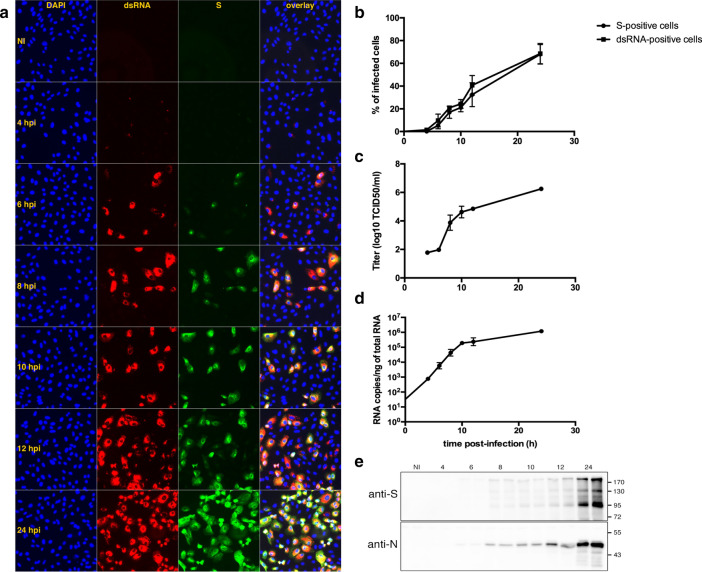
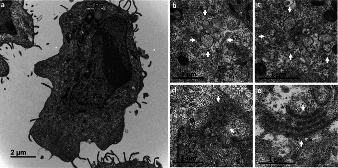
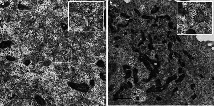
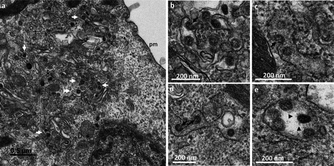
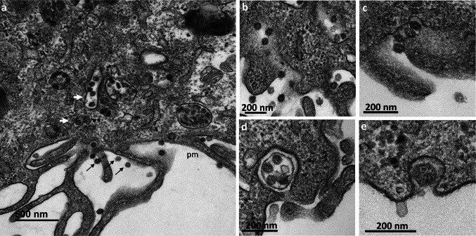
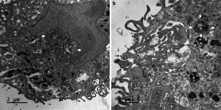
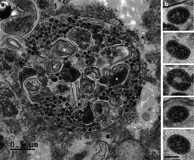
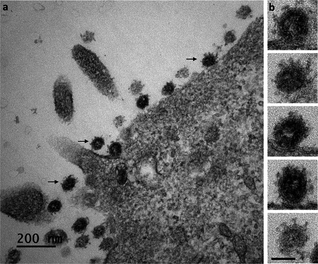
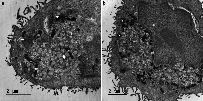
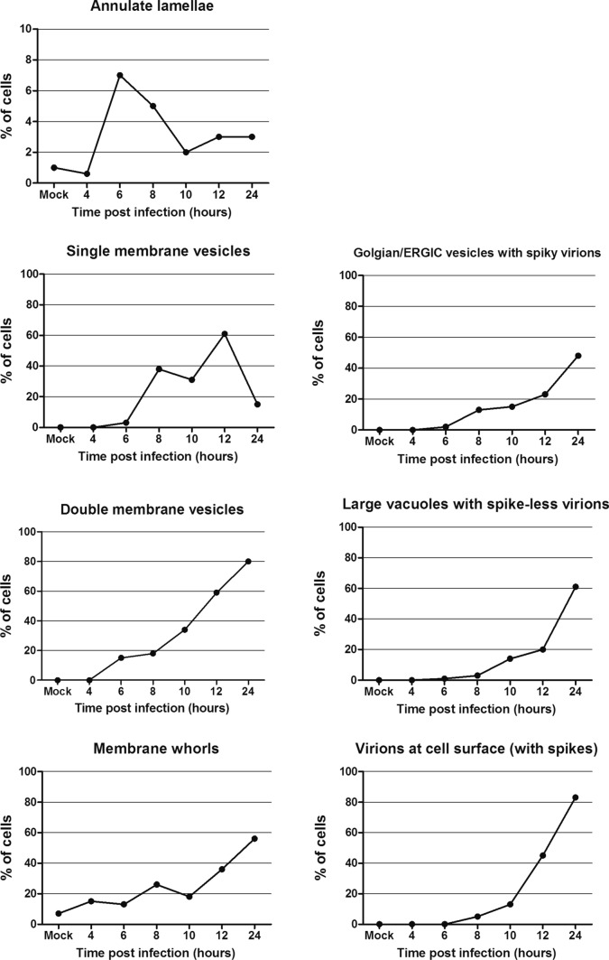
Many studies on SARS-CoV-2 have been performed over short-time scale, but few have focused on the ultrastructural characteristics of infected cells. We used TEM to perform kinetic analysis of the ultrastructure of SARS-CoV-2-infected cells. Early infection events were characterized by the presence of clusters of single-membrane vesicles and stacks of membrane containing nuclear pores called annulate lamellae (AL). A large network of host cell-derived organelles transformed into virus factories was subsequently observed in the cells. As previously described for other RNA viruses, these replication factories consisted of double-membrane vesicles (DMVs) located close to the nucleus. Viruses released at the cell surface by exocytosis harbored the typical crown of spike proteins, but viral particles without spikes were also observed in intracellular compartments, possibly reflecting incorrect assembly or a cell degradation process.

Keywords: Covid-19; Electron microscopy; SARS-CoV-2; Virus/cell interactions.


Figures

Fig. 1
Fig. 1
Kinetics of infection analyzed by different methods. (a) Vero cells grown on glass coverslips were infected, fixed at the indicated time points and processed for immunofluorescence labeling of S (green) and dsRNA (red). Nuclei were stained with DAPI (blue). (b) Cells positive for S and dsRNA were counted and the percentage of infected cells was plotted against time. (c) The quantity of virus secreted in the cell supernatant was assessed by the TCID50 method. (d) Intracellular genome quantification was performed by qRT-PCR. (e) Intracellular N and S viral proteins were detected by immunoblot
Fig. 2
Fig. 2
Early ultrastructural changes encountered in Vero cells at 6 h post-infection with SARS-CoV-2. (a) At 4 hpi, no ultrastructural modifications relative to mock-infected cells, shown here, were visible. (b,c) At 6 hpi, the first discrete virus-induced structures appeared, consisting of clusters (delimited by the white arrows) of several single-membrane vesicles. These clusters contained 5–10 single-membrane vesicles per cell section, and were frequently observed in the perinuclear area (n, nucleus). (d,e) At 6 hpi, annulate lamellae (AL) were frequently observed in infected cells, either as nuclear (n) expansions (d, white arrows) or isolated in the cytoplasm (e, white arrows), suggesting that the formation of these structures may have been promoted by virus infection. AL consist of stacks of highly ordered endoplasmic reticulum-derived membranes, arranged in parallel and containing nuclear pore complexes, that can be visualized in tangential (e) or cross (d) sections
Fig. 3
Fig. 3
Replication network established in Vero cells at 8 h post-infection with the SARS-CoV-2. (a,b) A massive replication network consisting of large clusters of numerous double-membrane vesicles (DMVs, presented at high magnification in the insets) was observed. These clusters of DMVs were frequently surrounded by mitochondria (m)
Fig. 4
Fig. 4
Morphogenesis of intracellular SARS-CoV-2 virions in Vero cells at 8 h post-infection. (a) Virus particles (white arrows) were observed in intracellular vesicles related to the Golgi apparatus or the endoplasmic reticulum/Golgi intermediate compartment (ERGIC). No virus was released at the plasma membrane (pm) at this time point. (b–d) The budding of the viruses (white arrows) toward the lumen of the ERGIC/Golgi vesicles was visualized in some cell sections. (e) At this time point, the intracellular viruses were detected in small vesicles, each containing only a few viral particles. These viral particles displayed prominent spikes, clearly visible at the surface (black arrowhead)
Fig. 5
Fig. 5
Release of the SARS-CoV-2 virions by exocytosis from the Vero cells at 10 h post-infection. (a) Virions (thin black arrows) were frequently observed at the plasma membrane (pm). Numerous virus-carrying vesicles (white arrows) presumably in transit to the plasma membrane were also visualized. (b–e) These virus-carrying vesicles fused with the plasma membrane to release their contents into the extracellular space by exocytosis. Similar ultrastructural events were observed at 12 hpi
Fig. 6
Fig. 6
Intracellular accumulation of SARS-CoV-2 particles in large vacuoles within Vero cells at 24 hpi. (a,b) Viruses were still detected at the cell surface (thin black arrows), but large numbers of viral particles were observed accumulated in very large intracellular vacuoles (white arrows) of various sizes, predominantly located in the perinuclear region. At this time point, an accumulation of myelin-like membrane whorls or autophagic-like packaged membranes (white asterisk in b) was also observed in the cells, separated from or associated with the viral particles
Fig. 7
Fig. 7
The SARS-CoV-2 particles accumulating in large intracellular vacuoles in Vero cells at 24 hpi have no spikes. (a) The numerous viral particles accumulating in these large intracellular compartments were often electron-dense and presented a smooth surface. Membrane whorls (white asterisks) were sometimes also associated with the viral particles within these large intracellular vacuoles. (b) At high magnification, the viral particles present in these large intracellular vacuoles were clearly observed to have no spikes (bar = 50 nm)
Fig. 8
Fig. 8
The SARS-CoV-2 particles released from the Vero cells at 24 hpi carry spikes. (a) All the viral particles released at the cell surface (thin black arrows) were surrounded by characteristic, “club-shaped”, spikes. (b) This was confirmed by an analysis of these viral particles at high magnification, revealing their distinctive crown-like appearance (bar = 50 nm)
Fig. 9
Fig. 9
Invasive replication network in Vero cells at 24 h post-infection with SARS-CoV-2. (a,b) At this time point, SARS-CoV-2-infected cells displayed an intense reticulovesicular network consisting mostly of large numbers of DMVs, which occupied almost all the cytoplasm and seemed to push against the nuclear compartment (n), with many mitochondria (m) recruited at the edge of this network. Virion assembly continued next to this network (white arrows in a), leading to release at the plasma membrane (thin black arrows in a). DMVs were often associated with myelin-like membrane whorls or autophagic-like packaged membranes (white asterisk in b)
Fig. 10
Fig. 10
Quantitative analysis of the ultrastructural features in infected Vero cells, compared to mock-infected cells. For each point of the kinetics, 100 cell sections have been analyzed. Identification of at least one element in a cell section counted as one in the total account. For the annulate lamellae graph, due to the rarity of these structures and therefore the low associated cell percentage, Y axis displays only a 10% value. * At 4 hpi, 170 cell sections have been analyzed to find one cell containing annulate lamellae, leading to a <1% result

References

    1. van der Hoek L, Pyrc K, Jebbink MF, Vermeulen-Oost W, Berkhout RJM, Wolthers KC, Wertheim-van Dillen PM, Kaandorp J, Spaargaren J, Berkhout B. Identification of a new human coronavirus. Nat Med. 2009;10:368–773. doi: 10.1038/nm1024. - DOI - PMC - PubMed
    1. Woo PCY, Lau SKP, Tsoi H-W, Huang Y, Poon RWS, Chu C-M, Lee RA, Luk WK, Wong GK, Wong BH, Cheng VC, Tang BS, Wu AK, Yung RW, Chen H, Guan Y, Chan KH, Yuen KY. Clinical and molecular epidemiological features of coronavirus HKU1-associated community-acquired pneumonia. J Infect Dis. 2005;192:1898–1907. doi: 10.1086/497151. - DOI - PMC - PubMed
    1. Edridge AWD, Kaczorowska J, Hoste ACR, Bakker M, Klein M, Loens K, Jebbink MF, Matser A, Kinsella CM, Rueda P, Ieven M, Goossens H, Prins M, Sastre P, Deijs M, van der Hoek L (2020) Seasonal coronavirus protective immunity is short-lasting. Nat Med. doi: 10.1038/s41591-020-1083-1 (online ahead of print) - PubMed
    1. Ksiazek TG, Erdman D, Goldsmith CS, Zaki SR, Peret T, Emery S, Ong S, Urbani C, Comer JA, Lim W, Rollin PE, Dowell SF, Ling AE, Humphrey CD, Shieh WJ, Guarner J, Paddock CD, Rota P, Fields B, DeRisi J, Yang JY, Cox N, Hughes JM, LeDuc JW, Bellini WJ, Anderson LJ. A novel coronavirus associated with severe acute respiratory syndrome. N Engl J Med. 2003;348:1953–1966. doi: 10.1056/NEJMoa030781. - DOI - PubMed
    1. Zaki AM, van Boheemen S, Bestebroer TM, Osterhaus ADME, Fouchier RAM. Isolation of a novel coronavirus from a man with pneumonia in Saudi Arabia. N Engl J Med. 2012;367:1814–1820. doi: 10.1056/NEJMoa1211721. - DOI - PubMed

MeSH terms

Substances

LinkOut - more resources