. 2021 Jun;28(3):581-588.
doi: 10.1007/s10140-020-01880-4. Epub 2021 Jan 15.

Safety and image quality of MR-conditional external fixators for 1.5 Tesla extremity MR

Affiliations

Safety and image quality of MR-conditional external fixators for 1.5 Tesla extremity MR

David H Ballard et al. Emerg Radiol. 2021 Jun.

Erratum in

Abstract

Purpose: To evaluate the safety and image quality of extremity MR examinations performed with two MR conditional external fixators located in the MR bore.

Materials and methods: Single-center retrospective study of a prospectively maintained imaging dataset that evaluated MR examinations of extremities in patients managed with external fixations instrumentation and imaged on a single 1.5T MR scanner. The fixation device was one of two MR-conditional instrumentation systems: DuPuy Synthes (aluminum, stainless steel, carbonium and Kevlar) or Dolphix temporary fixation system (PEEK-CA30). Safety events were recorded by the performing MR radiologic technologist. A study musculoskeletal radiologist assessed all sequences to evaluate for image quality, signal- and contrast-to-noise ratios (SNR/CNR), and injury patterns/findings.

Results: In the 13 men and 9 women with a mean age of 42 years (range 18 to 72 years), most patients (19/22 patients; 86%) were involved with trauma resulting in extremity injury requiring external fixation. MR examinations included 19 knee, 2 ankle, and 1 elbow examinations. There were no adverse safety events, heating that caused patient discomfort, fixation dislodgement/perturbment, or early termination of MR examinations. All examinations were of diagnostic quality. Fat-suppressed proton density sequences had significantly higher SNR and CNR compared to STIR (p = 0.01 to 0.04). The lower SNR of STIR and increased quality of fat-suppressed proton density during the study period led to the STIR sequence being dropped in standard MR protocol.

Conclusion: MR of the extremity using the two study MR conditional external fixators within the MR bore is safe and feasible.

Keywords: External fixator; MR artifact; MR safety; Susceptibility artifact; Tibial plateau fracture.


Conflict of interest statement

Conflict of interest

The authors have no potential conflicts of interest to disclose.

Figures

Figure 1.
Figure 1.
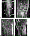
Image quality of STIR vs proton density. MR performed after external fixation of Schatzker V tibial plateau fracture to assess for associated injuries. A. and B. Frontal (A) and lateral (B) right knee radiographs demonstrate the lateral component of the tibial plateau fracture (dashed arrow) along with a moderate sized lipohemarthrosis and delineate the numerous titanium external fixation pins anchored to the femur and tibia. C-D. Coronal STIR (C) and fat-saturated proton density (D) MR acquisitions. Although proton density is more prone to susceptibility artifact (dashed boxes), the finding of a medial meniscus tear is only well seen on proton density and not SITR (solid arrow).
Figure 2.
Figure 2.
Local susceptibility artifact from external fixators. 63-year-old man ejected from a motor vehicle collision resulting in right-sided Schatzker V tibial plateau fracture. A. Frontal radiograph of the right knee demonstrates the right tibial plateau fracture (dashed box) along with the multiple titanium external fixator pins and constructs spanning the knee and anchored to the femur and tibia (thick arrows). In the coronal (B) and sagittal (C) proton density along with fat-suppressed sagittal proton density acquisitions, note the susceptibility artifact signal void of the external fixation device in the field of view (thick arrows); note how this results in localized failure of fat suppression in D. Injuries included tear of the posterior horn of the medial meniscus (B, solid box) and tear of the posterior cruciate ligament (C and D, thin arrows).
Figure 3.
Figure 3.
Injury patterns in complex fracture. 49-year-old man with a fall from approximately 3 feet resulting in right-sided proximal fibular and Schatzker V tibial plateau fractures, managed with titanium external fixation instrumentation. Proton-density coronal (A and B) and sagittal (C) MR acquisitions demonstrate diffuse laxity and wavy trajectory of the lateral collateral ligament (thin arrows in A and B) secondary to a displaced fibular head fracture (thick arrows in C). Despite this laxity and wavy appearance of the lateral collateral ligament, there was no intrinsic signal disruption to indicate tear and the ligament was confirmed to be intact at surgery.
Figure 4.
Figure 4.
Ligament and tendon injuries extending into fracture. 42-year-old man who was an unrestrained driver in a motor vehicle collision. Frontal radiograph of the left knee demonstrates a complex Schatzker VI tibial plateau fracture which extend into the intercondylar eminence (box) along with external fixators. Proton-density fat suppressed sagittal (B), coronal (C), and axial (D) sequences demonstrate partial thickness tears affecting the anterior (black dashed arrow, B) and posterior (thin white arrows, B and C) cruciate ligaments along with the patellar tendon (thick white arrows, B and D). Note that the posterior cruciate ligament tear and the patellar tendon tear are continuous with associated fracture lines. This is demonstrated in the coronal acquisition (C) for the posterior cruciate ligament tear-fracture continuum (thin white arrows) and in the axial acquisition (D) for the patellar tendon tear-fracture continuum (thin white arrows).

References

    1. Cinque ME, Godin JA, Moatshe G, et al. (2017) Do Tibial Plateau Fractures Worsen Outcomes of Knee Ligament Injuries? A Matched Cohort Analysis. Orthop J Sports Med 5:2325967117723895. 10.1177/2325967117723895 - DOI - PMC - PubMed
    1. Delamarter RB, Hohl M, Hopp E (1990) Ligament injuries associated with tibial plateau fractures. Clin Orthop Relat Res 226–233 - PubMed
    1. Karantanas AH (2014) What’s new in the use of MRI in the orthopaedic trauma patient? Injury 45:923–933. 10.1016/j.injury.2014.01.012 - DOI - PubMed
    1. Yacoubian SV, Nevins RT, Sallis JG, et al. (2002) Impact of MRI on treatment plan and fracture classification of tibial plateau fractures. J Orthop Trauma 16:632–637. 10.1097/00005131-200210000-00004 - DOI - PubMed
    1. Gillig JD, Goode RD, Campfield B, et al. (2018) Safety and Complications Associated With MRI-Conditional External Fixators in Patients With Tibial Plateau Fractures: A Case Series. J Orthop Trauma 32:521–525. 10.1097/BOT.0000000000001246 - DOI - PubMed

LinkOut - more resources