. 2021 Apr 12;60(16):8678-8682.
doi: 10.1002/anie.202013193. Epub 2021 Mar 4.

Cryo-EM Resolves Molecular Recognition Of An Optojasp Photoswitch Bound To Actin Filaments In Both Switch States

Affiliations

Cryo-EM Resolves Molecular Recognition Of An Optojasp Photoswitch Bound To Actin Filaments In Both Switch States

Sabrina Pospich et al. Angew Chem Int Ed Engl. .

Abstract

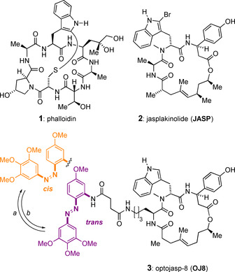
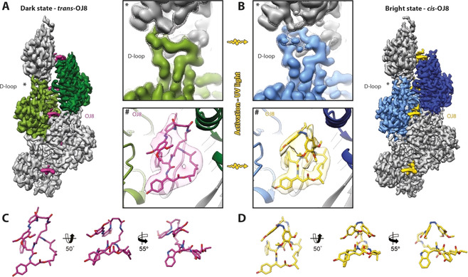
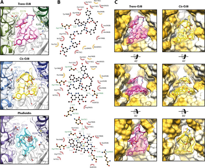
Actin is essential for key processes in all eukaryotic cells. Cellpermeable optojasps provide spatiotemporal control of the actin cytoskeleton, confining toxicity and potentially rendering F-actin druggable by photopharmacology. Here, we report cryo electron microscopy (cryo-EM) structures of both isomeric states of one optojasp bound to actin filaments. The high-resolution structures reveal for the first time the pronounced effects of photoswitching a functionalized azobenzene. By characterizing the optojasp binding site and identifying conformational changes within F-actin that depend on the optojasp isomeric state, we refine determinants for the design of functional F-actin photoswitches.

Keywords: actin filaments; drug design; electron microscopy; photoswitch; protein structures.


Conflict of interest statement

The authors declare no conflict of interest.

Figures

Figure 1
Figure 1
Chemical structures of F‐actin stabilizing natural product toxins and of optojasp‐8. Conditions: a) =380 nm; b) =520 nm or ΔT (t 1/2=15 min in PBS buffer at 37 °C). [6]
Figure 2
Figure 2
High‐resolution structures of OJ8‐stabilized F‐actin. A,B) Cryo‐EM structures of OJ8‐stabilized F‐actin in the (A) dark state, transOJ8 (magenta), and (B) bright state, cisOJ8 (yellow). To highlight the double stranded helical arrangement of F‐actin, the central and subsequent actin subunit are colored in shades of green (dark state) and blue (bright state), respectively. The central D‐loop (*) and OJ8 binding site (#) are enlarged for direct comparison of the structures before and after activation of OJ8 with UV light (380 nm). (Top) While the D‐loop solely adopts the open conformation with transOJ8 bound, it is mixed with a considerably higher population of the closed D‐loop state after irradiation. The map is additionally shown at a lower threshold (mesh) to highlight the second, less populated D‐loop conformation of the cisOJ8 structure. (Bottom) The azobenzene photoswitch stacks onto the macrocycle and changes its configuration from trans to cis upon UV‐irradiation. C,D) Atomic wire models of trans‐ and cisOJ8. For details also see Figure S2 and Movies S1,2.
Figure 3
Figure 3
Interactions at the binding site of OJ8 in comparison to phalloidin. A) The binding site of trans3 (magenta) and cis3 (yellow) in comparison to the one of phalloidin (cyan, PDB: 6T1Y, EMDB: 10363, [9] ). Due to the scorpion tail‐like fold of the photoswitch, OJ8 resembles the three‐dimensional arrangement of phalloidin more closely than the flat conformation of JASP. The conformation of cis3 is remarkably similar to phalloidin (see text). B) 2D ligand‐protein interaction diagrams of the ligands shown in (A). Both hydrophobic contacts (red arcs with rays) and putative hydrogen bonds (dashed green lines) are depicted. Chain IDs are stated in brackets for protein residues. Residues that only participate in interactions with either trans3 (top) or cis3 (center) are highlighted in orange. Note that all residues involved in the binding of phalloidin (bottom), also interact with cis3. C) Surface representation of the OJ8 binding site from three different perspectives. While trans3 adopts an elongated conformation, partially filling the cavity between the actin strands (into the plane of projection), the conformation of cis3 is more compact, thereby increasing contacts to the subunit displayed in the upper right corner (SU D, pointed end direction), also see Movie S1.

Similar articles

Cited by

References

    1. Svitkina T., Cold Spring Harbor Perspect. Biol. 2018, 10, a018267. - PMC - PubMed
    1. Merino F., Pospich S., Funk J., Wagner T., Küllmer F., Arndt H.-D., Bieling P., Raunser S., Nat. Struct. Mol. Biol. 2018, 25, 528–537. - PubMed
    1. Combeau C., Carlier M. F., J. Biol. Chem. 1988, 263, 17429–17436. - PubMed
    1. Merino F., Pospich S., Raunser S., Semin. Cell Dev. Biol. 2020, 102, 51–64. - PMC - PubMed
    1. Crews P., Manes L. V., Boehler M., Tetrahedron Lett. 1986, 27, 2797–2800.

Publication types

LinkOut - more resources