. 2021 Jun 1;162(6):1864-1875.
doi: 10.1097/j.pain.0000000000002191.

A peptide encoded within a 5' untranslated region promotes pain sensitization in mice

Affiliations

A peptide encoded within a 5' untranslated region promotes pain sensitization in mice

Paulino Barragan-Iglesias et al. Pain. .

Abstract

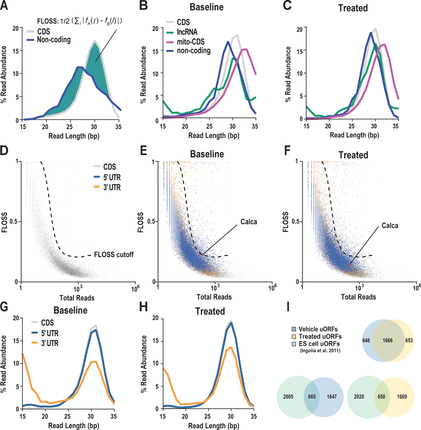
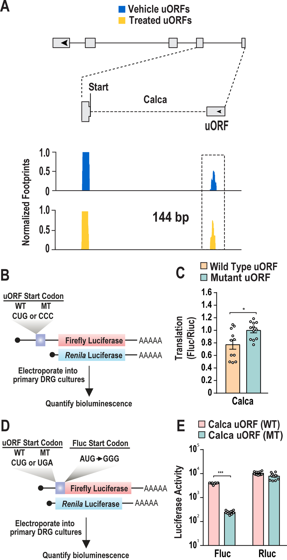
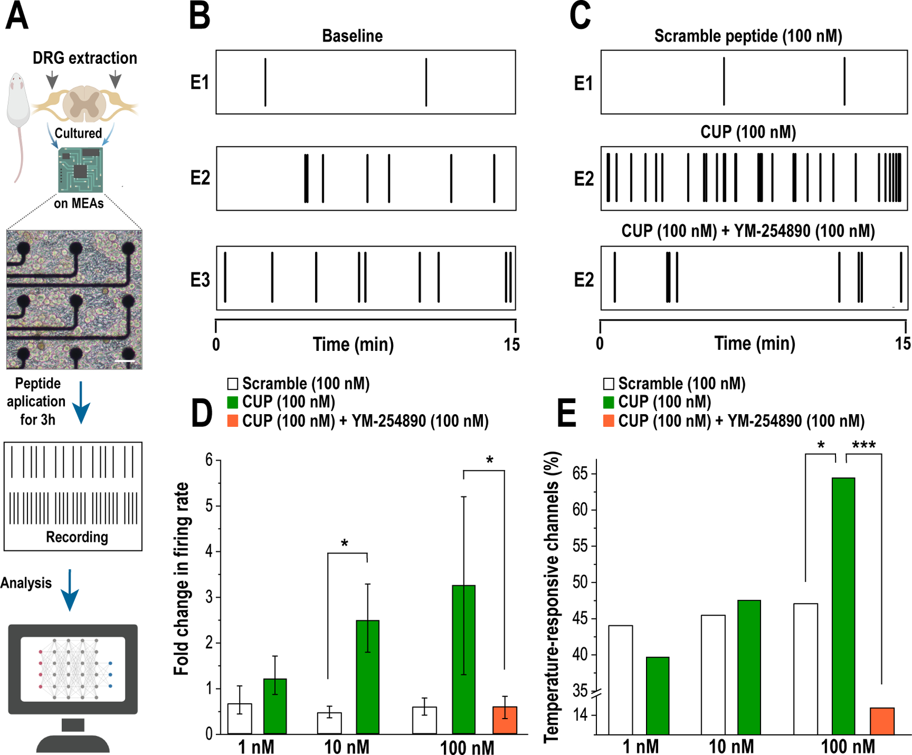
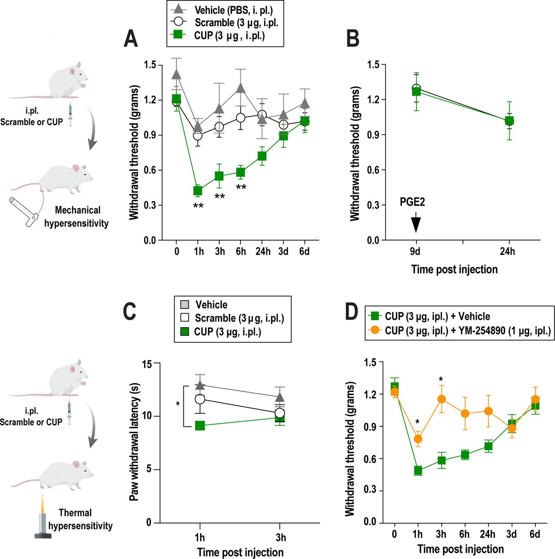
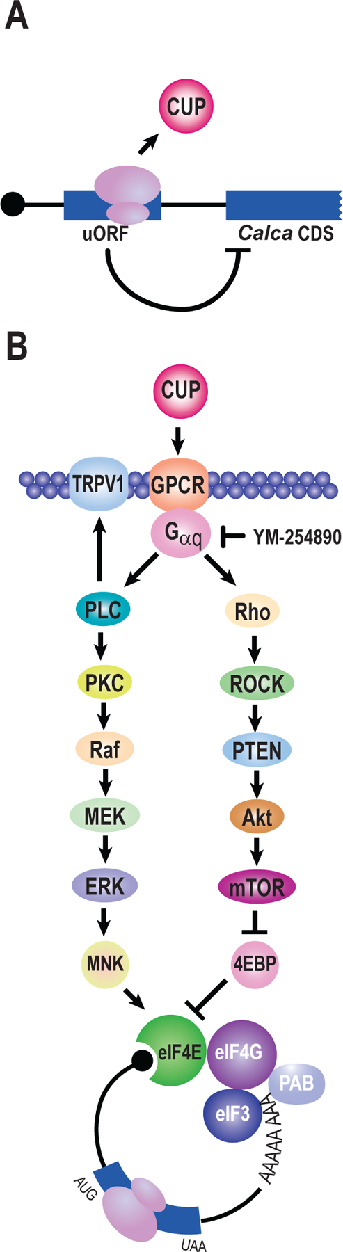
Translational regulation permeates neuronal function. Nociceptors are sensory neurons responsible for the detection of harmful stimuli. Changes in their activity, termed plasticity, are intimately linked to the persistence of pain. Although inhibitors of protein synthesis robustly attenuate pain-associated behavior, the underlying targets that support plasticity are largely unknown. Here, we examine the contribution of protein synthesis in regions of RNA annotated as noncoding. Based on analyses of previously reported ribosome profiling data, we provide evidence for widespread translation in noncoding transcripts and regulatory regions of mRNAs. We identify an increase in ribosome occupancy in the 5' untranslated regions of the calcitonin gene-related peptide (CGRP/Calca). We validate the existence of an upstream open reading frame (uORF) using a series of reporter assays. Fusion of the uORF to a luciferase reporter revealed active translation in dorsal root ganglion neurons after nucleofection. Injection of the peptide corresponding to the calcitonin gene-related peptide-encoded uORF resulted in pain-associated behavioral responses in vivo and nociceptor sensitization in vitro. An inhibitor of heterotrimeric G protein signaling blocks both effects. Collectively, the data suggest pervasive translation in regions of the transcriptome annotated as noncoding in dorsal root ganglion neurons and identify a specific uORF-encoded peptide that promotes pain sensitization through GPCR signaling.


Conflict of interest statement

Declaration interests

The authors declare no competing interests.

Figures

Figure 1.
Figure 1.. Ribosome profiling identifies uORFs in 5’ untranslated regions of mRNA in cultured DRG neurons
(A) A schematic illustrates the calculation of FLOSS values based on the discrepancy (solid green) between a transcript, region of a transcript, or transcript class (non-coding RNA blue) versus the coding region of mRNA (grey). The FLOSS equation is provided. (B-C) Read length distributions for transcript sequences of CDS (grey), lncRNAs (green), mitochondrial mRNAs (magenta), and non-coding RNAs (blue) after vehicle or plasticity treatments. (D-F) FLOSS distributions for coding regions (grey) of all protein coding gene mRNAs overlaid with 5’ (blue) and 3’ (orange) UTRs under baseline conditions (E) or after addition of the plasticity treatment (F). The 5’ UTR of Calca is highlighted in (E) and (F). The FLOSS cutoff is indicated as a dashed-line and indicates regions of the plot with a high potential for being coding. (G-H) Read length distribution for protein coding sequence (grey), 5’UTR (blue), and 3’UTR (orange) after an exposure to vehicle or following plasticity treatment respectively. (I) Venn diagrams depict the number of uORFs detected in DRG cultures treated with vehicle or plasticity treatment versus those found in mouse embryonic stem cells [42].
Figure 2.
Figure 2.. Translation in untranslated regions occurs on transcripts linked to pain
(A) Ribosome protected footprints situated in the 5’UTR of Calca. (B) A schematic of dual luciferase reporters for translational modulation of downstream coding regions by the Calca uORF. The Calca 5’UTR was cloned upstream of a firefly luciferase construct. Translation from the Calca uORF is discontinuous to that from the downstream luciferase ORF. Vectors were electroporated into primary DRG cultures prior to quantification of reporter gene activity. (C) Dual luciferase assays for the Calca uORF. The Calca uORF represses downstream translation of the main ORF by 20%. This repression is eliminated through mutation of the Calca uORF start codon (CUG) to (CCC). Data are plotted as mean ± s.e.m. Student’s unpaired t-test: CGRP wild type uORF vs mutant uORF: *P = 0.0135. n = 12 cultures. (D) Detection of uORF translation through the use of a translational fusion to a luciferase reporter. The Calca uORF was cloned in frame with luciferase. To eliminate utilization of the start codon (ATG) of the luciferase reporter, it was mutated to GGG. Vectors were electroporated into primary DRG cultures prior to luciferase quantification. (E) Dual luciferase assays for uORF translation. Luciferase activity is observed in the presence of the uORF start codon (CUG) indicating translation of the Calca uORF. Luciferase activity was abolished on mutating the start codon (CUG) to stop codon (UGA). n = 12 cultures. Data are plotted as mean ± s.e.m. Student’s unpaired t-test. Calca wild-type leader vs mutant leader sequence: *P = 0.00001.
Figure 3.
Figure 3.. CUP promotes sensory neuron excitability
(A) A schematic of the multi-electrode array system. White scale bar corresponds to 50 μm. (B) Baseline recordings of spontaneous activity for three individual electrodes. (C) Activities following addition of 100 nM of scramble (E1) peptide, CUP (E2) or the combination of CUP in the presence of YM-254890 (E3) after a three-hour incubation. (D) CUP (1–100 nM) peptide led to increased spontaneous firing rates after a three-hour incubation. Spontaneous firing rate is attenuated in the presence of the Gq inhibitor YM-254890. Two-way ANOVA: F(2, 4) = 3.48, P = 0.03. Bonferroni’s test. *P = 0.032. Scramble vs CUP at 10 nM: #P = 0.031. CUP vs CUP+YM-254890 at 100 nM: *P = 0.045. (E) CUP (100 nM) increased the percentage of temperature-responsive MEA channels. Percentage of temperature-responsive MEA channels is decreased in the presence of YM-254890. Two-sample test of proportions. Scramble vs CUP at 100 nM: *P = 0.026. CUP vs CUP+YM-254890 at 100 nM: ***P < 0.001.
Figure 4.
Figure 4.. CUP induces pronociceptive responses to mechanical and thermal stimulation
(A) Intraplantar administration of CUP at 3 μg, but not scrambled peptide at the same dose, induces mechanical hypersensitivity. Two-way repeated measures (RM) ANOVA: F(1, 22) = 13.56, P = 0.0013. Bonferroni’s multiple comparison test. Scramble vs CUP at 1 h: **P = 0.0018, at 3hr: *P = 0.0370, at 6 h: **P = 0.0062. (B) Intraplantar administration of CUP (3 μg) did not induce changes in mechanical sensitivity at day 9 following injection of prostaglandin E2 (PGE2, 100 ng, i.pl.). (C) Changes in pain sensitivity to thermal stimulation are detected after i.pl. scramble or CUP administration. Two-way RM ANOVA with RM: F(1, 11) = 6.30, P=0.0215. Bonferroni’s multiple comparisons test: vehicle vs CUP at 1h: *P=0.0467. (D) Intraplantar administration of YM-254890 (a Gq inhibitor; 1 μg) attenuates CUP-induced mechanical hypersensitivity. Two-way RM ANOVA: F (1, 22) = 6.130, P=0.0215. Bonferroni’s multiple comparisons test. CUP vs CUP + YM-254890 at 1h: *P=0.0467; at 3h: *P=0.0280.
Figure 5.
Figure 5.. A schematic of Calca translation and potential mechanisms of nociceptive sensitization by CUP
(A) A uORF present in Calca results in the production of a peptide termed CUP. Utilization of the uORF represses translation of the primary reading frame. (B) Potential mechanisms of sensitization by CUP. CUP sensitizes the nociceptive system through Gq signaling. First, activation of Gq results in phosphorylation and sensitization of TRPV1 [12]. Gq signaling promotes phospholipase C (PLC) activation [81]. PLC generates diacylglycerol (DAG) and inositol trisphosphate (IP3). A key consequence of these secondary messengers is activation of protein kinase C (PKC). PKC stimulates multiple pathways including MAPK signaling [69]. PKC activates Raf which activates Raf, MEK, ERK, and ultimately MNK. MNK phosphorylates eIF4E and promotes nociception [59]. Conversely, Gq also stimulates the Rho kinase [20]. Rho activates the Rho-associated protein kinase (ROCK). Rock activates PTEN, a key upstream regulatory component of PI3K/mTOR signaling [50]. PTEN controls Akt which ultimately stimulates mTOR. mTOR promotes cap-dependent translation through negative regulation of 4EBPs which sequester eIF4E.

Similar articles

Cited by

References

    1. Amara SG, Jonas V, Rosenfeld MG, Ong ES, Evans RM. Alternative RNA processing in calcitonin gene expression generates mRNAs encoding different polypeptide products. Nature 1982;298:240–244. doi:10.1038/298240a0. - DOI - PubMed
    1. Andreev DE, O’Connor PB, Fahey C, Kenny EM, Terenin IM, Dmitriev SE, Cormican P, Morris DW, Shatsky IN, Baranov PV. Translation of 5’ leaders is pervasive in genes resistant to eIF2 repression. Elife 2015;4:e03971. doi:10.7554/eLife.03971. - DOI - PMC - PubMed
    1. Avona A, Burgos-Vega C, Burton MD, Akopian AN, Price TJ, Dussor G. Dural Calcitonin Gene-Related Peptide Produces Female-Specific Responses in Rodent Migraine Models. J Neurosci 2019;39:4323–4331. doi:10.1523/JNEUROSCI.0364-19.2019. - DOI - PMC - PubMed
    1. Babendure JR, Babendure JL, Ding JH, Tsien RY. Control of mammalian translation by mRNA structure near caps. RNA 2006;12:851–861. doi:10.1261/rna.2309906. - DOI - PMC - PubMed
    1. Baird TD, Palam LR, Fusakio ME, Willy JA, Davis CM, McClintick JN, Anthony TG, Wek RC. Selective mRNA translation during eIF2 phosphorylation induces expression of IBTKalpha. Mol Biol Cell 2014;25:1686–1697. doi:10.1091/mbc.E14-02-0704. - DOI - PMC - PubMed

Publication types

Substances