. 2021 Feb 22;49(3):1708-1723.
doi: 10.1093/nar/gkaa1218.

Crystal structures of the EVE-HNH endonuclease VcaM4I in the presence and absence of DNA

Affiliations

Crystal structures of the EVE-HNH endonuclease VcaM4I in the presence and absence of DNA

Michal Pastor et al. Nucleic Acids Res. .

Abstract

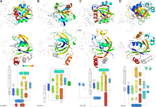
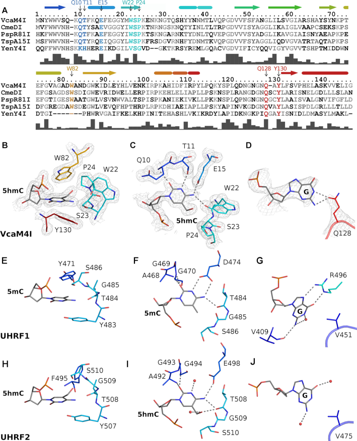
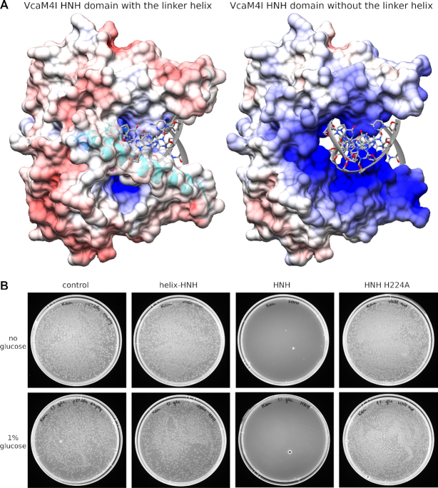
Many modification-dependent restriction endonucleases (MDREs) are fusions of a PUA superfamily modification sensor domain and a nuclease catalytic domain. EVE domains belong to the PUA superfamily, and are present in MDREs in combination with HNH nuclease domains. Here, we present a biochemical characterization of the EVE-HNH endonuclease VcaM4I and crystal structures of the protein alone, with EVE domain bound to either 5mC modified dsDNA or to 5mC/5hmC containing ssDNA. The EVE domain is moderately specific for 5mC/5hmC containing DNA according to EMSA experiments. It flips the modified nucleotide, to accommodate it in a hydrophobic pocket of the enzyme, primarily formed by P24, W82 and Y130 residues. In the crystallized conformation, the EVE domain and linker helix between the two domains block DNA binding to the catalytic domain. Removal of the EVE domain and inter-domain linker, but not of the EVE domain alone converts VcaM4I into a non-specific toxic nuclease. The role of the key residues in the EVE and HNH domains of VcaM4I is confirmed by digestion and restriction assays with the enzyme variants that differ from the wild-type by changes to the base binding pocket or to the catalytic residues.


Figures

Graphical Abstract
Graphical Abstract
Crystal structures of VcaM4I restriction endonuclease provide the first structural description of the EVE domain-modified DNA interaction. Each protomer of the dimeric enzyme is composed of EVE domain responsible for modification detection and HNH domain responsible for DNA cleavage. The EVE domains flip the modified cytosine bases and bind them in dedicated hydrophobic pockets.
Figure 1.
Figure 1.
Overall structure of the VcaM4I–dsDNA complex. One protomer of the VcaM4I dimer is colored: the N-terminal EVE domain (residues 1–147) is in yellow, the helical linker (residues 148–178) in cyan, and the C-terminal HNH domain (residues 179–309) in green. The other protomer and the two dsDNA molecules bound to the EVE domains are in gray. The dimer axis runs vertically in the top panel and towards the reader in the bottom one. The 5-methylcytosine residues flipped out of the DNA stack are indicated in magenta.
Figure 2.
Figure 2.
Structure of the N-terminal EVE domain of VcaM4I in comparison to other PUA-superfamily domains. The figure compares (A) the N-terminal EVE domain of VcaM4I with (B) the SRA domain of UHRF2 (PDB ID 4pw5 (36)), (C) the EVE domain of THY28 (PDB 5j3e, unpublished), (D) and the 6-methyladenine binding YTH domain of McrB from T. gammatolerans (PDB 6p0g (18)). Top: cartoon representation of the domain structures colored from N- to C-terminus (blue to red) in two orientations. The N- and C- terminal fragments outside of the core 5-stranded β-sheet are colored in light gray. The dsDNA complexes of VcaM4I, UHRF2 and McrB were determined experimentally. The complex of THY28 is a model based on the 5j3e coordinates, altered by flipping of a single base and substituting it with 5hmC. DNA polarity was consistent except in the McrB–DNA complex. A revised model presented in the figure with consistent DNA strand orientation is at least as compatible with the diffraction data as the published model. Bottom: topology diagrams of the domains generated with the PDBSUM program (47), with minor manual adjustments necessary for direct comparison with the structures. The core secondary structure elements of the domains are labelled.
Figure 3.
Figure 3.
DNA binding to the VcaM4I EVE domain. (A) Electrostatic potential was generated with APBS (48) and mapped to the VcaM4I surface with CHIMERA (49). Negatively charged surface regions are red, positively charged ones blue. (B) Amino acid conservation scores for the EVE domain were generated with ConSurf with default parameters (50) and mapped with CHIMERA (49). The highest conservation is in the region of the flipped base. The wedge intercalates into dsDNA with Q128 residue, the pocket accommodates the flipped modified base (shown in magenta).
Figure 4.
Figure 4.
Specific interactions of the VcaM4I EVE domain with DNA. (A) Alignment of EVE domains of modification dependent restriction endonucleases. The bars under the alignment indicate the degree of sequence conservation. (B) ‘Side’ view of the VcaM4I EVE pocket demonstrating the sandwiching of the flipped modified base. (C) ‘Top’ view of the VcaM4I modified base binding pocket, showing the specific interactions of its Watson-Crick edge with the protein. (D) Interactions of the estranged base with Q128 of the VcaM4I EVE domain. The model and the composite omit electron density map are based on the 5hmC ssDNA complex for panels B-C (1.5 rmsd contour level) and on dsDNA complex for panel D (1.3 rmsd contour level). (E–G) Equivalent views of the interactions of the SRA domain of UHRF1 with 5mC containing DNA (8). (H–J) Equivalent views of the interactions of the SRA domain of UHRF2 with 5hmC containing DNA (36). Color coding of secondary structure elements (helices represented by cylinders, β-strands by arrows) and functional residues is consistent with Figure 2 (the N- and C-termini were included in the rainbow-colored region to indicate their sequence locations).
Figure 5.
Figure 5.
Phage T4gt plaque forming unit (PFU) assay on cells carrying wild-type and mutant VcaM4I expressed in the presence of IPTG. Cells carrying Q10V, W82R and Y130W variants formed poor cell lawns (mutants are toxic to the host in the presence of IPTG) and the three mutants could not be tested for the in vivo restriction activity.
Figure 6.
Figure 6.
ββα-Me core of the VcaM4I HNH domain in comparison with related endonucleases. Top row presents the ββα-Me regions of (A) VcaM4I, (B) TagI (PDB 6ghs, (3)), (C) EcoKMcrA (PDB 6ghc (4)), (D) Hpy99I (PDB 3fc3 (38)) and (E) colicin E9 (PDB 1v15 (40)). The composite omit density map in (A) was based on the structure of VcaM4I with 5hmC-modified ssDNA and contoured at 1.5 rmsd. The coordination of the metal ions is indicated with faint lines. Additional faint line in (D) indicates the distance between the potential nucleophilic water and the scissile phosphate. The H103A mutation present in the structure used for (E) was in silico mutated back to the active site histidine but a catalytically unproductive conformer was chosen. (F) Structure-based sequence alignment of VcaM4I and similar HNH domains. The alignment was corrected manually. The faint regions indicate the lack of direct structural correspondence. The active site residues are marked with asterisk. Gray asterisk indicates the metal ligand present in some endonucleases which lost its function in VcaM4I and TagI. The bars under the alignment indicate the degree of sequence conservation.
Figure 7.
Figure 7.
Model of the VcaM4I HNH domain dimer bound to DNA. The model is based on the co-crystal structure of colicin E9 with DNA (40). For each subunit, the substrate strand was modelled separately based on a superposition of the core catalytic ββα-motif in VcaM4I and colicin E9. Although this was not imposed by the modelling, the two strands base-pair (with Watson-Crick hydrogen bond distances shorter than expected). The red arrows indicate the sites of cleavage. The model is consistent with the experimental observation that VcaM4I generates fragments with single nucleotide 3′-overhangs.
Figure 8.
Figure 8.
VcaM4I catalytic activity is regulated by the presence of the linker helix. (A) The depiction of the surface of the VcaM4I HNH domain dimer in the presence and absence of the linker helix. The DNA was not present in the structure but modelled as described in Figure 7. The electrostatic potential was calculated by APBS (48) with modelled Mg2+ ions in the active sites and mapped on the domain dimer surface with CHIMERA (49). (B) The VcaM4I HNH domain with and without the linker helix, as well as its catalytic mutant were expressed in E. coli. The domain without the helix and the inactivating mutation is toxic to the cells due to nonspecific endonuclease activity.
Figure 9.
Figure 9.
DNA binding by MDREs. DNA fragments bound to the catalytic HNH domains were modelled. The DNA binding modes to the PUA (exemplified by SRA and EVE) and NEco domains were determined experimentally except for TagI, which was modelled based on the UHRF1-DNA complex (PDB ID 3clz, (9)). For EcoKMcrA and ScoMcrA, the complexes of the isolated domains with bound DNA were mapped back onto the DNA free full-length enzyme structures. The EcoKMcrA structure was symmetrized based on the relative domain orientation in one of the protomers. In all cases there is no way to connect the DNA fragments bound to the catalytic and modification binding sites. There is also no easy way to extend the HNH domain bound DNA due to the clashes with the other domains. The repositioning of the non-catalytic parts of the enzymes would be necessary for the connection of the fragments and/or DNA extension.

References

    1. Horton J.R., Wang H., Mabuchi M.Y., Zhang X., Roberts R.J., Zheng Y., Wilson G.G., Cheng X.. Modification-dependent restriction endonuclease, MspJI, flips 5-methylcytosine out of the DNA helix. Nucleic Acids Res. 2014; 42:12092–12101. - PMC - PubMed
    1. Kazrani A.A., Kowalska M., Czapinska H., Bochtler M.. Crystal structure of the 5hmC specific endonuclease PvuRts1I. Nucleic Acids Res. 2014; 42:5929–5936. - PMC - PubMed
    1. Kisiala M., Copelas A., Czapinska H., Xu S.Y., Bochtler M.. Crystal structure of the modification-dependent SRA-HNH endonuclease TagI. Nucleic Acids Res. 2018; 46:10489–10503. - PMC - PubMed
    1. Czapinska H., Kowalska M., Zagorskaite E., Manakova E., Slyvka A., Xu S.Y., Siksnys V., Sasnauskas G., Bochtler M.. Activity and structure of EcoKMcrA. Nucleic Acids Res. 2018; 46:9829–9841. - PMC - PubMed
    1. Siwek W., Czapinska H., Bochtler M., Bujnicki J.M., Skowronek K.. Crystal structure and mechanism of action of the N6-methyladenine-dependent type IIM restriction endonuclease R.DpnI. Nucleic Acids Res. 2012; 40:7563–7572. - PMC - PubMed

Publication types

LinkOut - more resources