. 2021 Apr;1868(4):118966.
doi: 10.1016/j.bbamcr.2021.118966. Epub 2021 Jan 13.

Regulation of SIRT2 by Wnt/β-catenin signaling pathway in colorectal cancer cells

Affiliations

Regulation of SIRT2 by Wnt/β-catenin signaling pathway in colorectal cancer cells

Chang Li et al. Biochim Biophys Acta Mol Cell Res. 2021 Apr.

Abstract

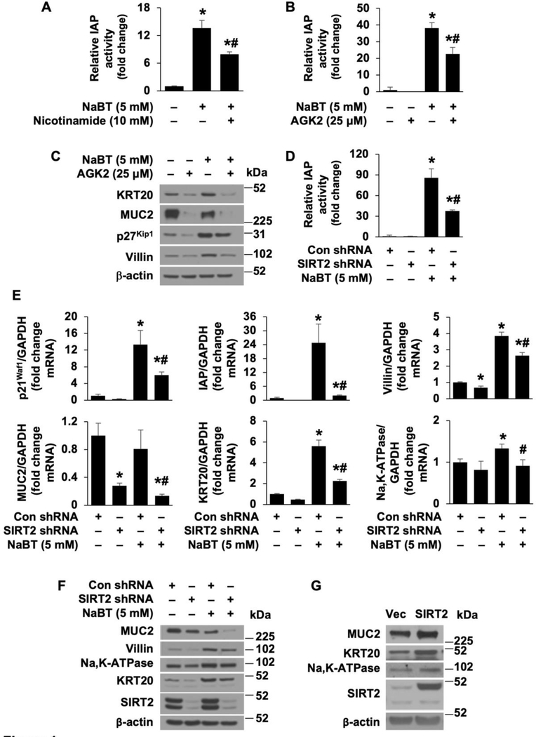
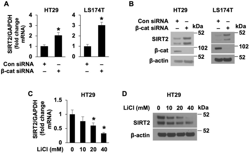
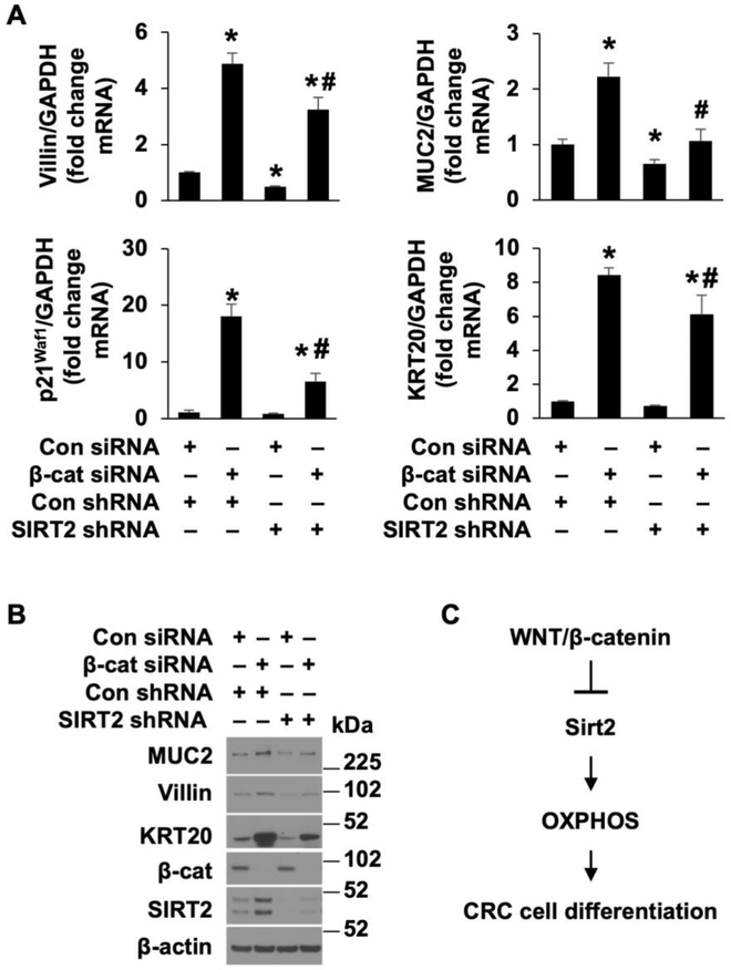
Activation of the Wnt/β-catenin pathway is one of the hallmarks of colorectal cancer (CRC). Sirtuin 2 (SIRT2) protein has been shown to inhibit CRC proliferation. Previously, we reported that SIRT2 plays an important role in the maintenance of normal intestinal cell homeostasis. Here, we show that SIRT2 is a direct target gene of Wnt/β-catenin signaling in CRC cells. Inhibition or knockdown of Wnt/β-catenin increased SIRT2 promoter activity and mRNA and protein expression, whereas activation of Wnt/β-catenin decreased SIRT2 promoter activity and expression. β-Catenin was recruited to the promoter of SIRT2 and transcriptionally regulated SIRT2 expression. Wnt/β-catenin inhibition increased mitochondrial oxidative phosphorylation (OXPHOS) and CRC cell differentiation. Moreover, inhibition of OXPHOS attenuated the differentiation of CRC cells induced by Wnt/β-catenin inhibition. In contrast, inhibition or knockdown of SIRT2 decreased, while overexpression of SIRT2 increased, OXPHOS activity and differentiation in CRC cells. Consistently, inhibition or knockdown or SIRT2 attenuated the differentiation induced by Wnt/β-catenin inhibition. These results demonstrate that SIRT2 is a novel target gene of the Wnt/β-catenin signaling and contributes to the differentiation of CRC cells.

Keywords: Cell differentiation; Cell signaling; Protein expression.


Conflict of interest statement

DISCLOSURES: The authors have no potential conflicts of interest.

Declaration of interests

The authors declare that they have no known competing financial interests or personal relationships that could have appeared to influence the work reported in this paper.

Figures

Figure 1.
Figure 1.. SIRT2 is required for NaBT-induced CRC cell differentiation.
A. HT29 cells were treated with NaBT with or without the sirtuin inhibitor nicotinamide for 48 h. IAP activity was assayed. (n=3, data represent mean ± SD; *p<0.01 vs control; #p<0.01 vs NaBT alone). B and C. HT29 cell cells were treated with NaBT with or without the SIRT2 inhibitor AGK2 for 48 h. IAP activity assay (B). (n=3, data represent mean ± SD; *p<0.01 vs control; #p<0.01 vs NaBT alone). Western blot analysis using antibodies as indicated (C). D-F. HT29 cells, stably transfected with control or SIRT2 shRNA, were treated with NaBT for 48 h. Cell lysates were used for IAP activity assay (D). (n=3, data represent mean ± SD; *p<0.01 vs con shRNA; #p<0.01 vs NaBT plus Con shRNA). Total RNA was extracted and p21Waf1, IAP, villin, MUC2, KRT20, and Na,K-ATPase mRNA levels were determined by real-time RT-PCR (E). (n=3, data represent mean ± SD; *p<0.01 vs con shRNA; #p<0.01 vs NaBT plus Con shRNA). Western blot analysis using antibodies as indicated (F). G. Doxycycline-induced overexpression of SIRT2 in HT29 cells. Western blot analysis was performed.
Figure 2.
Figure 2.. SIRT2 promotes oxidative catabolism to increases CRC cell differentiation.
A and B. HT29 cells stably transfected with control shRNA or SIRT2 shRNA (A) or with overexpression of SIRT2 (B) was assessed for oxygen consumption using the Seahorse Bioscience XFe96 extracellular flux analyzer. The normalized OCR was shown. (n=10, data represent mean± S.D.; *P <0.05 compared with control shRNA or control vector). C and D. HT29 cell cells were treated with OXPHOS inhibitors oligomycin (1 uM) or rotenone (100 nM) in the presence or absence of NaBT for 48 h. IAP activity assay (C). (n=3, data represent mean ± SD; *p<0.01 vs control; #p<0.01 vs NaBT alone). Western blotting analysis using antibodies as indicated (D).
Figure 3.
Figure 3.. Inhibition of Wnt/β-catenin signaling increases SIRT2 protein expression.
A and B. HT29 and LS174T cells were treated with Wnt inhibitor ICG001 at various dosages for 48 h (A) or at 40 μM at various times (B). Total RNA was extracted and SIRT2 mRNA levels were determined by real-time RT-PCR. (n=3, data represent mean ± SD; *p<0.01 vs control). C. HT29 and LS174T cells were treated with ICG001 at various dosages for 48 h. Western blot analysis was performed using antibodies against SIRT2 and β-actin.
Figure 4.
Figure 4.. Regulation of SIRT2 expression by Wnt/β-catenin signaling.
A and B. Knockdown of β-catenin results in the induction of SIRT2 expression. HT29 and LS174T cells were transfected with β-catenin siRNA or nontargeting control siRNA. After 48 h incubation, SIRT2 expression was determined by real-time RT-PCR (A) or western blot analysis (B), respectively. (n=3, data represent mean ± SD; * p < 0.05 vs. control). C and D. Activation of Wnt/β-catenin pathway suppressed SIRT2 expression. HT29 and LS174T cells were treated with LiCl at with various dosages or 40 mM of NaCl as control for 24 h. SIRT2 mRNA levels were assessed by real-time RT-PCR (C). (n=3, data represent mean ± SD; * p < 0.05 vs. control). Western blot analysis using the antibodies as indicated (D).
Figure 5.
Figure 5.. Transcriptional regulation of SIRT2 expression by Wnt/β-catenin signaling.
A. SIRT2 promoter cloning. B. Basal SIRT2 promoter activity in Caco-2 cells. Caco-2 cells were transfected with SIRT2 promoter deletion constructs and relative luciferase activity was measured. C. Inhibition of Wnt/β-catenin by treatment with ICG001 increases SIRT2 promoter activity. SIRT2 −2800 promoter construct were transfected into Caco-2 cells and then treated with or without ICG001 (20 uM). Luciferase activity was measured 24 h after treatment. D. Overexpression of β-catenin decreased SIRT2 promoter activity. Caco-2 cells were transfected with construct containing −2800 bp SIRT2 promoter together with either the control vector, or Flag-tagged β-catenin. After incubation for 48 h, cells were lysed and luciferase activity determined. Results were normalized for transfection efficiency using the pRL-Tk-luc plasmid (Promega). (n=3, data shown as mean ± SD; *, P<0.05 vs control). E. Promoter sequences of the SIRT2 gene with a putative TCF/β-catenin binding site. F, ChIP assays were used to elucidate the binding of β-catenin to the SIRT2 promoter. Chromatin DNA fragments were precipitated using normal rabbit IgG and anti–β-catenin antibody as indicated.
Figure 6.
Figure 6.. Knockdown of β-catenin increases OXPHOS activity and induces CRC cell differentiation.
A and B. HT29 and LS174T cells were transfected with control siRNA or β-catenin siRNA. (A) Western blot analysis was performed using antibodies as indicated. (B) MUC2, villin and KRT20 mRNA expression was determined by real-time RT-PCR (n=3, data represent mean ± SD; *p<0.01 vs control siRNA). C and D. HT29 and LS174T cells transfected with control siRNA or β-catenin siRNA was assessed for oxygen consumption using the Seahorse Bioscience XFe96 extracellular flux analyzer. The normalized OCR was shown. (n=10, data represent mean± S.D.; *P <0.05 compared with control siRNA). E. HT29 cells, transfected with control or β-catenin siRNA, were treated with oligomycin (1 μM) for 48 h. MUC2, villin and KRT20 mRNA levels were determined by real-time RT-PCR (E). (n=3, data represent mean ± SD; *p<0.05 vs con siRNA; #p<0.05 vs β-catenin siRNA).
Figure 7.
Figure 7.. β-catenin regulation of CRC cell differentiation through, at least in part, the regulation of SIRT2 expression.
HT29 cells, stably transfected with control or SIRT2 shRNA, were transfected with control or β-catenin siRNA. A and B. Forty-eight h after transfection, cells were lysed and total RNA was extracted and villin, MUC2, p21Waf1 and KRT20 mRNA levels were determined by real-time RT-PCR (A). (n=3, data represent mean ± SD; *p<0.05 vs con siRNA; #p<0.05 vs β-catenin siRNA). Western blot analysis using antibodies as indicated (B). C. Schematic diagram summarizing the overall findings.

References

    1. Bahrami A, Amerizadeh F, ShahidSales S, Khazaei M, Ghayour-Mobarhan M, Sadeghnia HR, Maftouh M, Hassanian SM, Avan A, Therapeutic Potential of Targeting Wnt/beta-Catenin Pathway in Treatment of Colorectal Cancer: Rational and Progress, Journal of cellular biochemistry, (2017). - PubMed
    1. Schatoff EM, Leach BI, Dow LE, Wnt Signaling and Colorectal Cancer, Current colorectal cancer reports, 13 (2017) 101–110. - PMC - PubMed
    1. Gordon MD, Nusse R, Wnt signaling: multiple pathways, multiple receptors, and multiple transcription factors, J Biol Chem, 281 (2006) 22429–22433. - PubMed
    1. Nusse R, Clevers H, Wnt/beta-Catenin Signaling, Disease, and Emerging Therapeutic Modalities, Cell, 169 (2017) 985–999. - PubMed
    1. Dow LE, O’Rourke KP, Simon J, Tschaharganeh DF, van Es JH, Clevers H, Lowe SW, Apc Restoration Promotes Cellular Differentiation and Reestablishes Crypt Homeostasis in Colorectal Cancer, Cell, 161 (2015) 1539–1552. - PMC - PubMed

Publication types