. 2021 Jan 13;22(2):720.
doi: 10.3390/ijms22020720.

Topical Application of Mesenchymal Stem Cell Exosomes Alleviates the Imiquimod Induced Psoriasis-Like Inflammation

Affiliations

Topical Application of Mesenchymal Stem Cell Exosomes Alleviates the Imiquimod Induced Psoriasis-Like Inflammation

Bin Zhang et al. Int J Mol Sci. .

Abstract

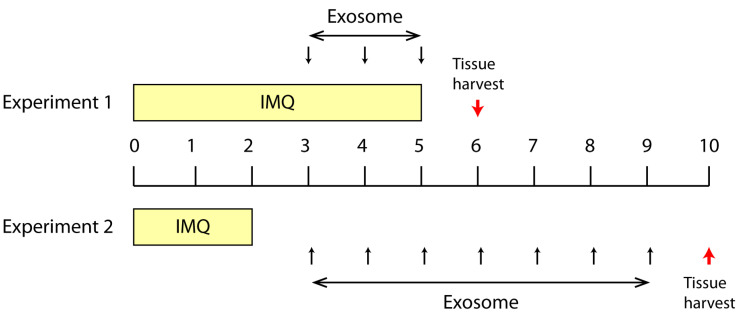
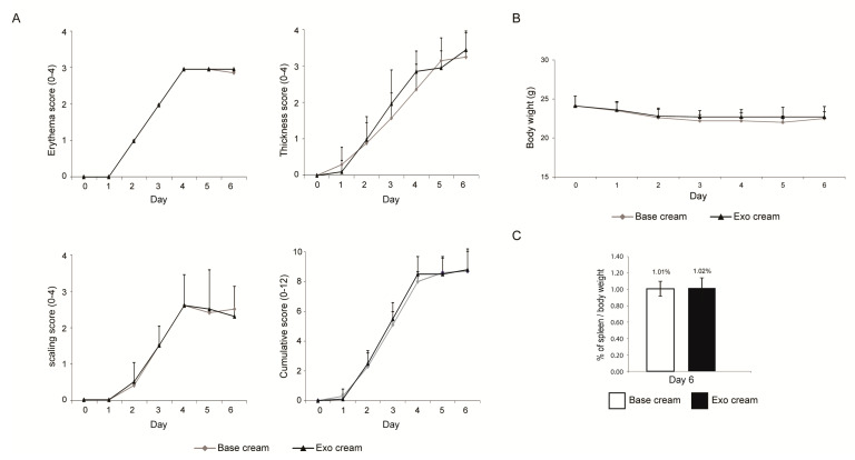
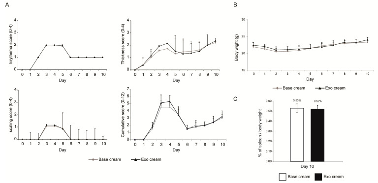
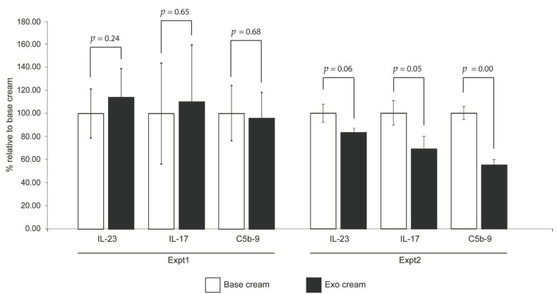
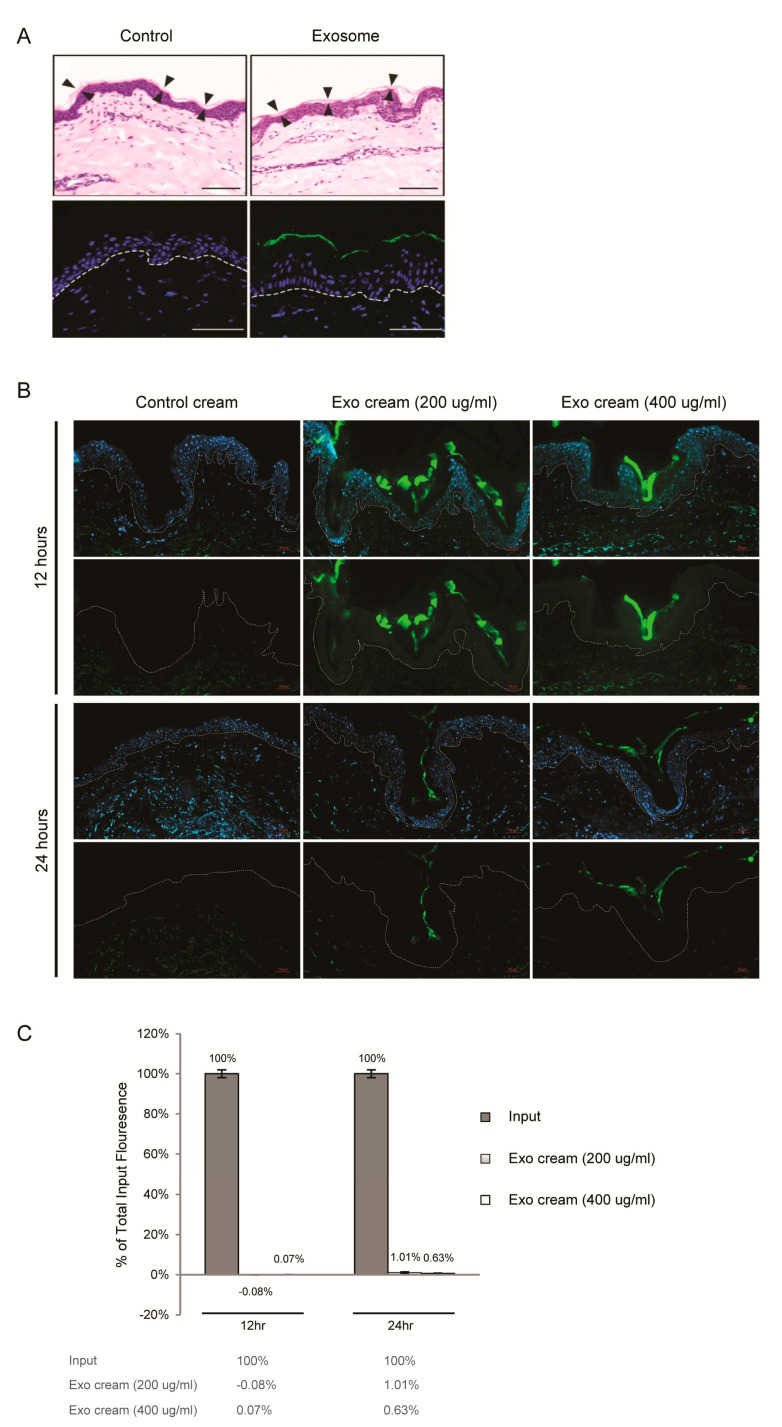
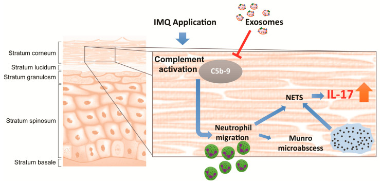
Severe psoriasis, a chronic inflammatory skin disease is increasingly being effectively managed by targeted immunotherapy but long-term immunotherapy poses health risk and loss of response. Therefore, there is a need for alternative therapy strategies. Mesenchymal stem/stromal cell (MSC) exosomes are widely known for their potent immunomodulatory properties. Here we investigated if topically applied MSC exosomes could alleviate psoriasis-associated inflammation. Topically applied fluorescent exosomes on human skin explants were confined primarily to the stratum corneum with <1% input fluorescence exiting the explant over a 24-h period. Nevertheless, topically applied MSC exosomes in a mouse model of imiquimod (IMQ) psoriasis significantly reduced IL-17 and terminal complement activation complex C5b-9 in the mouse skin. MSC exosomes were previously shown to inhibit complement activation, specifically C5b-9 complex formation through CD59. Infiltration of neutrophils into the stratum corneum is characteristic of psoriasis and neutrophils are a major cellular source of IL-17 in psoriasis through the release of neutrophil extracellular traps (NETs). We propose that topically applied MSC exosomes inhibit complement activation in the stratum corneum and this alleviates IL-17 release by NETS from neutrophils that accumulate in and beneath the stratum corneum.

Keywords: exosome; mesenchymal stem cell; psoriasis.


Conflict of interest statement

The authors declare no conflict of interest. The funders had no role in the design of the study; in the collection, analyses, or interpretation of data; in the writing of the manuscript, or in the decision to publish the results. S.K.L. is a founder of Paracrine Therapeutics Pte Ltd. and Vesiderm Pte Ltd.

Figures

Figure 1
Figure 1
Summary of two treatment protocols in the Imiquimod (IMQ) induced psoriasis mouse model. In Expt.1, IMQ was topically applied from Day 0 to Day 5 and exosome cream was applied from Day 3 to Day 5. The experiment was terminated on day 6. In Expt. 2, IMQ was topically applied from Day 0 to Day 2 and exosome cream was applied from Day 3 to Day 9. The experiment was terminated on Day 10. The red arrow indicated the study termination day. This experiment was performed twice with two independently prepared batches of exosomes.
Figure 2
Figure 2
Skin phenotype score, body weight and ratio of spleen to body weight. (A) Erythema, scaling, and thickness of the back skin were scored independently daily from Day 0 to Day 6 (Expt. 1) on a scale from 0 to 4, and the cumulative score for erythema, scaling, and thickness over time was determined daily on a scale from 0–12. (B) The body weight (Day 0–6) and (C) ratio of spleen to body weight (Day 6) were also determined. Scoring (erythema, scaling, thickness, and cumulative score), body weights and ratio of spleen to body weight were analyzed using Student’s t-test. p values < 0.05 were considered as statistically significant.
Figure 3
Figure 3
Skin phenotype score, body weight and ratio of spleen to body weight. (A) Erythema, scaling, and thickness of the back skin were scored independently daily from Day 0 to Day10 (Expt. 2) on a scale from 0 to 4, and the cumulative score for erythema, scaling, and thickness over time was determined daily on a scale from 0–12. (B) The body weight (Day 0–10) and (C) ratio of spleen to body weight (Day 10) were also determined. Two independent animal experiments using different batches of exosome preparations were performed. The scores from the two independent studies were combined. Scoring (erythema, scaling, thickness, and cumulative score), body weights and ratio of spleen to body weight were analyzed using Student’s t-test. p values < 0.05 were considered as statistically significant.
Figure 4
Figure 4
Cytokine induction and complement activation. The shaved back skin of each mouse was removed on day 6 (Expt. 1) or day 10 (Expt. 2), and assayed for IL-17, IL-23 and C5b-9 terminal complement complex (TCC). The levels of cytokines and TCC in “exo cream” (with MSC exsomes) was normalized to that of the “base cream” (vehicle control). Expt. 1 was performed with one independent animal experiment, and Expt. 2 was performed with two independent studies and combined. Statistical significance was determined by Student’s t test. p values < 0.05 were considered as statistically significant.
Figure 5
Figure 5
Dermal penetrance of MSC exosomes (A) Topical application of MSC exosomes (aqueous) on human skin explant culture. Human skin explant was topically treated with PBS (Control) or Alexa Fluor 488-labeled exosomes, washed, frozen in OCT medium and sectioned. (Upper panel) H&E staining with arrowheads indicating boundary of the stratum corneum. Scale bar = 100 μm. (Lower panel) Fluorescence imaging of sections counterstained with DAPI (blue) to show nuclei. White broken lines denote epidermal-dermal junction. Scale bar = 50 μm. (B) Topical application of exosome oil-in-water emulsion cream on skin explant culture. Human skin explant culture was topically treated with AF488-labeled PBS (control cream) or AF488-labeled exosome cream (200 μg/mL or 400 μg/mL) for 12 and 24 h, washed, frozen in OCT medium and sectioned. Fluorescence imaging of sections counterstained with DAPI (blue, upper panel) or without DAPI (lower panel). White broken lines denote epidermal-dermal junction. Scale bar = 50 μm (C) Relative AF488 signal in culture medium of skin explants to input fluorescence. After subtracting the AF488 signal in the label control cream, the fluorescence signal in the culture of explants treated with exosome cream (200 μg/mL or 400 μg/mL) was normalized to their respective input signals to calculate the percentage of total input fluorescence in culture medium of skin explants.
Figure 6
Figure 6
Illustration showing topically applied MSC exosomes localize to the stratum corneum, where they inhibit complement activation caused by the IMQ application, leading to attenuation of NETosis and subsequent release of IL-17 through neutrophil extracellular traps (NET). The T arrow indicated the inhibition of C5b-9 complex assembly by exosomes.

References

    1. Menter A., Gottlieb A., Feldman S.R., Van Voorhees A.S., Leonardi C.L., Gordon K.B., Lebwohl M., Koo J.Y., Elmets C.A., Korman N.J., et al. Guidelines of care for the management of psoriasis and psoriatic arthritis: Section 1. Overview of psoriasis and guidelines of care for the treatment of psoriasis with biologics. J. Am. Acad. Derm. 2008;58:826–850. doi: 10.1016/j.jaad.2008.02.039. - DOI - PubMed
    1. Armstrong A.W., Read C. Pathophysiology, Clinical Presentation, and Treatment of Psoriasis: A Review. JAMA. 2020;323:1945–1960. doi: 10.1001/jama.2020.4006. - DOI - PubMed
    1. Brandon A., Mufti A., Gary Sibbald R. Diagnosis and Management of Cutaneous Psoriasis: A Review. Adv. Ski. Wound Care. 2019;32:58–69. doi: 10.1097/01.ASW.0000550592.08674.43. - DOI - PubMed
    1. Rendon A., Schäkel K. Psoriasis Pathogenesis and Treatment. Int. J. Mol. Sci. 2019;20:1475. doi: 10.3390/ijms20061475. - DOI - PMC - PubMed
    1. Kim W.B., Jerome D., Yeung J. Diagnosis and management of psoriasis. Can. Fam. Physician. 2017;63:278–285. - PMC - PubMed