. 2021 Jan 13;13(2):273.
doi: 10.3390/cancers13020273.

Aberrant Dyskerin Expression Is Related to Proliferation and Poor Survival in Endometrial Cancer

Affiliations

Aberrant Dyskerin Expression Is Related to Proliferation and Poor Survival in Endometrial Cancer

Rafah Alnafakh et al. Cancers (Basel). .

Abstract

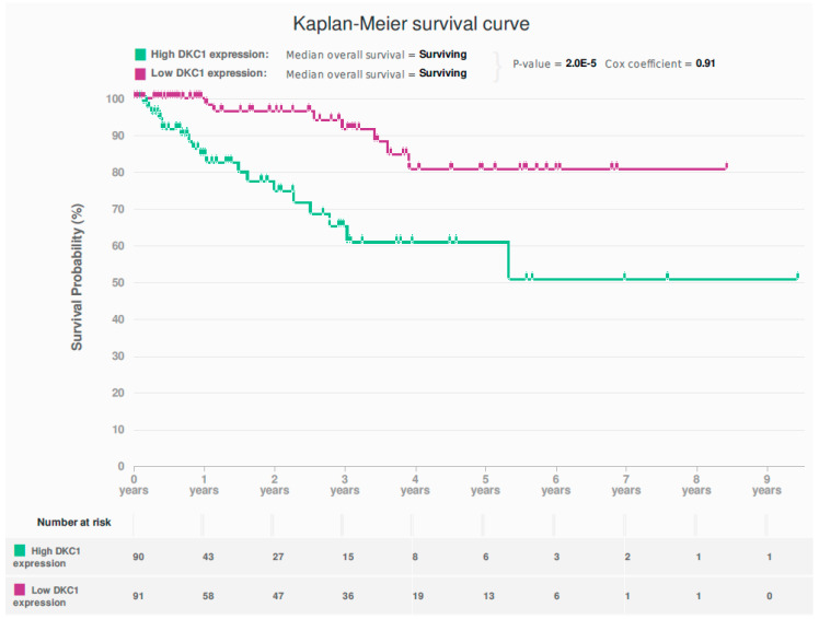
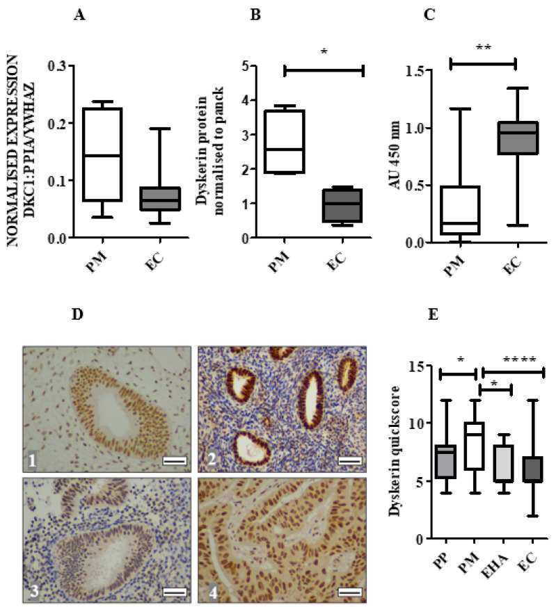
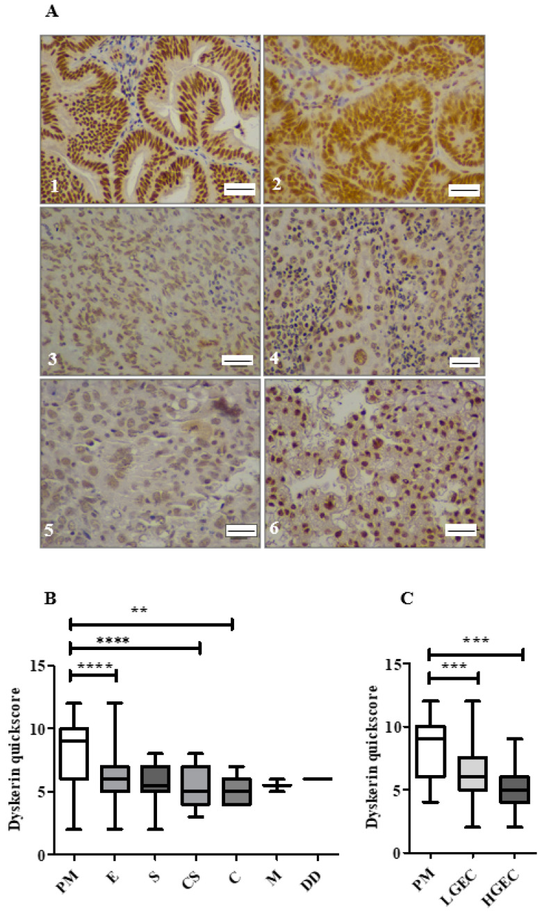
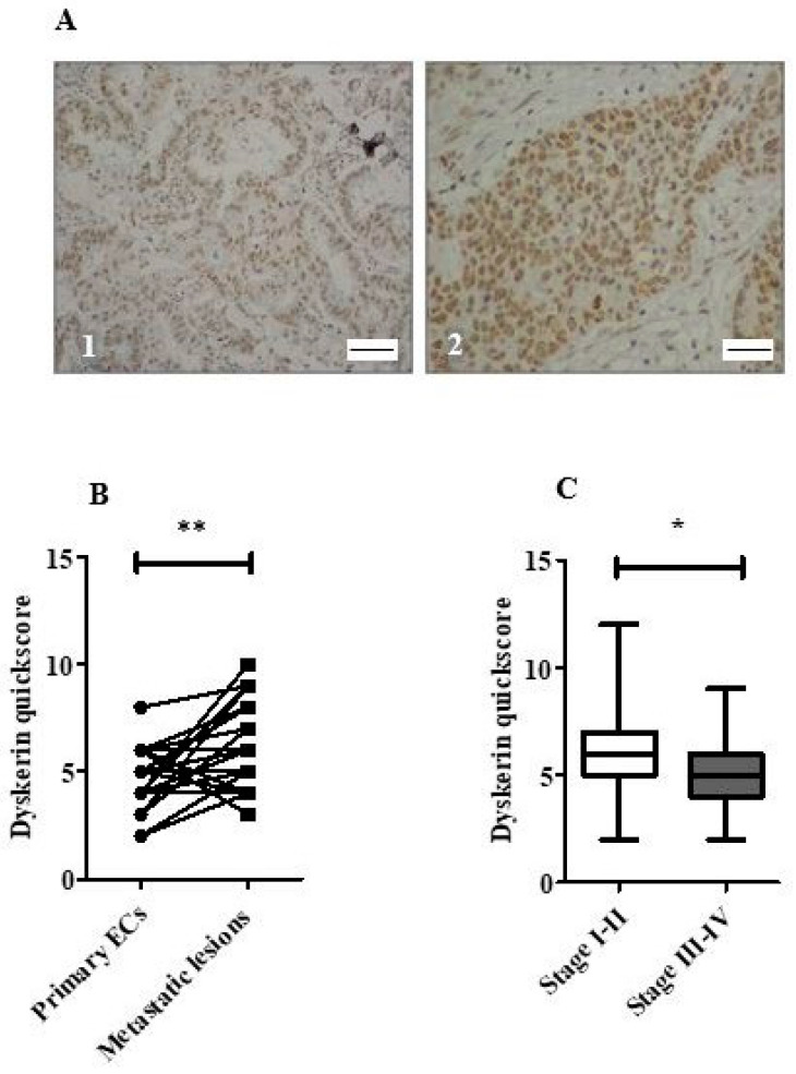
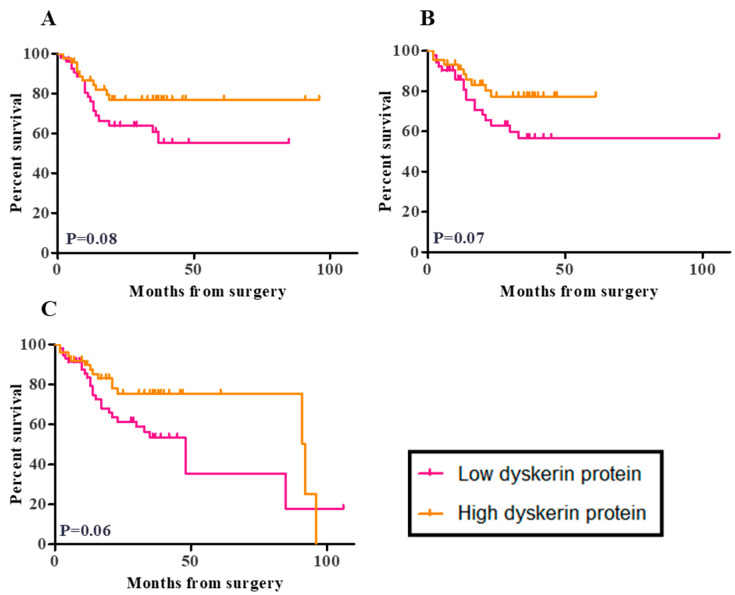
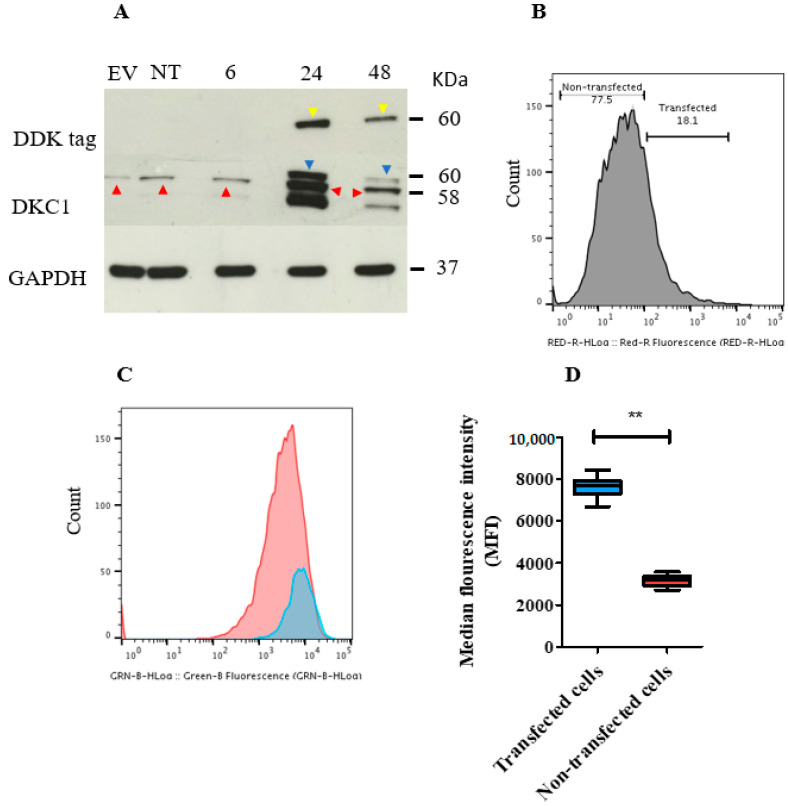
Dyskerin is a core-component of the telomerase holo-enzyme, which elongates telomeres. Telomerase is involved in endometrial epithelial cell proliferation. Most endometrial cancers (ECs) have high telomerase activity; however, dyskerin expression in human healthy endometrium or in endometrial pathologies has not been investigated yet. We aimed to examine the expression, prognostic relevance, and functional role of dyskerin in human EC. Endometrial samples from a cohort of 175 women were examined with immunohistochemistry, immunoblotting, and qPCR. The EC cells were transfected with Myc-DDK-DKC1 plasmid and the effect of dyskerin overexpression on EC cell proliferation was assessed by flow cytometry. Human endometrium expresses dyskerin (DKC1) and dyskerin protein levels are significantly reduced in ECs when compared with healthy postmenopausal endometrium. Low dyskerin immunoscores were potentially associated with worse outcomes, suggesting a possible prognostic relevance. Cancer Genome Atlas (TCGA) ECs dataset (n = 589) was also interrogated. The TCGA dataset further confirmed changes in DKC1 expression in EC with prognostic significance. Transient dyskerin overexpression had a negative effect on EC cell proliferation. Our data demonstrates a role for dyskerin in normal endometrium for the first time and confirms aberrant expression with possible prognostic relevance in EC. Interventions aimed at modulating dyskerin levels may provide novel therapeutic options in EC.

Keywords: DKC1; dyskerin; endometrial cancer; proliferation; telomerase; telomeres.


Conflict of interest statement

The authors declare no conflict of interest.

Figures

Figure 1
Figure 1
Kaplan-Meier survival curve for the association between DKC1 mRNA levels and overall survival (p = 2 × 10⁻⁵, Cox-regression = 0.91) in The Cancer Genome Atlas (TCGA) dataset (endometrioid and serous endometrial cancer) {n = 477}.
Figure 2
Figure 2
DKC1 mRNA and dyskerin protein in human endometrium. (A) DKC1 mRNA is normalised to geometric means of PPIA and YWHAZ and measured by qPCR in endometrial tissue samples: healthy postmenopausal (PM) (n = 6) and endometrial cancer (EC) (n = 22). Mann-Whitney test. (B) The amount of dyskerin protein was evaluated by immuno-blotting in healthy PM (n = 4) and EC (n = 4), Glyceraldehyde 3-Phosphate Dehydrogenase (GAPDH) was used to ensure equal loading of protein. Dyskerin protein levels in epithelial cells of tissue samples were analysed by normalising to pancytokeratin (panck). Mann-Whitney test, * p < 0.05. (C) Telomerase activity (TA) in healthy endometrial PM (n = 6) and EC (n = 32) was measured using a Telomere Repeat Amplification Protocol (TRAP) assay, Mann-Whitney test, ** p < 0.01. AU: arbitrary units (D) Representative microphotographs illustrating dyskerin IHC staining at the cellular level in endometrial samples in (1) normal proliferative phase (PP) endometrium, (2) healthy PM endometrium, (3) endometrial hyperplasia with cytological atypia (EHA) and (4) EC. Positive staining appears brown. Magnification 400×. Scale bar 50 μm. (E) Immunostaining quickscores for dyskerin protein in the human endometrium, healthy PP (n = 16), PM (n = 30), EHA (n = 15), EC (n = 109). Kruskal-Wallis test, * p < 0.05, **** p < 0.0001.
Figure 3
Figure 3
Immunostaining of dyskerin in endometrial cancer subtypes (n = 109). (A) Representative microphotographs of dyskerin in human ECs. (13) grade 1–3 endometrioid carcinoma, (4) serous subtype, (5) Carcinosarcoma and (6) clear cell carcinoma. Positive staining appears brown. Magnification 400×. Scale bar 50 μm. (B) Dyskerin immunoscores in healthy PM (n = 30) and various EC subtypes including endometrioid (E) (n = 65), Serous (S) (n = 12), carcinosarcoma (CS) (n = 19), clear cell carcinoma (C) (n = 10), mixed cell adenocarcinoma (M) (n = 2) and dedifferentiated EC (DD) (n = 1). ** p < 0.01, **** p < 0.0001. Kruskal-Wallis test. (C) Dyskerin immunoscores in human endometrial epithelium of healthy PM (n = 30), LGEC (n = 53) and HGEC (n = 56). *** p < 0.001. Kruskal-Wallis test.
Figure 4
Figure 4
Dyskerin immunostaining in endometrial cancers. (A) Representative microphotographs illustrating dyskerin immunohistochemical staining in primary endometrial cancer (EC) (1) and matched metastatic lesion (2). Positive staining appears in brown. Magnification 400×, Scale bar 50 μm (B) Difference in dyskerin immunoscores in primary EC samples versus matched metastatic lesions (n = 30) each, ** p < 0.01. (C) Difference in dyskerin immunoscores between early-stage ECs (FIGO stage I–II) (n = 63) and advanced stage ECs (FIGO stage III–IV) (n = 43). Mann-Whitney test, * p < 0.05.
Figure 5
Figure 5
Kaplan Meier survival curves for the correlation between dyskerin immunoscores and patient outcome. (A) Disease-free survival (DFS), the median DFS time is undefined for low dyskerin and high dyskerin endometrial cancer groups. Hazard ratio (HR) = 1.92, 95% CI of the ratio (0.9200–4.006) (B) Cancer-specific survival (CSS), the median CSS time was undefined for low dyskerin and high dyskerin endometrial cancer groups. HR = 1.991, 95% CI of HR (0.9300–4.261) and (C) Overall survival (OS) in endometrial cancer samples (n = 109). Median OS time: Low dyskerin protein 8.00 months, High dyskerin protein 2.00 months. Low dyskerin/high dyskerin median survival Ratio: 0.5217, 95% CI of ratio (0.004444–1.039) HR = 1.841, 95% CI of HR (0.9667–3.506). A quickscore of 6 was chosen as the cut-off point. The p values relevant to the difference between low and high dyskerin protein levels in endometrial cancer groups that is visually represented in Kaplan Meier survival curves from the log-rank test.
Figure 6
Figure 6
Transient overexpression of DKC1 in ISK cells. The plasmid and the empty vector (EV) used were tagged with the synthetic DYKDDDDK (DDK) protein to discern the transfected cells by using an anti-DDK antibody. (A) Immunoblot showing the level of dyskerin protein in DKC1 and EV transfected and non-transfected (NT) ISK cells. Cells were harvested 6, 24, and 48 h following transfection. Endogenous and exogenous dyskerin bands were present at the molecular weight of 58 and 60 KDa (red and blue arrows, respectively). DDK bands (yellow arrows) were observed at 60 KDa. Glyceraldehyde 3-Phosphate Dehydrogenase (GAPDH) bands were at 37 KDa. (B) Flow cytometric histogram showing the level of DDK tag protein in ISK cells. Cells positively stained with anti-DDK tag antibody represent transfected cells. (C) Cell proliferation was analysed using flow cytometry. ISK cells were stained with CellTrace Carboxyfluorescein Diacetate Succinimidyl Ester (CFSE) and fluorochrome-conjugated DDK Tag Antibody. Transfected cells (blue curve) and non-transfected cells (red curve). Higher proliferation is suggested when the curve was shifted to the left. (D) The difference in median fluorescence index (MFI) between transfected (T) and non-transfected ISK cells. ** p < 0.01, Wilcoxon signed-rank test.

References

    1. De Lange T. Shelterin: The protein complex that shapes and safeguards human telomeres. Genes Dev. 2005;19:2100–2110. doi: 10.1101/gad.1346005. - DOI - PubMed
    1. Griffith J.D., Comeau L., Rosenfield S., Stansel R.M., Bianchi A., Moss H., De Lange T. Mammalian Telomeres End in a Large Duplex Loop. Cell. 1999;97:503–514. doi: 10.1016/S0092-8674(00)80760-6. - DOI - PubMed
    1. Van Steensel B., Smogorzewska A., de Lange T. TRF2 protects human telomeres from end-to-end fusions. Cell. 1998;92:401–413. doi: 10.1016/S0092-8674(00)80932-0. - DOI - PubMed
    1. Levy M.Z., Allsopp R.C., Futcher A., Greider C.W., Harley C.B. Telomere end-replication problem and cell aging. J. Mol. Biol. 1992;225:951–960. doi: 10.1016/0022-2836(92)90096-3. - DOI - PubMed
    1. Von Zglinicki T., Saretzki G., Döcke W., Lotze C. Mild hyperoxia shortens telomeres and inhibits proliferation of fibroblasts: A model for senescence? Exp. Cell Res. 1995;220:186–193. doi: 10.1006/excr.1995.1305. - DOI - PubMed

LinkOut - more resources