. 2021 Jan 13;22(2):739.
doi: 10.3390/ijms22020739.

Arsenite Inhibits Tissue-Type Plasminogen Activator Synthesis through NRF2 Activation in Cultured Human Vascular Endothelial EA.hy926 Cells

Affiliations

Arsenite Inhibits Tissue-Type Plasminogen Activator Synthesis through NRF2 Activation in Cultured Human Vascular Endothelial EA.hy926 Cells

Tsuyoshi Nakano et al. Int J Mol Sci. .

Abstract

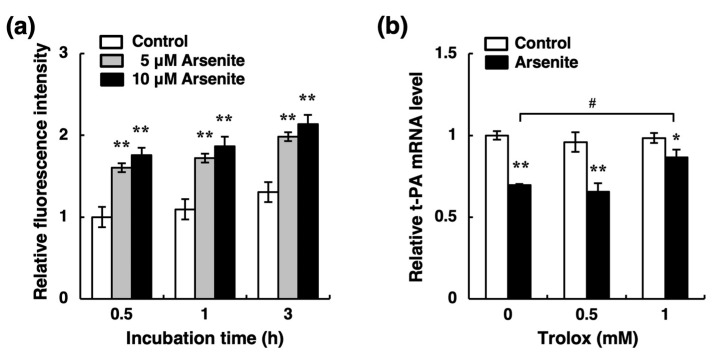
Chronic arsenic exposure is known to be related to the progression of atherosclerosis. However, the pathogenic mechanisms of arsenic-induced atherosclerosis have not been fully elucidated. Because disruption of the blood coagulation/fibrinolytic system is involved in the development of arteriosclerosis, we investigated the effect of arsenite on fibrinolytic activity in human vascular endothelial EA.hy926 cells in the present study. Fibrinolysis depends on the balance between tissue-type plasminogen activator (t-PA) and plasminogen activator inhibitor 1 (PAI-1) secreted from vascular endothelial cells. We found that arsenite reduced fibrinolytic t-PA activity by inhibiting its synthesis without affecting PAI-1 production. The inhibitory effect of arsenite on t-PA expression was partially recovered by the reactive oxygen species (ROS) scavenger Trolox. The nuclear factor erythroid 2 related factor 2 (NRF2) pathway is known to be activated by arsenite via ROS production. We confirmed that arsenite activated the NRF2 pathway, and arsenite-induced inhibition of fibrinolytic t-PA activity was abrogated in NRF2-knockdown EA.hy926 cells. These results suggest that arsenite inhibits the fibrinolytic activity of t-PA by selectively suppressing its synthesis via activation of the NRF2 pathway in vascular endothelial cells.

Keywords: arsenite; atherosclerosis; endothelial cell; fibrinolysis; nuclear factor erythroid 2 related factor 2; tissue-type plasminogen activator.


Conflict of interest statement

The authors declare no conflict of interest.

Figures

Figure 1
Figure 1
Effect of arsenite on the cytotoxicity of human vascular endothelial EA.hy926 cells. (a) Morphological appearance of endothelial EA.hy926 cells after exposure to arsenite at 1, 2, 5, 10, or 20 µM for 24 h. Scale bar = 400 µm. (b) The cell viability of endothelial EA.hy926 cells after exposure to arsenite at 1, 2, 5, 10, or 20 µM for 24 h. The data are reported as the mean ± S.D. of four samples. The data were analyzed using one-way ANOVA, followed by the Bonferroni/Dunn test.
Figure 2
Figure 2
Effect of arsenite on the fibrinolytic activity of endothelial EA.hy926 cells. (a) Fibrin zymography of conditioned medium collected from endothelial EA.hy926 cells after exposure to arsenite. The cells were incubated at 37 °C for 24 h with arsenite at 1, 2, 5, or 10 µM. (b) Fibrin zymography of conditioned medium collected from endothelial EA.hy926 cells incubated with arsenite under cell-free conditions. The cells were incubated at 37 °C for 24 h in the absence of arsenite, and then the conditioned medium was incubated at 37 °C for 24 h in the presence of arsenite at 1, 2, 5, 10, 20, or 50 µM under cell-free conditions.
Figure 3
Figure 3
Effects of arsenite on the expression and secretion of fibrinolytic proteins in endothelial EA.hy926 cells. The accumulation of t-PA (a) and PAI-1 (b) in the conditioned medium of endothelial EA.hy926 cells. The cells were incubated with arsenite at 1, 2, 5, or 10 µM for 24 h. The data are reported as the mean ± S.D. of four samples. The data were analyzed using one-way ANOVA, followed by the Bonferroni/Dunn test. * Significantly different from the control, p < 0.05. The mRNA expression of t-PA (c) and PAI-1 (d) in endothelial EA.hy926 cells. The cells were incubated with arsenite at 1, 2, 5, or 10 µM for 24 h. The data are reported as the mean ± S.D. of three samples. The data were analyzed using one-way ANOVA, followed by the Bonferroni/Dunn test. Significantly different from the control, * p < 0.05; ** p < 0.01. The mRNA expression of t-PA (e) and PAI-1 (f) in endothelial EA.hy926 cells. The cells were incubated with arsenite at 10 µM for 6, 12, 24, or 48 h. The data are reported as the mean ± S.D. of three samples. The data were analyzed using Student’s t-test. Significantly different from the corresponding control, * p < 0.05; ** p < 0.01.
Figure 4
Figure 4
Effects of arsenate on the mRNA expression of t-PA (a) and PAI-1 (b) in endothelial EA.hy926 cells. The cells were incubated with arsenate at 1, 2, 5, or 10 µM for 24 h. The data are reported as the mean ± S.D. of three samples. The data were analyzed using one-way ANOVA, followed by the Bonferroni/Dunn test.
Figure 5
Figure 5
Possible involvement of reactive oxygen species (ROS) production in the inhibition of t-PA mRNA expression by arsenite in endothelial EA.hy926 cells. (a) Effect of arsenite on the levels of intracellular ROS in endothelial EA.hy926 cells. The cells were pretreated with 2′,7′-dichlorodihydrofluorescein diacetate (H2DCFDA) at 100 µM for 1 h and then treated with arsenite at 5 or 10 µM for 0.5, 1, or 3 h. The data are reported as mean ± S.D. of six samples. The data were analyzed using one-way ANOVA, followed by the Bonferroni/Dunn test. ** Significantly different from the corresponding control, p < 0.01. (b) Effect of Trolox, a radical scavenger, on arsenite-induced suppression of t-PA mRNA expression in endothelial EA.hy926 cells. The cells were treated with arsenite at 10 µM in the presence of Trolox at 0.5 or 1 mM for 24 h. The data are reported as the mean ± S.D. of three samples. The data were analyzed using one-way ANOVA, followed by the Bonferroni/Dunn test. Significantly different from the corresponding control, * p < 0.05; ** p < 0.01. # Significantly different from the arsenite-treated cells without Trolox, p < 0.05.
Figure 6
Figure 6
Effect of arsenite on NRF2 pathway activation in endothelial EA.hy926 cells. (a) Intranuclear NRF2 levels in endothelial EA.hy926 cells. The cells were incubated with arsenite at 10 µM for 6, 12, or 24 h. (+) indicates treated with arsenite; (−) indicates treated without arsenite. (b) The expression levels of NQO1 mRNA in endothelial EA.hy926 cells. The cells were incubated with arsenite at 10 µM for 6, 12, or 24 h. The data are reported as the mean ± S.D. of three samples. The data were analyzed using Student’s t-test. ** Significantly different from the corresponding control, p < 0.01. (c) The expression levels of NQO1 mRNA in endothelial EA.hy926 cells. The cells were incubated with arsenite at 1, 2, 5, or 10 µM for 24 h. The data are reported as the mean ± S.D. of three samples. The data were analyzed using one-way ANOVA, followed by the Bonferroni/Dunn test. ** Significantly different from the control, p < 0.01. (d) Intranuclear NRF2 levels in endothelial EA.hy926 cells. The cells were incubated with arsenite at 10 µM in the presence of Trolox at 0.5 and 1 mM for 24 h. (e) Ratio of NRF2/Lamin A in arsenite-treated groups to that in the corresponding control groups in (d).
Figure 7
Figure 7
Possible involvement of the NRF2 pathway in the inhibition of t-PA expression by arsenite in endothelial EA.hy926 cells. (a) The expression level of NRF2 mRNA in endothelial EA.hy926 cells transfected with control siRNA (siCON) or NRF2 siRNA (siNRF2). The cells were incubated with arsenite at 10 µM for 24 h. The data are reported as the mean ± S.D. of three samples. (b) The protein level of NRF2 in siCON or siNRF2 transfected endothelial EA.hy926 cells after exposure to arsenite at 10 µM for 24 h. (+) indicates treated with arsenite; (−) indicates treated without arsenite. (c) The expression levels of NQO1 mRNA in siCON and siNRF2 transfected endothelial EA.hy926 cells after exposure to arsenite at 10 µM for 24 h. The data are reported as the mean ± S.D. of three samples. The data were analyzed using one-way ANOVA, followed by the Bonferroni/Dunn test. ## Significantly different from siCON transfected cells treated with arsenite, p < 0.01. (d) Fibrin zymography of conditioned medium collected from siCON or siNRF2 transfected endothelial EA.hy926 cells after exposure to arsenite at 10 µM for 24 h. (e) The accumulation of t-PA in the conditioned medium of siCON or siNRF2 transfected endothelial EA.hy926 cells. The cells were incubated in the presence or absence of arsenite at 10 µM for 24 h. The data are reported as the mean ± S.D. of four samples. The data were analyzed using one-way ANOVA, followed by the Bonferroni/Dunn test. * Significantly different from the corresponding control, p < 0.05. ## Significantly different from the corresponding siCON transfected cells, p < 0.01. (f) The expression level of t-PA mRNA in siCON or siNRF2 transfected endothelial EA.hy926 cells. The cells were incubated with arsenite at 10 µM for 24 h. The data are reported as the mean ± S.D. of three samples. The data were analyzed using one-way ANOVA, followed by the Bonferroni/Dunn test. ** Significantly different from the corresponding control, p < 0.01. # Significantly different from the corresponding siCON transfected cells, p < 0.05.
Figure 8
Figure 8
The intracellular signaling pathway that mediates arsenite-induced suppression of t-PA expression in vascular endothelial cells. Arsenite activates NRF2 transcriptional activity partly by enhancing ROS production. Activation of the NRF2 pathway reduces t-PA expression and secretion, resulting in a decrease in the fibrinolytic activity of t-PA. In this study, the mechanism underlying the inhibition of fibrinolysis by arsenite in human vascular endothelial cells was clarified.

References

    1. James K.A., Byers T., Hokanson J.E., Meliker J.R., Zerbe G.O., Marshall J.A. Association between lifetime exposure to inorganic arsenic in drinking water and coronary heart disease in Colorado residents. Environ. Health Perspect. 2015;123:128–134. doi: 10.1289/ehp.1307839. - DOI - PMC - PubMed
    1. Savage K.S., Tingle T.N., O’Day P.A., Waychunas G.A., Bird D.K. Arsenic speciation in pyrite and secondary weathering phases, Mother Lode Gold District, Tuolumne County, California. Appl. Geochem. 2000;15:1219–1244. doi: 10.1016/S0883-2927(99)00115-8. - DOI
    1. Podgorski J., Berg M. Global threat of arsenic in groundwater. Science. 2020;368:845–850. doi: 10.1126/science.aba1510. - DOI - PubMed
    1. Tseng W.P., Chu H.M., How S.W., Fong J.M., Lin C.S., Yeh S. Prevalence of skin cancer in an endemic area of chronic arsenicism in Taiwan. J. Natl. Cancer Inst. 1968;40:453–463. - PubMed
    1. Engel R.R., Hopenhayn-Rich C., Receveur O., Smith A.H. Vascular effects of chronic arsenic exposure: A review. Epidemiol. Rev. 1994;16:184–209. doi: 10.1093/oxfordjournals.epirev.a036150. - DOI - PubMed

LinkOut - more resources