Review
. 2021 Jan 13;13(1):105.
doi: 10.3390/v13010105.

The Unique, the Known, and the Unknown of Spumaretrovirus Assembly

Affiliations
Review

The Unique, the Known, and the Unknown of Spumaretrovirus Assembly

Dirk Lindemann et al. Viruses. .

Abstract

Within the family of Retroviridae, foamy viruses (FVs) are unique and unconventional with respect to many aspects in their molecular biology, including assembly and release of enveloped viral particles. Both components of the minimal assembly and release machinery, Gag and Env, display significant differences in their molecular structures and functions compared to the other retroviruses. This led to the placement of FVs into a separate subfamily, the Spumaretrovirinae. Here, we describe the molecular differences in FV Gag and Env, as well as Pol, which is translated as a separate protein and not in an orthoretroviral manner as a Gag-Pol fusion protein. This feature further complicates FV assembly since a specialized Pol encapsidation strategy via a tripartite Gag-genome-Pol complex is used. We try to relate the different features and specific interaction patterns of the FV Gag, Pol, and Env proteins in order to develop a comprehensive and dynamic picture of particle assembly and release, but also other features that are indirectly affected. Since FVs are at the root of the retrovirus tree, we aim at dissecting the unique/specialized features from those shared among the Spuma- and Orthoretrovirinae. Such analyses may shed light on the evolution and characteristics of virus envelopment since related viruses within the Ortervirales, for instance LTR retrotransposons, are characterized by different levels of envelopment, thus affecting the capacity for intercellular transmission.

Keywords: Env leader protein; Ortervirales; Pol protein packaging; RNA-mediated Pol tethering; assembly; foamy virus; particle budding; retrovirus evolution; spumavirus.


Conflict of interest statement

The authors declare no conflict of interest. The funders had no role in the design of the study; in the collection, analyses, or interpretation of data; in the writing of the manuscript, or in the decision to publish the results.

Figures

Figure A1
Figure A1
FFV capsomere and capsid formation by wt and C-terminal FFV Gag deletion mutants. (a) Schematic of the FFV C-terminal Gag deletion mutants with p4 and the GR-rich domain marked by white and red boxes, respectively. (b) HEK293T cells were transfected in 10 cm dishes with 12 μg truncated or wt Gag expressing plasmid. At 36 h post transfection, cytoplasmic extracts were prepared and ultracentrifuged through a 10%–60% continuous sucrose gradient at 32,000 rpm for 2 h. For each sample, 20 fractions (from 1 to 20) were collected from the top of the gradient and analyzed by immunoblotting by using an anti-Gag MA antiserum. Fractions 15–20 are not shown because no bands were visible. Fractions 1–2: unassembled Gag proteins; fractions 3–7: capsomeres; fractions 12–14 capsids. Shortened from detailed data in [92].
Figure 1
Figure 1
Structure of foamy virus (FV) virions. (a) Schematic representation of the prototype FV isolate (PFV) particle structure. pr: precursor protein; p: protein; gp: glycoprotein; (b) cryo-electron tomography of PFV virion and the corresponding radial density profile with the various peaks corresponding to the capsid (C), intermediate shell (I), viral membrane (M), glycoprotein (G), labeled and colored in red, green, blue, and yellow respectively. (ce) Ultrastructure of feline FV (FFV) virions by cryo-electron microscopy analysis. The MA layer (white arrowheads) follows the shape of the capsid in particles with central (c) and off-center (d) capsids as schematically shown in panel (e). Many particles in the population show internal angular capsid (the edge of the capsid is marked by black arrowheads) displaced from the center of the particle. Panel (a) adapted from [3,8]; panel (b) adapted from [7]; panel (ce) adapted from [6].
Figure 2
Figure 2
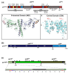
Schematic representation of the FV Gag, Pol, and Env protein organization. (a) Schematic illustration of the PFV Gag protein organization and selected functional motifs. Several functional motifs of PFV Gag are highlighted in differently colored boxes. Numbers indicate amino acid (aa) positions of the PFV Gag protein. The black arrow marks the cleavage site of pr71Gag for processing into p68Gag and p3Gag. Gray boxes represent the proline-rich (PR-rich) and glycine-arginine-rich (GR-rich) regions respectively. CC1 to CC4: coiled-coil domain 1 to 4; CTRS: cytoplasmic targeting and retention signal; NES: nuclear export sequence; L: late budding domain motif; A: assembly motif. (i) Cartoon representation of the three-dimensional (3D) structure of the PFV Gag N-terminal domain (NtD, aa 1–179) homodimer in complex with Env leader protein (Elp/LP) peptides. The Gag-NtD monomer-A is shown in pale blue and monomer-B in green. The helical Elp/LP peptides bound at the periphery of each head domain are colored magenta and gold with N and C termini indicated (ii) Cartoon representation of the 3D structure of the PFV-Gag central domain (CEN, aa 300–477) with its two subdomains NtDCEN and CtDCEN. The peptide backbone is shown in cyan. The secondary structure elements are numbered sequentially from the amino-terminus and the N and C termini are indicated. Helices α1 to α4 and α5 to α9 that comprise NtDCEN and CtDCEN, respectively, are indicated. (b) Schematic illustration of the PFV Pol protein organization. Numbers indicate aa positions of the PFV Pol protein. The black arrow marks the cleavage site of pr127Pol for processing into p85PR-RT-RH and p40IN. PR: protease domain; L: linker sequence; RT: reverse transcriptase domain; RH: RNase H domain; IN: integrase domain. (c) Schematic organization of PFV Env protein. The furin cleavage sites within the gp130Env precursor that are used for generation of the mature gp18LP, gp80SU, and gp48TM subunits are indicated by arrows. The individual subunits are shown as boxes in different shades of green. Hydrophobic sequences spanning the membrane in Elp/LP (h) and transmembrane (TM) (membrane-spanning domain, MSD) subunit are indicated. The aa sequence of the PFV Env Elp/LP subunit is shown in the enlargement below. The conserved WxxW and RxxR motif are highlighted in red, the lysine residues potentially ubiquitinated are highlighted in blue. The approximate positions of PFV Env N-glycosylation sites are marked by Y-shaped symbols. Panel (a,b,c) adapted from [3,8,22]; panel (ai) adapted from [25]; panel (aii) adapted from [24].
Figure 3
Figure 3
FV provirus organization, genomic and structural gene transcripts, and essential cis-acting viral RNA sequence elements. Schematic illustration of the PFV proviral DNA genome structure with long terminal repeats (LTRs) and open reading frames (ORFs) indicated as boxes. For ORFs encoding Gag, Pol, and Env precursor proteins, the regions encompassing the mature subunits generated by proteolytic processing are indicated by different colors and are labeled accordingly. Transcription initiation sites in the LTR and internal promoter (IP) and direction of transcription are indicated by dashed line arrows. Spliced and unspliced viral transcripts originating from the LTR and encompassing the viral RNA genome (vgRNA) or encoding the structural proteins Gag, Pol, and Env are schematically illustrated below and their respective coding capacity indicated to the right. Transcripts originating at the IP are omitted. Cis-acting sequence (CAS) elements localized within the full-length viral RNA genome, which are essential for viral replication, are indicated by black bars underneath the vgRNA. Individual functionally important or essential RNA sequence motifs are marked in the enlarged individual CAS elements below. Numbers represent nucleotide positions of the viral PFV RNA genome (HSRV2 isolate). At the bottom, individual regions within the vgRNA, which are essential for specific functions in viral replication as indicated to the right, are marked as differentially colored bars. U3: unique 3′ LTR region; R: repeat LTR region; U5: unique 5′ LTR region; ©: cap structure; An: poly A tail; mSD: major splice donor; PBS: primer binding site; PARM: protease activating RNA motif; cPPT: central poly-purine tract; 3′ PPT: 3′ poly-purine tract; pA: polyadenylation signal; A-D: purine-rich sequence motifs PPT A through D; RTr: reverse transcription; tas: transactivator of spumaviruses; bel-2: between envelope and LTR ORF-2. Adapted from [8].
Figure 4
Figure 4
Structure of FV envelope glycoprotein complex (GPC) and the GPC lattice of FV virions. (a) Surface features of PFV particles detected by negative-staining EM. Virions show a network of trimeric viral spike proteins on the particle surface and are predominantly arranged into rings of six subunits. Adjacent rings always share two completely integrated spikes. Images at higher magnification (b) reveal three separate densities (arrowheads) in the triangular spike. When grouped in hexameric rings, a stain-filled hole with a diameter of about 8 nm was formed (asterisk). Bars represent 50 nm (a) and 25 nm (b); (ce) subtomogram averaging of PFV glycoprotein. (c) 0.8 nm thick tomographic slice perpendicular to the glycoprotein long axis and its corresponding schematic of interlocked hexagonal assemblies of trimers. Numbers are indicated at the center of each hexagon and triangles represent the position of each trimer of Env in the hexagonal network. (d) Top view of intertwined hexagonal assemblies. (e) Side view of a single trimer. (fg) In Situ single particle 3D reconstruction of PFV glycoprotein by cryo-EM. Full (f) and cut-away (g) side views of a single PFV Env trimer (sharpened map) after 3-fold symmetry application (~9 A resolution at FSC = 0.143). The densities corresponding to the extracellular domains and the viral membrane are colored salmon and gray respectively in (f). The three central helices attributed to gp48 fusion peptide are represented by three green α helices of 22 residues long each. The transmembrane helices (TMHs) are represented by three inner (colored blue) and three outer (colored orange) α helices. In (g), the densities surrounding the three central helices and the three inner and outer TMHs are colored green, blue and orange respectively while the remaining of the spike is gray colored. Panel (a,b) adapted from [95]; panel (cg) adapted from [7].
Figure 5
Figure 5
Schematic illustration of FV replication and virion maturation. (a) Simplified schematic illustration of the FV replication cycle. FVs attach to target cells by interaction with cell surface heparan sulfate. Virions having already undergone RTr during assembly and release (up to 20% of total) contribute to the majority of productive infection events. Virions enter the cell by endocytosis and capsids are released into the cytoplasm by a pH-regulated fusion of viral and cellular lipid membranes mediated by FV Env and unknown cellular entry receptor(s). Intact capsids migrate along microtubules to the centrosome where they remain in a latent state until the host cell enters mitosis. This induces further disassembly and enables chromatin access of the FV pre-integration complex upon nuclear membrane breakdown. Following integration and FV Tas-mediated transcription regulation viral RNAs are transported to the cytoplasm or the ER where protein translation takes place. Further details of the late steps of FV replication are discussed and summarized throughout the manuscript. (b) Model of sequential events resulting in infectious FV capsid maturation. 1. FV Pol is incorporated predominantly at the precursor state involving binding of Gag (or capsomeres) and Pol to vRNA and potentially additional Gag–Pol protein interactions; 2. Binding of Pol to the PARM elements of the vgRNA and potentially oligomerization by the IN domain result in PR domain dimerization associated with PR activation; 3. Different possibilities exist for subsequent Gag and Pol precursor maturation. First either Gag (3.1) or Pol (3.3) is cleaved followed by the cleaving of the other component, or both proteins (3.2) are cleaved simultaneously. 4. Gag precursor processing activates RT activity either in the Pol precursor (4.1) or the mature PR-RT subunit (4.2); 5. This leads to RTr of the packaged vRNA genome in up to 20% of all released particles, which contribute to the great majority of FV infectivity. The processing pathways that are less likely in the opinion of the authors are indicated by lighter representation.
Figure 6
Figure 6
Maximum-likelihood phylogeny of viral reverse transcriptases. The tree includes sequences of 290 viruses belonging to all International Committee on Taxonomy of Viruses (ICTV)-recognized genera of reverse-transcribing viruses. The phylogeny was inferred using PhyML [115] with the LG + G + F substitution model and is rooted with sequences from nonviral retroelements (bacterial group II introns and eukaryotic LINE retroelements). Genomic organizations of selected representatives of reverse-transcribing viruses are shown next to the corresponding branches. Long terminal repeats are shown as black triangles. Note that members of the virus families display considerable variation in gene/domain content [116], which is not captured in this figure. Abbreviations: 6, 6-kDa protein; ATF, aphid transmission factor; CA/CP, capsid protein; CHR, chromodomain (present only in the integrase of particular clades of metaviruses of plants, fungi, and several vertebrates); gag, group-specific antigen; env, envelope genes; INT, integrase; LTR, long terminal repeat; MA, matrix protein; MP, movement protein; NC, nucleocapsid; nef, tat, rev, vif, vpr, and vpu, genes that express regulatory proteins via spliced mRNAs; P, polymerase; pol, polymerase gene; PR, protease; PreS, pre-surface protein (envelope); PX/TA, protein X/transcription activator; RH, RNase H; RT, reverse transcriptase; SU, surface glycoprotein; TM, transmembrane glycoprotein; TP, terminal protein domain; TT/SR, translation trans-activator/suppressor of RNA interference; VAP, virion-associated protein. HIV-1: Human immunodeficiency virus 1; DmeGypV: Drosophila melanogaster gypsy virus; CaMV: Cauliflower mosaic virus; DmeBelV: Drosophila melanogaster Bel virus; SceTy1V: Saccharomyces cerevisiae Ty1 virus; HBV: Hepatitis B virus. Image and caption adapted from [2].

References

    1. Khan A.S., Bodem J., Buseyne F., Gessain A., Johnson W., Kuhn J.H., Kuzmak J., Lindemann D., Linial M.L., Löchelt M., et al. Spumaretroviruses: Updated taxonomy and nomenclature. Virology. 2018;516:158–164. doi: 10.1016/j.virol.2017.12.035. - DOI - PMC - PubMed
    1. Krupovic M., Blomberg J., Coffin J.M., Dasgupta I., Fan H., Geering A.D., Gifford R., Harrach B., Hull R., Johnson W., et al. Ortervirales: New Virus Order Unifying Five Families of Reverse-Transcribing Viruses. J. Virol. 2018;92 doi: 10.1128/JVI.00515-18. - DOI - PMC - PubMed
    1. Lindemann D., Rethwilm A. Foamy virus biology and its application for vector development. Viruses. 2011;3:561–585. doi: 10.3390/v3050561. - DOI - PMC - PubMed
    1. Materniak-Kornas M., Tan J., Heit-Mondrzyk A., Hotz-Wagenblatt A., Löchelt M. Bovine Foamy Virus: Shared and Unique Molecular Features in Vitro and In Vivo. Viruses. 2019;11:1084. doi: 10.3390/v11121084. - DOI - PMC - PubMed
    1. Goff S.P. Retroviridae. In: Knipe D.M., Howley P.M., editors. Fields Virology. 6th ed. Volume 2. Lippincott Williams & Wilkins, a Wolters Kluwer Business; Philadelphia, PA, USA: 2013. pp. 1424–1473.

Publication types

LinkOut - more resources