

Review of Current Spinal Robotic Orthoses
- PMID: 33451142
- PMCID: PMC7828606
- DOI: 10.3390/healthcare9010070
Review of Current Spinal Robotic Orthoses
Abstract
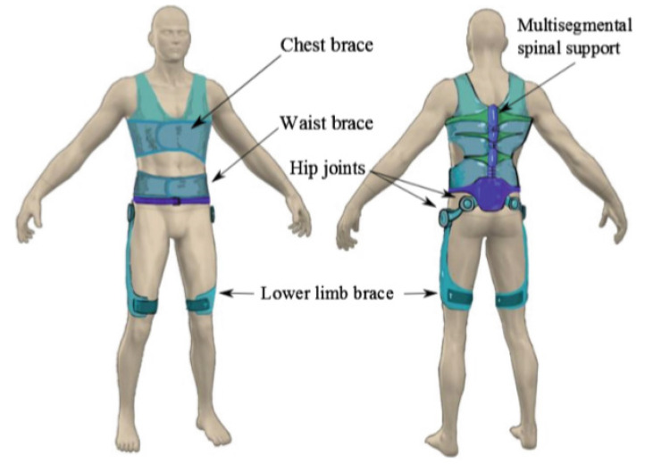
Osteoporotic spine fractures (OSF) are common sequelae of osteoporosis. OSF are directly correlated with increasing age and incidence of osteoporosis. OSF are treated conservatively or surgically. Associated acute pain, chronic disabilities, and progressive deformities are well documented. Conservative measures include a combination of initial bed rest, analgesia, early physiotherapy, and a spinal brace (orthosis), with the aim for early rehabilitation to prevent complications of immobile state. Spinal bracing is commonly used for symptomatic management of OSF. While traditional spinal braces aim to maintain the neutral spinal alignment and reduce the axial loading on the fractured vertebrae, they are well known for complications including discomfort with reduced compliance, atrophy of paraspinal muscles, and restriction of chest expansion leading to chest infections. Exoskeletons have been developed to passively assist and actively augment human movements with different types of actuators. Flexible, versatile spinal exoskeletons are designed to better support the spine. As new technologies enable the development of motorized wearable exoskeletons, several types have been introduced into the medical field application. We have provided a thorough review of the current spinal robotic technologies in this paper. The shortcomings in the current spinal exoskeletons were identified. Their limitations on the use for patients with OSF with potential improvement strategies were discussed. With our current knowledge of spinal orthosis for conservatively managed OSF, a semi-rigid backpack style thoracolumbar spinal robotic orthosis will reduce spinal bone stress and improve back muscle support. This will lead to back pain reduction, improved posture, and overall mobility. Early mobilization is an important part of management of patients with OSF as it reduces the chance of developing complications related to their immobile state for patients with OSF, which will be helpful for their recovery.
Keywords: active orthosis; exoskeleton; osteoporotic spine fracture; spinal orthosis; wearable robotics.
Conflict of interest statement
The authors declare that they have no competing interests.
Figures


Similar articles
-
Is postoperative bracing after pedicle screw fixation of spine fractures necessary? Study protocol of the ORNOT study: a randomised controlled multicentre trial.BMJ Open. 2018 Jan 13;8(1):e019596. doi: 10.1136/bmjopen-2017-019596. BMJ Open. 2018. PMID: 29331975 Free PMC article. Clinical Trial.
-
[Influence of spinal orthosis on gait and physical functioning in women with postmenopausal osteoporosis].Orthopade. 2012 Mar;41(3):200-5. doi: 10.1007/s00132-011-1867-6. Orthopade. 2012. PMID: 22139393 Clinical Trial. German.
-
Effect of a semi-rigid backpack type thoracolumbar orthosis on thoracic kyphosis angle and muscle performance in older adults with hyperkyphosis: a randomized controlled trial.Disabil Rehabil. 2023 May;45(9):1488-1497. doi: 10.1080/09638288.2022.2065541. Epub 2022 Apr 22. Disabil Rehabil. 2023. PMID: 35452347 Clinical Trial.
-
A systematic literature review of spinal brace/orthosis treatment for adults with scoliosis between 1967 and 2018: clinical outcomes and harms data.BMC Musculoskelet Disord. 2020 Feb 8;21(1):87. doi: 10.1186/s12891-020-3095-x. BMC Musculoskelet Disord. 2020. PMID: 32035480 Free PMC article.
-
Spinal Deformities and Advancement in Corrective Orthoses.Bioengineering (Basel). 2020 Dec 25;8(1):2. doi: 10.3390/bioengineering8010002. Bioengineering (Basel). 2020. PMID: 33375594 Free PMC article. Review.
Cited by
-
The Role of Orthoses in Chronic Axial Spinal Conditions.Curr Pain Headache Rep. 2024 Jun;28(6):501-506. doi: 10.1007/s11916-024-01233-7. Epub 2024 Feb 26. Curr Pain Headache Rep. 2024. PMID: 38407764 Review.
-
Emerging Medical Technologies and Their Use in Bionic Repair and Human Augmentation.Bioengineering (Basel). 2024 Jul 9;11(7):695. doi: 10.3390/bioengineering11070695. Bioengineering (Basel). 2024. PMID: 39061777 Free PMC article. Review.
References
-
- United Nations Population Division. [(accessed on 10 January 2021)]; Available online: https://www.un.org/en/development/desa/population/publications/pdf/agein....
-
- International Osteoporosis Federation Data and Publications: Facts and Statistics. [(accessed on 10 January 2021)]; Available online: https://www.osteoporosis.foundation/facts-statistics.
-
- Kim H.-J., Yi J.-M., Cho H.-G., Chang B.-S., Lee C.-K., Kim J.H., Yeom J.S. Comparative Study of the Treatment Outcomes of Osteoporotic Compression Fractures without Neurologic Injury Using a Rigid Brace, a Soft Brace, and no Brace: A Prospective Randomized Controlled Non-inferiority Trial. J. Bone Jt. Surg. Am. Vol. 2014;96:1959–1966. doi: 10.2106/JBJS.N.00187. - DOI - PubMed
Publication types
LinkOut - more resources
Full Text Sources
Other Literature Sources
Research Materials
