Case Reports
. 2021 Jan 16;21(1):25.
doi: 10.1186/s12883-021-02056-4.

Massive intracranial hemorrhage caused by intraventricular meningioma: case report

Affiliations
Case Reports

Massive intracranial hemorrhage caused by intraventricular meningioma: case report

Koichiro Sumi et al. BMC Neurol. .

Abstract

Background: Meningiomas are the most common benign intracranial tumors, and commonly comprise high-vascularizing but slow-growing tumors. On the other hand, meningiomas arising from the ventricular system are of rare occurrence, and spontaneous hemorrhage is an infrequent event.

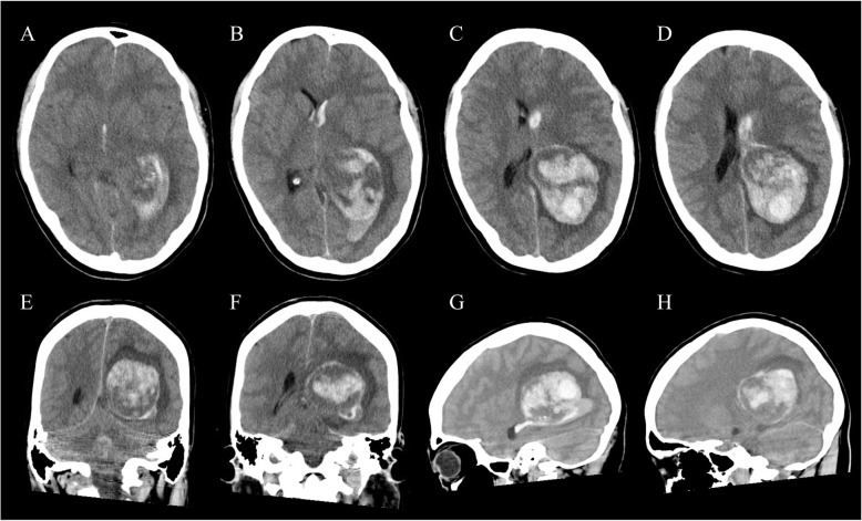
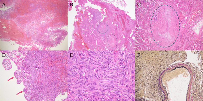
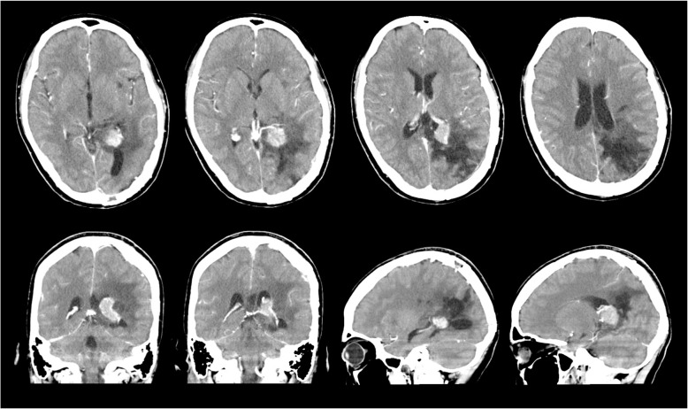
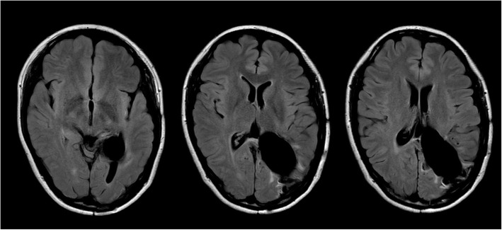
Case presentation: We describe here the rare clinical manifestations of a 28-year-old female with acute intracranial hemorrhage located in the trigone of the lateral ventricle who was initially thought to have suffered an acute cerebrovascular accident, but was subsequently confirmed to have a benign intraventricular meningioma. To clarify the clinical features of such a rare course of meningioma, we also present a short literature review of acute intracranial hemorrhage caused by intraventricular meningioma.

Conclusions: Ventricular meningioma presenting with hemorrhage such as acute stroke is a rare event, but recognition of such a pathogenesis is important. Although further accumulation of clinical data is needed, we suggest that early surgery should be undertaken in patients with lateral ventricular meningioma, even if it is not so large or asymptomatic.

Keywords: Fibrous meningioma; Intraventricular hemorrhage; Intraventricular tumor; Meningioma.


Conflict of interest statement

All authors have no affiliations with or involvement in any organization or entity with any financial interest, or non-financial interest, in the subject matter or materials discussed in this case report.

Figures

Fig. 1
Fig. 1
Non-contrast axial (a-d), coronal (e, f), and sagittal (g, h) CT scans on admission revealing a large heterogeneous mass lesion in the trigone of the left lateral ventricle with intraventricular hemorrhage extending to the right lateral ventricle
Fig. 2
Fig. 2
Histological findings of the 1st surgical specimen. Massive hemorrhage was observed within the tumor (a: HE stain, x 2). Two closed arteries circled by the blue dotted line were identified in the peri-tumoral abundant collagen regions (b: HE stain, x 2). High magnification of one of the closed arteries (c: HE stain, x 40). The tumor involved the choroid plexus (d: HE stain, x 10). This tumor is consistent with fibrous type meningioma composed of spindle-shaped tumor cells with a fascicular or storiform arrangement (e: HE stain, x 20). In addition, dilated abnormal vessels can be seen, of which the walls are thin and the internal elastic membrane appears fragile (f: Elastic-van Gieson stain, x 20)
Fig. 3
Fig. 3
Postcontrast CT scan on day 38 after first surgery showing an enhanced small residual tumor in the trigone of the left lateral ventricle and enlarged feeding artery
Fig. 4
Fig. 4
Postoperative axial fluid-attenuated inversion recovery MRI at 15 months after 2nd surgery showing total removal of the meningioma in the trigone of the left lateral ventricle

Similar articles

Cited by

References

    1. Wakai S, Yamakawa K, Manaka S, Takakura K. Spontaneous intracranial hemorrhage caused by brain tumor: its incidence and clinical significance. Neurosurgery. 1982;10:437–44. doi: 10.1227/00006123-198204000-00004. - DOI - PubMed
    1. Martinez-Lage JF, Poza M, Martinez M, Esteban JA, Antunez MC, Sola J. Meningiomas with haemorrhagic onset. Acta Neurochir (Wien) 1991;110:129–32. doi: 10.1007/BF01400680. - DOI - PubMed
    1. Lang I, Jackson A, Strang FA. Intraventricular hemorrhage caused by intraventricular meningioma: CT appearance. AJNR. 1995;16:378–81. - PMC - PubMed
    1. Lee EJ, Choi KH, Kang SW, Lee IW. Intraventricular hemorrhage caused by lateral ventricular meningioma: a case report. Korean J Radiol. 2001;2:105–7. doi: 10.3348/kjr.2001.2.2.105. - DOI - PMC - PubMed
    1. Bosnjak R, Derham C, Popović M, Ravnik J. Spontaneous intracranial meningioma bleeding: clinicopathological features and outcome. J Neurosurg. 2005;103:473–84. doi: 10.3171/jns.2005.103.3.0473. - DOI - PubMed

Publication types

MeSH terms

LinkOut - more resources