

Asymmetric response to ranibizumab in mixed choroidal neovascularization in a neovascular age-related macular degeneration diagnosed on OCT angiography - case report
- PMID: 33451290
- PMCID: PMC7811211
- DOI: 10.1186/s12886-021-01810-z
Asymmetric response to ranibizumab in mixed choroidal neovascularization in a neovascular age-related macular degeneration diagnosed on OCT angiography - case report
Abstract
Background: To present a case report of a patient with a mixed choroidal neovascular membrane (CNV) with an asymmetric response to ranibizumab diagnosed on optical coherence tomography angiography (OCTa).
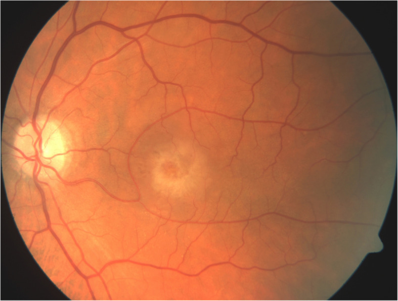
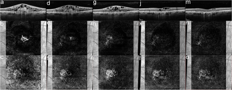
Case presentation: A 61-year-old male was referred to our department in September 2017 due to decreased vision in his left eye. Best-corrected visual acuity (BCVA) was 43 Early Treatment Diabetic Retinopathy Study (ETDRS) letters in the left eye. Macular edema was present in the left eye, and a mixed CNV was identified on the OCTa. Therapy with intravitreal ranibizumab was commenced. After 5 ranibizumab injections, the BCVA was 42 ETDRS letters, and considerable intraretinal edema was still present. OCTa showed a resolution of the type 2 lesion of the mixed CNV; however, the type 1 lesion had continued to grow. The patient was then switched to intravitreal aflibercept. After 3 monthly aflibercept injections, the BCVA improved to 53 ETDRS letters, and a reduction of the edema was observed on the optical coherence tomography (OCT). OCTa showed a decrease in both the area and vessel density in the type 1 lesion of the CNV. Therapy with aflibercept was continued; however, while the intraretinal edema continued to improve, atrophy developed in the macula and the BCVA worsened to 43 ETDRS letters.
Conclusions: Ranibizumab nonresponse in a neovascular age-related macular degeneration is not uncommon. However, to our knowledge, this is the first described case of an asymmetric response to ranibizumab in a mixed CNV. While the type 2 lesion of the CNV reacted swiftly to the ranibizumab therapy, the type 1 lesion continued to grow. As with some other cases of ranibizumab resistance, switching to aflibercept proved effective.
Keywords: Age‐related macular degeneration; Anti-VEGF; Case report; Mixed CNV; Resistance.
Conflict of interest statement
The authors declare that they have no conflict of interest.
Figures
References
-
- Bressler NM, Bressler SB, et al. Chapter 66 - Neovascular (exudative or “wet”) age-related macular degeneration A2 - Ryan, Stephen J. In: Sadda SR, Hinton DR, Schachat AP, Sadda SR, Wilkinson CP, Wiedemann P, et al., editors. Retina Fifth Ed. London: W.B. Saunders; 2013. p. 1183–212.
-
- Stepanov A, Nemcansky J, Veith M, Manethova K, Stredova M, Pencak M, et al. Two-year results of a combined regimen of aflibercept treatment in three types of choroidal neovascular membrane in the wet form of age-related macular degeneration: Real-life evidence in the Czech Republic. Eur J Ophthalmol. 2020 doi: 10.1177/1120672120971190. - DOI - PubMed
Publication types
MeSH terms
Substances
LinkOut - more resources
Full Text Sources
Other Literature Sources
Medical
Research Materials
