

Bioactive glasses and electrospun composites that release cobalt to stimulate the HIF pathway for wound healing applications
- PMID: 33451366
- PMCID: PMC7811269
- DOI: 10.1186/s40824-020-00202-6
Bioactive glasses and electrospun composites that release cobalt to stimulate the HIF pathway for wound healing applications
Abstract
Background: Bioactive glasses are traditionally associated with bonding to bone through a hydroxycarbonate apatite (HCA) surface layer but the release of active ions is more important for bone regeneration. They are now being used to deliver ions for soft tissue applications, particularly wound healing. Cobalt is known to simulate hypoxia and provoke angiogenesis. The aim here was to develop new bioactive glass compositions designed to be scaffold materials to locally deliver pro-angiogenic cobalt ions, at a controlled rate, without forming an HCA layer, for wound healing applications.
Methods: New melt-derived bioactive glass compositions were designed that had the same network connectivity (mean number of bridging covalent bonds between silica tetrahedra), and therefore similar biodegradation rate, as the original 45S5 Bioglass. The amount of magnesium and cobalt in the glass was varied, with the aim of reducing or removing calcium and phosphate from the compositions. Electrospun poly(ε-caprolactone)/bioactive glass composites were also produced. Glasses were tested for ion release in dissolution studies and their influence on Hypoxia-Inducible Factor 1-alpha (HIF-1α) and expression of Vascular Endothelial Growth Factor (VEGF) from fibroblast cells was investigated.
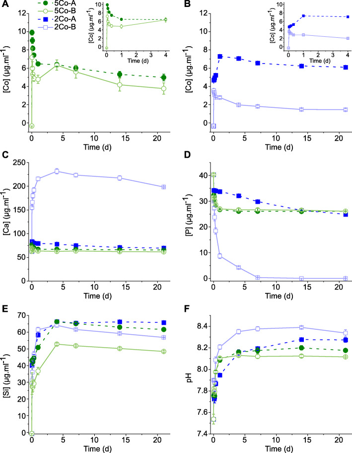
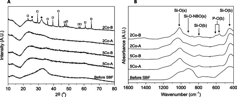
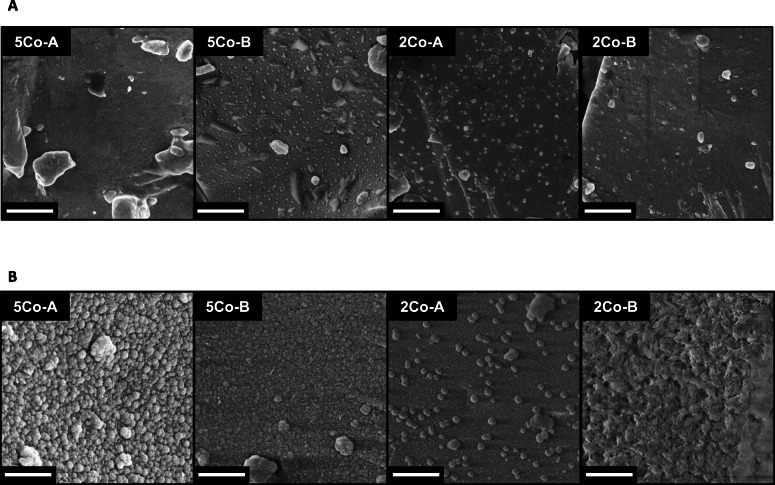
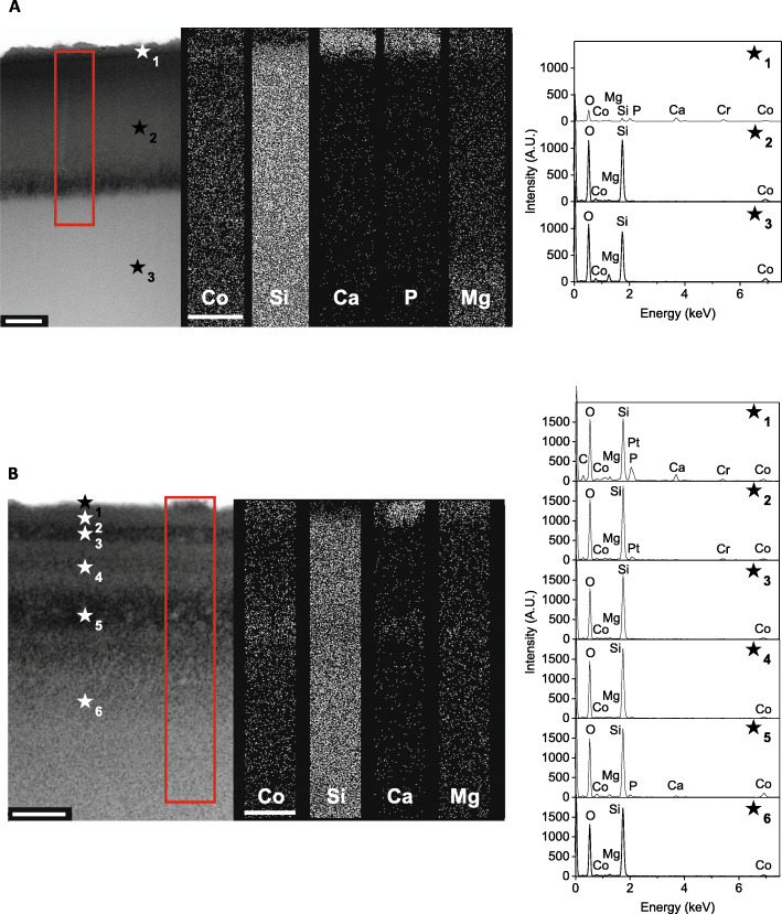
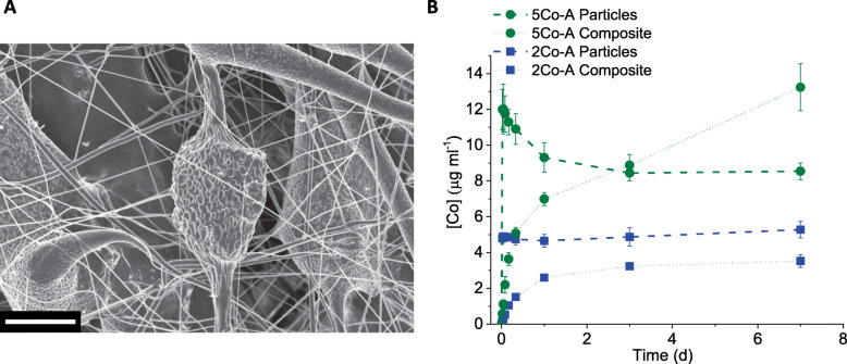
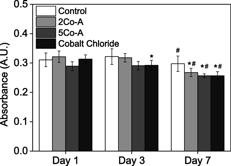
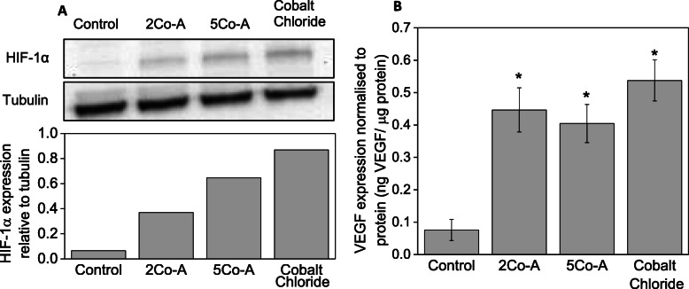
Results: Dissolution tests showed the silica rich layer differed depending on the amount of MgO in the glass, which influenced the delivery of cobalt. The electrospun composites delivered a more sustained ion release relative to glass particles alone. Exposing fibroblasts to conditioned media from these composites did not cause a detrimental effect on metabolic activity but glasses containing cobalt did stabilise HIF-1α and provoked a significantly higher expression of VEGF (not seen in Co-free controls).
Conclusions: The composite fibres containing new bioactive glass compositions delivered cobalt ions at a sustained rate, which could be mediated by the magnesium content of the glass. The dissolution products stabilised HIF-1α and provoked a significantly higher expression of VEGF, suggesting the composites activated the HIF pathway to stimulate angiogenesis.
Keywords: Bioactive composites; Bioactive glass; Cobalt; HIF pathway; Wound healing.
Conflict of interest statement
The authors declare that they have no competing interests.
Figures
References
-
- Hench LL. Opening paper 2015- some comments on bioglass: four eras of discovery and development. Biomedical Glasses. 2015;1(1):1–11. doi: 10.1515/bglass-2015-0001. - DOI
-
- Jones JR, Brauer DS, Hupa L, Greenspan DC. Bioglass and bioactive glasses and their impact on healthcare. Int J Appl Glas Sci. 2016;7(4):423–434. doi: 10.1111/ijag.12252. - DOI
-
- Hench LL, Splinter RJ, Allen WC, Greenlee TK. Bonding mechanisms at the interface of ceramic prosthetic materials. J Biomed Mater Res Symp. 1971;2(1):117–141. doi: 10.1002/jbm.820050611. - DOI
Grants and funding
LinkOut - more resources
Full Text Sources
Other Literature Sources
