. 2021 Jun;11(6):1562-1581.
doi: 10.1158/2159-8290.CD-20-1109. Epub 2021 Jan 15.

A CRISPR/Cas9-Engineered ARID1A-Deficient Human Gastric Cancer Organoid Model Reveals Essential and Nonessential Modes of Oncogenic Transformation

Affiliations

A CRISPR/Cas9-Engineered ARID1A-Deficient Human Gastric Cancer Organoid Model Reveals Essential and Nonessential Modes of Oncogenic Transformation

Yuan-Hung Lo et al. Cancer Discov. 2021 Jun.

Abstract

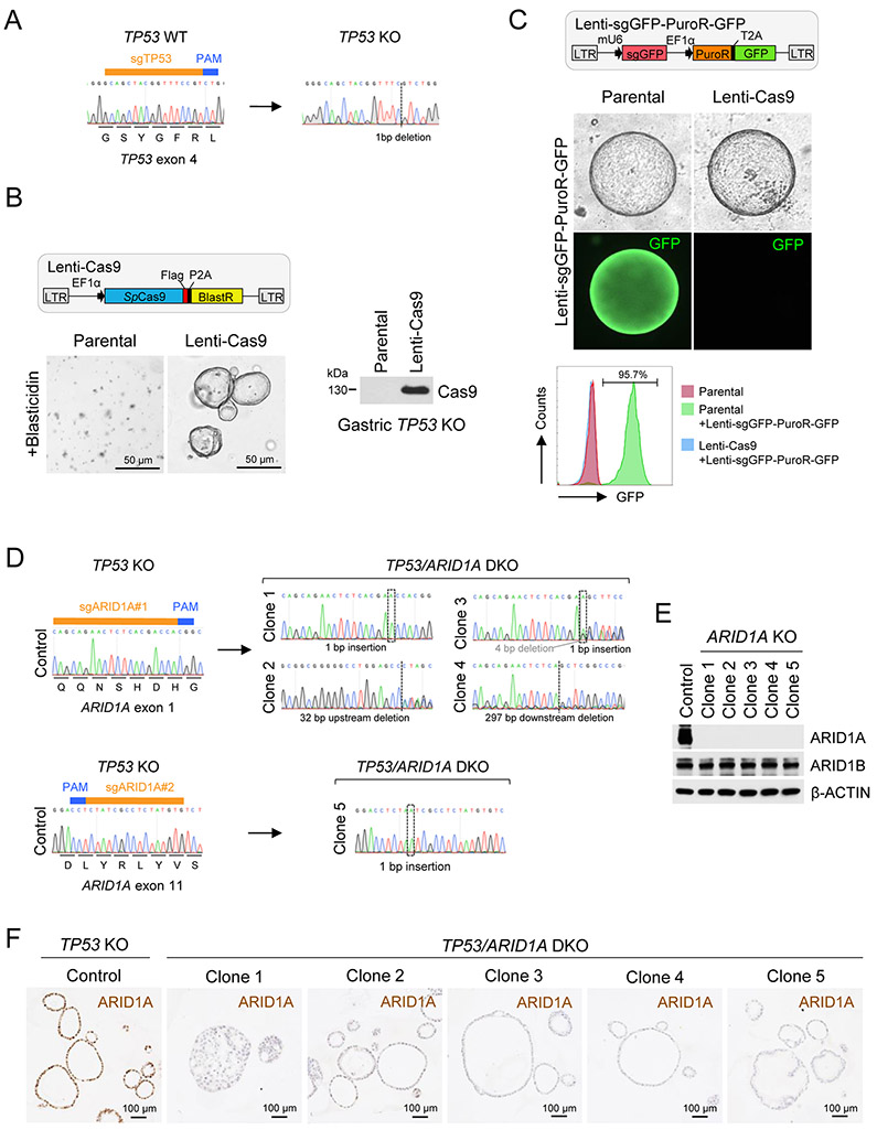
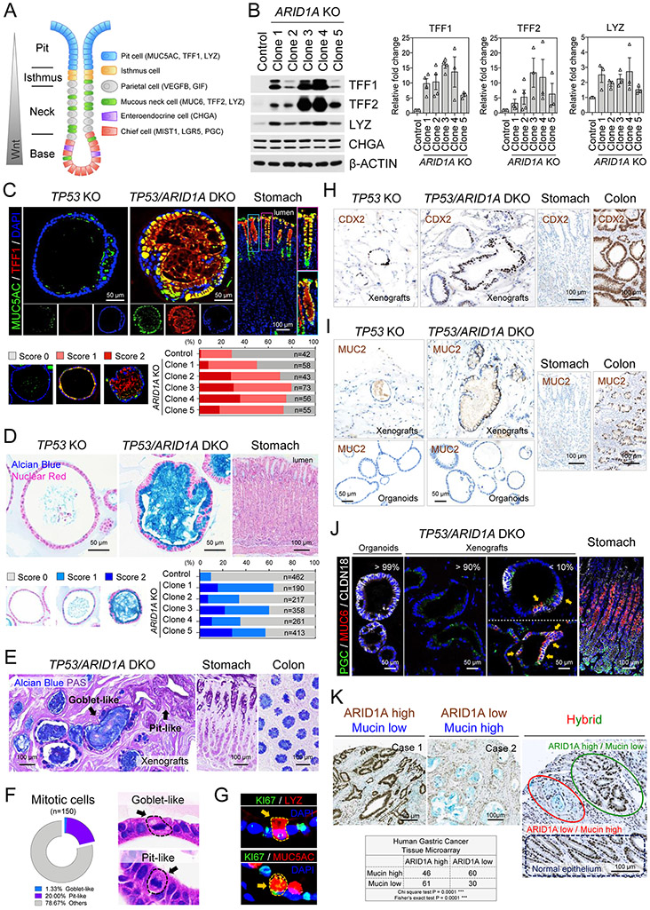
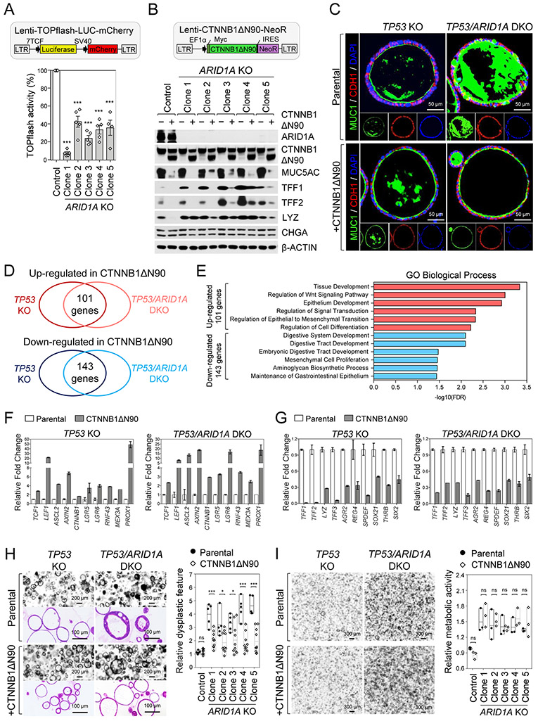
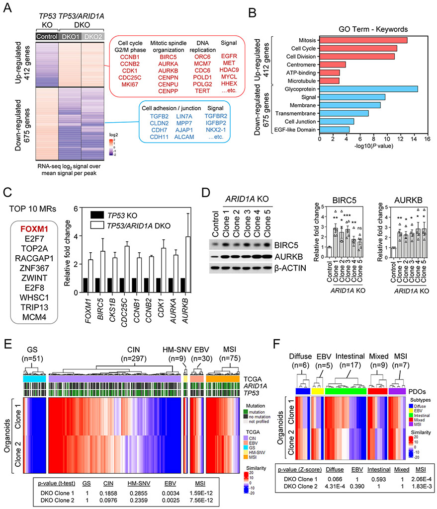
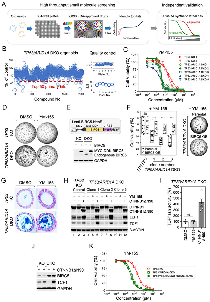
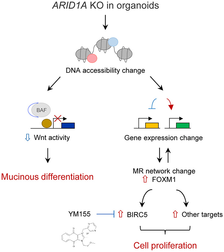
Mutations in ARID1A rank among the most common molecular aberrations in human cancer. However, oncogenic consequences of ARID1A mutation in human cells remain poorly defined due to lack of forward genetic models. Here, CRISPR/Cas9-mediated ARID1A knockout (KO) in primary TP53-/- human gastric organoids induced morphologic dysplasia, tumorigenicity, and mucinous differentiation. Genetic WNT/β-catenin activation rescued mucinous differentiation, but not hyperproliferation, suggesting alternative pathways of ARID1A KO-mediated transformation. ARID1A mutation induced transcriptional regulatory modules characteristic of microsatellite instability and Epstein-Barr virus-associated subtype human gastric cancer, including FOXM1-associated mitotic genes and BIRC5/survivin. Convergently, high-throughput compound screening indicated selective vulnerability of ARID1A-deficient organoids to inhibition of BIRC5/survivin, functionally implicating this pathway as an essential mediator of ARID1A KO-dependent early-stage gastric tumorigenesis. Overall, we define distinct pathways downstream of oncogenic ARID1A mutation, with nonessential WNT-inhibited mucinous differentiation in parallel with essential transcriptional FOXM1/BIRC5-stimulated proliferation, illustrating the general utility of organoid-based forward genetic cancer analysis in human cells. SIGNIFICANCE: We establish the first human forward genetic modeling of a commonly mutated tumor suppressor gene, ARID1A. Our study integrates diverse modalities including CRISPR/Cas9 genome editing, organoid culture, systems biology, and small-molecule screening to derive novel insights into early transformation mechanisms of ARID1A-deficient gastric cancers.See related commentary by Zafra and Dow, p. 1327.This article is highlighted in the In This Issue feature, p. 1307.


Conflict of interest statement

Conflict of Interest Statement

The authors declare no potential conflicts of interest

Figures

Figure 1.
Figure 1.. Establishment of clonal TP53/ARID1A knockout human gastric organoid lines.
A, The TP53 indel created by CRISPR/Cas9 cleavage was determined by Sanger sequencing. B, Establishment of a stable Cas9-expressing engineered TP53 KO human gastric organoid line. After antibiotic (Blasticidin) selection, Cas9 expression was confirmed by immunoblot analysis. C, Highly efficient CRISPR/Cas9 cleavage in Cas9-expressing TP53 KO organoids. A lentiviral construct containing a GFP guide RNA targeting the GFP reporter in the same construct was delivered into control TP53 KO and the Cas9-expressing TP53 KO organoids. After antibiotic (Puromycin) selection, GFP-positive cells were quantified by flow cytometry. D, Five different TP53/ARID1A DKO clones were established. ARID1A indels were determined by Sanger sequencing. E, Immunoblot analysis of ARID1A and ARID1B expression. F, IHC staining of ARID1A in TP53 KO versus TP53/ARID1A DKO organoids.
Figure 2.
Figure 2.. CRISPR KO of ARID1A promotes gastric malignancy.
A, TP53 KO (control) organoids were typically well-organized morphologically; however, TP53/ARID1A DKO organoids exhibited different degrees of architectural complexity. H&E staining. Quantitation revealed increased epithelial stratification (green bar), structural complexity (blue bar), and loss of polarity (red bar) in all five TP53/ARID1A DKO clones. B, Immunofluorescence staining of the apical-specific marker ZO1 (red) showed disruptions in apicobasal polarity in a subset of TP53/ARID1A DKO organoid cells. The arrow (orange) indicates loss of polarity with inappropriate basolateral ZO1 expression. Cell membrane was stained with CTNNB1 (green). Nuclei were stained with DAPI (blue). C, ARID1A-deficient organoids exhibit hyperproliferation. TP53 KO and TP53/ARID1A DKO organoids were grown from 20,000 single FACS-sorted BFP+ cells. Brightfield images were taken after cell sorting. Quantification of organoid size is shown (n=400 per group). D, Quantification of EdU-positive proliferating cells in TP53 KO and TP53/ARID1A DKO organoids from independent experiments (N=3) at day 6 after passage. E, Quantification of metabolic activity from independent experiments (N=6) was determined by Alamar blue assay at day 12 after passage. Relative metabolic activity was normalized to TP53 KO organoids (Control). Dots indicate independent experiments. The horizontal bar indicates mean. The error bar represents SEM. *P<0.05, ***P<0.005. ns, not significant. F, ARID1A-deficient organoids exhibited efficient in vivo tumor formation upon subcutaneous xenografting into NSG mice. TP53/ARID1A DKO xenografts formed larger tumors compare with TP53 KO xenografts. H&E staining. G, A significant negative correlation between ARID1A expression and tumor grade was identified in a human gastric cancer tissue microarray (total 197 patients). ARID1A expression was assessed by IHC.
Figure 3.
Figure 3.. ARID1A knockout induces mucinous metaplasia.
A, Schematic illustration of gastric epithelium. Different cell lineages and specific lineage markers are indicated. B, Western blot of mucin-producing pit cell and mucous neck cell markers, TFF1, TFF2 and LYZ, reveals upregulation in TP53/ARID1A DKO organoids. Quantification of expression from independent experiments (N>3) was shown. Dots indicate independent experiments. C, Immunofluorescence staining of MUC5AC (green) and TFF1 (red) in engineered organoids and the donor primary gastric tissues. Nuclei were stained with DAPI (blue). Quantification of MUC5AC-positive organoids is shown. D, Mucin production in engineered organoids and donor primary gastric tissues detected by Alcian blue staining. Nuclei were counterstained by nuclear fast red. Quantification of Alcian blue-positive organoids indicate increased mucin in all five TP53/ARID1A DKO organoids lines. E, TP53/ARID1A DKO xenografts in a subcutaneously xenografted NSG mice retain their mucin-secreting phenotype in vivo. Alcian blue and PAS staining. Goblet-like (Alcian blue -positive) and pit-like (PAS positive) cells were indicated. F, Quantification of mitotic cells. Goblet-like and pit-like cells with mitotic figures were shown. H&E staining. G, Immunofluorescence staining of TP53/ARID1A DKO organoids showing LYZ-positive (red) or MUC5-positive (red) proliferating cells (KI67+, green). H, IHC staining of CDX2 in xenografts and the donor primary gastric tissues. Colon tissues were used as positive control. I, IHC staining of MUC2 in organoids, xenografts and the donor primary gastric tissues. Colon tissues were used as positive control. J, Immunofluorescence staining of CLDN18 (white), MUC6 (red) and PGC (green) in TP53/ARID1A DKO organoids, xenografts and the donor primary gastric tissues. Cells within SPEM features (MUC6 and PGC double positive) are marked by arrows. K, A significant negative correlation between ARID1A (brown) IHC expression and mucin (blue, Alcian blue) production was identified in a human gastric cancer tissue microarray (total 197 patients).
Figure 4.
Figure 4.. Loss of ARID1A inhibits canonical Wnt/β-catenin activity.
A, Wnt/β-catenin-induced activity was decreased in TP53/ARID1A DKO organoids infected by lentivirus containing TOPflash Wnt reporter and mCherry followed by luciferase assay on 20,000 sorted mCherry-positive cells. Quantification of luciferase activity from independent experiments (N=5) is shown. Luciferase activity was normalized to TP53 KO organoids (Control). B, The mucin-producing phenotype was genetically rescued by lentiviral expression of an N-terminal truncated gain-of-function β-catenin mutant (CTNNB1ΔN90). After virus transduction and antibiotic (Neomycin) selection, protein expression in the engineered organoids was analyzed by Western blot as indicated. C, Immunofluorescence staining of apically-restricted transmembrane MUC1 (green) and membrane protein CDH1 (red) demonstrates that CTNNB1ΔN90 reduces mucin production and architectural complexity of TP53/ARID1A DKO organoids. D, Venn diagram indicates overlap of genes that are significantly increased (101 genes) or decreased (143 genes) at least 2-fold in organoids with CTNNB1ΔN90 alleles. E, Gene ontology analysis identified top key terms significantly associated with transcriptional profiles in CTNNB1ΔN90 organoids. F, Wnt/β-catenin target genes were upregulated in CTNNB1ΔN90 organoids. G, Gastric mucous cell and intestinal goblet cell markers were significantly downregulated in CTNNB1ΔN90 organoids. The expression of transcription factors SPDEF, SOX21, THRB, SIX2 was shown. H, Phenotypic changes induced by ARID1A loss were partially restored by lentivirus CTNNB1ΔN90. H&E staining and brightfield images. Relative stratification was quantified by counting the number of cells per length of perimeter of individual organoids. I, Constitutive Wnt signaling activation by CTNNB1ΔN90 did not rescue ARID1A KO-mediated proliferation. Single cells (20,000/40 μL Matrigel) from TP53 KO and TP53/ARID1A DKO organoids with and without lentivirus CTNNB1ΔN90 underwent Alamar blue quantification of cell viability at day 12. Relative cell viability was normalized to control TP53 KO organoids (Control). Three independent experiments (N=3) were performed. In A, H and I, dots indicate independent experiments, horizontal bars indicate mean and error bars represent SEM. *P<0.05, ***P<0.005. ns, not significant.
Figure 5.
Figure 5.. ARID1A loss-associated gene master regulatory modules identify a FOXM1/BIRC5 node and recapitulate TCGA MSI and EBV human gastric cancers.
A, Heatmap of significant differentially expressed genes with at least 2-fold change in each TP53/ARID1A DKO lines, compared with TP53 KO control line. A total of 412 up-regulated genes and 675 down-regulated genes were identified. Selected genes and signaling pathways are listed. B, Gene ontology analysis identified top key terms significantly associated with transcriptional profiles in TP53/ARID1A DKO organoids. C, Top 10 master regulators from ARACNe and VIPER prediction that were activated in TP53/ARID1A DKO organoids versus control TP53 KO are reported. Several FOXM1 targets, including BIRC5, CKS1B, CDC25C, CCNB1, CCNB2, CDK1, AURKA and AURKB were significantly upregulated in ARID1A-deficient cells. D, Western immunoblotting analysis demonstrated that FOXM1 targets, BIRC5 and AURKB, were upregulated in TP53/ARID1A DKO organoids. Quantification of BIRC5 and AURKB expression from independent experiments (N>3) was shown. Dots indicate independent experiments. The horizontal bar indicates mean. The error bar represents SEM. E, Comparison of master transcriptional regulators in ARID1A KO organoids to TCGA STAD gastric cancer patient cases indicated significant similarities between organoids and TCGA MSI and EBV subtypes. The p-value computed by t-test (one sample) with the alternative hypothesis of true mean of the similarity score is greater than zero. Red and blue colors indicate high and low similarity concurrence, respectively. F, Comparison of master transcriptional regulators in ARID1A-deficient organoids to gastric cancer patient-derived organoids (PDOs) indicated significant similarities between engineered TP53/ARID1A DKO organoids and MSI subtype PDOs.
Figure 6.
Figure 6.. ARID1A deletion confers therapeutic vulnerability to BIRC5/survivin inhibition.
A, High-throughput small molecule and bioactive screening in engineered organoids. B, Histogram of high-throughput screening of an FDA-approved small molecule compound library (2,036 compounds) in TP53/ARID1A DKO organoids. Organoids were dissociated into smaller clusters, re-plated into 384-well plates, and cultured for 5 days before drug treatment. Cell viability was quantified 3 days after compound treatment. The signal-to-background (S/B) ratio and Z’ indicated robust assay performance. The top 50 primary hits are indicated below the dashed red line and were selected for counter screening. C, YM-155, a BIRC5/survivin inhibitor, exhibited ARID1A-specific synthetic lethality. Fully-titrated counter screening for YM-155 was performed in two TP53 KO lines versus five additional TP53/ARID1A DKO clones. D, Brightfield images after organoid treatment with YM-155 (IC50, 0.03 μM) for 3 days. YM-155 selectively inhibited growth of TP53/ARID1A DKO but not TP53 KO organoids. E, Establishment of stable BIRC5 over-expressing BIRC5/TP53 KO and BIRC5/TP53/ARID1A DKO organoid lines. After antibiotic (Neomycin) selection, BIRC5 expression was confirmed by immunoblot analysis. F, Constitutive expression of BIRC5 rescued the YM-155-associated sensitivity in TP53/ARID1A DKO organoids. Organoids were treated with YM-155 (IC50, 0.03 μM) for 3 days. Three independent experiments (N=3) were performed. G, YM-155 treatment did not alter mucin production in TP53/ARID1A DKO organoids. Alcian blue staining. Nuclei were counterstained by nuclear fast red. H, Western immunoblotting analysis indicated that a gain-of-function β-catenin mutant (CTNNB1ΔN90) was sufficient to induce Wnt/β-catenin targets, LEF1 and TCF1; however, YM-155 treatment did not affect Wnt/β-catenin activity. I, YM-155 IC50 treatment (0.03 μM) did not affect Wnt/β-catenin-induced TOPflash reporter activity. Quantification of luciferase activity from independent experiments (N=4) is shown. Luciferase activity was normalized to DMSO treatment. A gain-of-function β-catenin mutant (CTNNB1ΔN90) organoid line was used as the positive control. J, Lentiviral expression of CTNNB1ΔN90 did not rescue the BIRC5 expression, Western blot. K, Lentiviral expression of CTNNB1ΔN90 did not rescue the selective YM-155 sensitivity of ARID1A-deficient cells. Fully-titrated YM-155 treatment was performed in TP53 KO versus TP53/ARID1A DKO and TP53/ARID1A DKO plus CTNNB1ΔN90 organoid clones. Alamar blue, three independent experiments (N=3).
Figure 7.
Figure 7.. Model of ARID1A loss-mediated oncogenic transformation in early human gastric cancer.
ARID1A loss induces functionally independent transformation pathways during early gastric tumorigenesis in which non-essential Wnt-regulated mucinous differentiation operates in parallel with versus essential YM-155-sensitive FOXM1/BIRC5-regulated cell proliferation.

Comment in

References

    1. Flavahan WA, Gaskell E, Bernstein BE. Epigenetic plasticity and the hallmarks of cancer. Science. 2017;357:eaal2380. - PMC - PubMed
    1. Hodges C, Kirkland JG, Crabtree GR. The many roles of BAF (mSWI/SNF) and PBAF complexes in cancer. Cold Spring Harb Perspect Med. 2016;6:a026930. - PMC - PubMed
    1. Mashtalir N, D’Avino AR, Michel BC, Luo J, Pan J, Otto JE, et al. Modular Organization and Assembly of SWI/SNF Family Chromatin Remodeling Complexes. Cell. 2018;175:1272–88. - PMC - PubMed
    1. Wu JI, Lessard J, Crabtree GR. Understanding the Words of Chromatin Regulation. Cell. 2009;136:200–6. - PMC - PubMed
    1. Ho L, Crabtree GR. Chromatin remodelling during development. Nature. 2010;463:474–84. - PMC - PubMed

Publication types