. 2021 Jan 15;11(1):1616.
doi: 10.1038/s41598-020-80816-x.

Regulation of mitochondrial dynamics in 2-methoxyestradiol-mediated osteosarcoma cell death

Affiliations

Regulation of mitochondrial dynamics in 2-methoxyestradiol-mediated osteosarcoma cell death

Magdalena Gorska-Ponikowska et al. Sci Rep. .

Abstract

Osteosarcoma (OS) is one of the most malignant tumors of childhood and adolescence. Research on mitochondrial dynamics (fusion/fission) and biogenesis has received much attention in last few years, as they are crucial for death of cancer cells. Specifically, it was shown that increased expression of the cytoplasmic dynamin-related protein 1 (Drp1) triggers mitochondrial fission (division), which activates BAX and downstream intrinsic apoptosis, effectively inhibiting OS growth. In the presented study, human OS cells (metastatic 143B OS cell line) were incubated with 2-methoxyestradiol (2-ME) at both physiologically and pharmacologically relevant concentrations. Cell viability was determined by the MTT assay. Confocal microscopy and western blot methods were applied to examine changes in Drp1 and BAX protein levels. Mitochondrial Division Inhibitor 1, MDIVI-1, was used in the study to further examine the role of Drp1 in 2-ME-mediated mechanism of action. To determine quantitative and qualitative changes in mitochondria, electron microscopy was used. 2-ME at all used concentrations increased mitochondrial fission and induced autophagy in OS cells. At the concentration of 1 µM 2-ME increased the area density of mitochondria in OS cells. Subsequent, upregulated expression of Drp1 and BAX proteins by 2-ME strongly suggests the activation of the intrinsic apoptosis pathway. We further observed 2-ME-mediated regulation of glycolytic state of OS cells. Therefore, we suggest that changes of mitochondrial dynamics may represent a novel mechanism of anticancer action of 2-ME. This finding may open new approaches to improve the efficacy of chemotherapy in the treatment of OS, however, it has to be confirmed by in vivo studies.


Conflict of interest statement

The authors declare no competing interests.

Figures

Figure 1
Figure 1
Electron microscopic analysis of osteosarcoma 143B cells treated with 2-ME. The cells were incubated in serum- and amino acid-free medium with 2-ME at the concentration of 10 nM, 100 nM and 1 µM 2-ME for 8 h, fixed and processed for transmission electron microscopy. (A) Morphometric analysis of mitochondria area density. 2-ME at the concentration of 1 µM significantly increased the number of mitochondria/µm2 of cytoplasm. Statistical analyses were performed using GraphPad Prism (v. 6.0; San Diego, CA, USA). After checking for the outlier values with the Grubbs’s test, the normal distribution of data sets was established with the Shapiro–Wilk test and Student’s t-test was used to assess statistical significance of the differences that was set at p < 0.05. Data present mean ± SD. * p < 0.05 vs control value. (B) Control cells contain moderate number of mitochondria (M), cell nucleus (N) with abundant euchromatin. (C) 2-ME, 1 µM. Abundant large mitochondria, lamellar body formation (LM), cisterna of rough endoplasmic reticulum (RER). (D) 2-ME, 100 nM. Numerous mitochondria in the cytoplasm, cytoskeletal filaments (cs). (E) 2-ME, 10 nM. Abundant mitochondria with thin, elongated cristae and electron-light matrix accompanied by RER. Magnifications: (A)—20,000×, (B–D)—10,000× .
Figure 2
Figure 2
2-ME induces mitochondrial fission and authophagy in osteosarcoma 143B cells. The cells were incubated in serum- and amino acid-free medium with 10 nM, 100 nM and 1 µM 2-ME for 8 h, fixed and processed for transmission electron microscopy. (A) 2-ME, 1 µM. Mitochondrial fussion (FM) and division of mitochondria (DM). (B) 2-ME, 1 µM. Very large autophagic vacuole (Av), surrounded by double membrane (arrowheads) contains numerous lamellar bodies (thin arrows); presence of many mitochondria (M). (C) 2-ME, 100 nM. Numerous mitochondria, mitochondrial fission (FM), abundant glycogen rosettes (thick arrows). (D) 2-ME, 10 nM. Abundant mitochondria and glycogen rosettes, N—cell nucleus. Magnifications: (AC)—20,000×, (D)—10,000×.
Figure 3
Figure 3
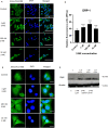
2-ME increases Drp1 protein expression in OS 143B cells. Representative immunofluorescence confocal images of OS 143B cells stained with Alexa Fluor 488-labeled anti-Drp1 antibody (green) and co-stained with nuclear stain (DAPI, blue). OS 143B cells were treated with 10 nM, 100 nM and 1 µM 2-ME for 8 h. Magnifications: (A) 200× (B) 1300 × Scale bars: A. 100 µm, B. 20 µm. (C) The fluorescence intensity of Drp1 after treatment with 2-ME. The images were analyzed and merged using the ImageJ software. The fluorescence intensity was presented in relative fluorescence units (RFUs). (D) Treatment with 2-ME at 10 nM, 100 nM, and 1 μM concentrations upregulates Drp1 protein level in OS 143B cells evaluated by Western blotting. Densitometric analysis of ratio Drp1/β-actin was performed using Quantity One 4.5.2 software. The presented immunoblots representative from one membrane are shown. Values are the mean ± SD of three independent experiments. ****p < 0.0001 versus control cells.
Figure 4
Figure 4
MDIVI-1 exerts cytotoxic effect in OS 143B cells. 2-ME regulates the ATP intracellular level in OS 143B cells. (A) MDIVI –1 significantly decreases OS 143B cell viability in a dose-dependent manner at concentrations higher than 10–6. OS 143B cells were treated with serial dilutions of MDIVI-1 within concentrations range of 300–0.1 µM for 24 h. The cell viability was determined by MTT assay. (B) Incubation of OS 143B cells with 5 µM MDIVI-1 1 for 6 h does not affect cell viability. (C) Changes in cellular level of ATP after 8 h-treatment of OS 143B cells with 10 nM, 100 nM, and 1 µM 2-ME. Values are the mean ± SD of three independent experiments. *p < 0.01, **p < 0.001, ***p < 0.0001, ****p < 0.00001 versus control cells.
Figure 5
Figure 5
MDIVI-1 decreases the 2-ME-mediated upregulation of Drp1, BAX and cytochrome C. (A) Pre-treatment with 5 µM MDIVI-1 for 6 h decreases 2-ME-mediated upregulation of cytochrome C (A), BAX (B) and Drp-1 (C) proteins level in OS 143B cells evaluated by Western blotting. Densitometric analysis of ratio cytochrome C/β-actin, BAX/β-actin, Drp1/β-actin were performed using Quantity One 4.5.2 software. The presented immunoblots representative from one membrane are shown.
Figure 6
Figure 6
2-ME increases BAX protein expression in OS 143B cells. Representative immunofluorescence confocal images of OS 143B cells stained with Alexa Fluor 488-labeled anti-BAX antibody (green) and co-stained with nuclear stain (DAPI, blue). OS 143B cells were treated with 10 nM, 100 nM and 1 µM 2-ME for 8 h. Magnifications: (A) 200× , (B) 1300× Scale bars: (A) 100 µm, (B) 20 µm. (C) The fluorescence intensity of BAX protein after treatment with 2-ME. The images were analyzed and merged using the ImageJ software. The fluorescence intensity was presented in relative fluorescence units (RFUs). Values are the mean ± SD of three independent experiments. ****p < 0.0001 versus control cells.

Similar articles

Cited by

References

    1. Yen J-H, Huang HS, Chuang CJ, Huang S-T. Activation of dynamin-related protein 1 - dependent mitochondria fragmentation and suppression of osteosarcoma by cryptotanshinone. J. Exp. Clin. Cancer Res. 2019;38:42. doi: 10.1186/s13046-018-1008-8. - DOI - PMC - PubMed
    1. Jackson M, et al. Mitochondrial genome and functional defects in osteosarcoma are associated with their aggressive phenotype. PLoS ONE. 2018;13:e0209489. doi: 10.1371/journal.pone.0209489. - DOI - PMC - PubMed
    1. Ciruzzi MS. To mdivi-1 or not to mdivi-1: is that the question? Arch. Argent. Pediatr. 2018;116:365–367. - PubMed
    1. Westermann B. Bioenergetic role of mitochondrial fusion and fission. Biochim. Biophys. Acta - Bioenerg. 2012;1817:1833–1838. doi: 10.1016/j.bbabio.2012.02.033. - DOI - PubMed
    1. Ikeda Y, et al. Endogenous Drp1 mediates mitochondrial autophagy and protects the heart against energy stress. Circ. Res. 2015;116:264–278. doi: 10.1161/CIRCRESAHA.116.303356. - DOI - PubMed

Publication types

MeSH terms

Substances