. 2021 Jan 15;11(1):1518.
doi: 10.1038/s41598-021-81170-2.

PMCA4 inhibition does not affect cardiac remodelling following myocardial infarction, but may reduce susceptibility to arrhythmia

Affiliations

PMCA4 inhibition does not affect cardiac remodelling following myocardial infarction, but may reduce susceptibility to arrhythmia

Nicholas Stafford et al. Sci Rep. .

Abstract

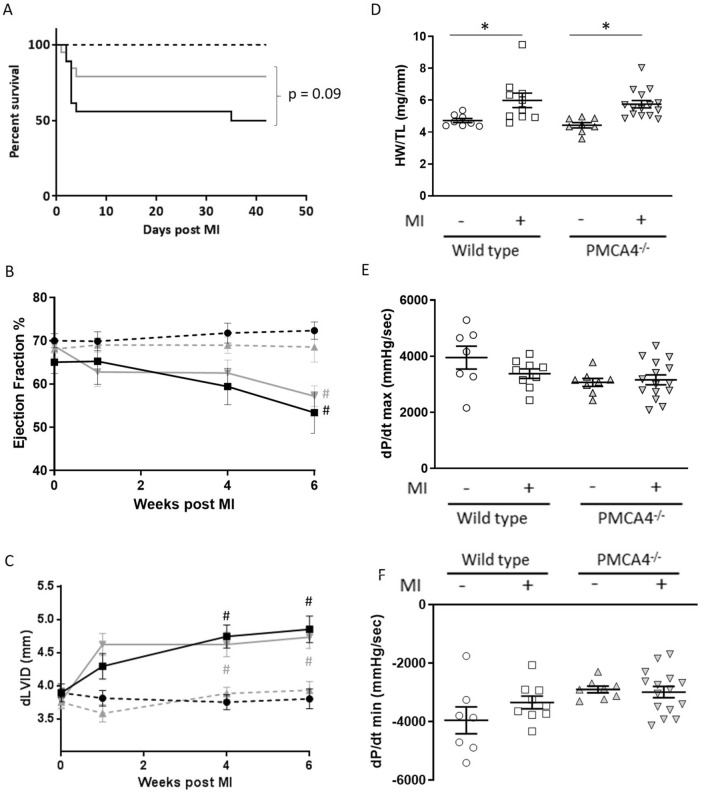
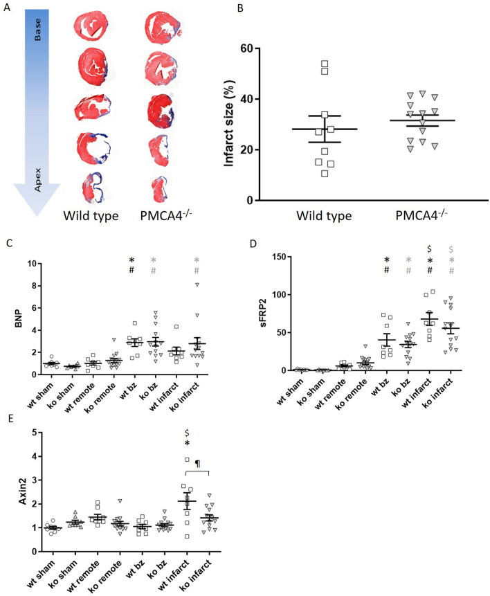
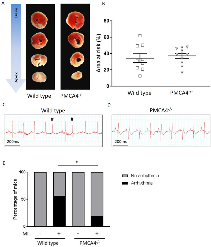
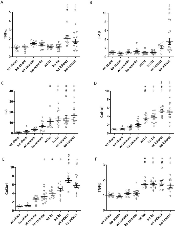
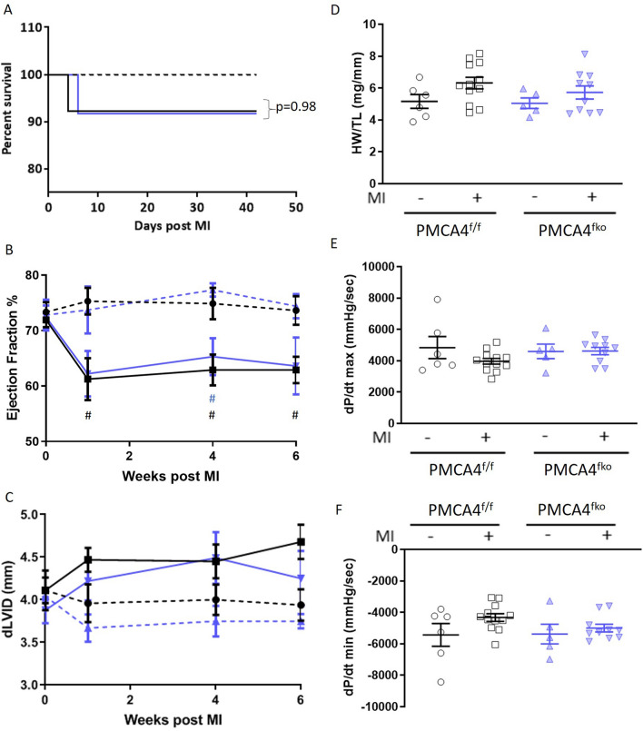
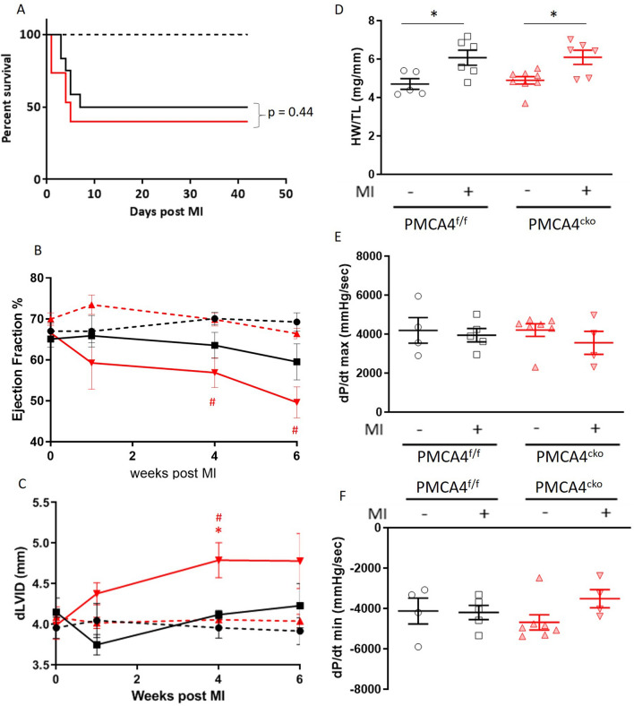
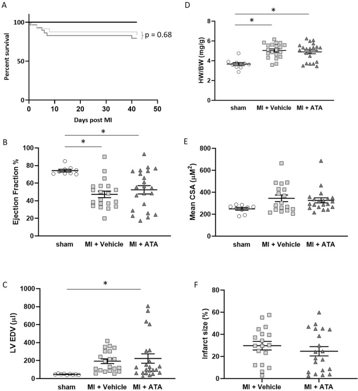
Ischaemic heart disease is the world's leading cause of mortality. Survival rates from acute myocardial infarction (MI) have improved in recent years; however, this has led to an increase in the prevalence of heart failure (HF) due to chronic remodelling of the infarcted myocardium, for which treatment options remain poor. We have previously shown that inhibition of isoform 4 of the plasma membrane calcium ATPase (PMCA4) prevents chronic remodelling and HF development during pressure overload, through fibroblast mediated Wnt signalling modulation. Given that Wnt signalling also plays a prominent role during remodelling of the infarcted heart, this study investigated the effect of genetic and functional loss of PMCA4 on cardiac outcomes following MI. Neither genetic deletion nor pharmacological inhibition of PMCA4 affected chronic remodelling of the post-MI myocardium. This was the case when PMCA4 was deleted globally, or specifically from cardiomyocytes or fibroblasts. PMCA4-ablated hearts were however less prone to acute arrhythmic events, which may offer a slight survival benefit. Overall, this study demonstrates that PMCA4 inhibition does not affect chronic outcomes following MI.


Conflict of interest statement

The authors declare no competing interests.

Figures

Figure 1
Figure 1
PMCA 4 global deletion does not affect cardiac structure or function post-MI. (A) Kaplan–Meier survival plots for wild type (black) and PMCA4−/− mice (grey) followed for 6 weeks post MI (solid lines) or sham (dashed lines) surgery. Starting populations; WT sham n = 8, WT MI n = 18, PMCA4−/− sham n = 8, PMCA4−/− MI n = 19. (B) Ejection fraction and (C) Diastolic left ventricular diameter tracked for 6 weeks post-MI or sham surgery by echocardiography. # p < 0.05 versus sham control at matched time point (D) Heart weight/tibia length ratio 6 weeks post-MI or sham surgery. (E) dP/dt max and (F) dP/dt min 6 weeks post MI or sham surgery. WT sham n = 8, WT MI n = 9, PMCA4−/− sham n = 8, PMCA4−/− MI n = 15 * p < 0.05.
Figure 2
Figure 2
PMCA 4 global deletion does not affect chronic infarct size, remodelling or Wnt signalling post MI. (A) Representative masson’s trichrome stained sections from wildtype and PMCA4−/− hearts following 6 weeks MI. Images were acquired using a 3D Histech Panoramic slide scanner and CaseViewer software version 2.4.0. (B) Quantification of infarct size measurements 6 weeks post-MI. WT n = 9, PMCA4−/− n = 13. Regional gene expression was examined in WT (clear points) and PMCA4−/− (grey points) hearts 6 weeks post sham or MI surgery for (C) Hypertrophic marker BNP (D) Wnt inhibitor sFRP2 and (E) Wnt transcriptional marker Axin2. bz = infarct border zone. Shams n = 8, WT MI n = 8, PMCA4−/− MI n = 13. *p < 0.05 versus sham, # p < 0.05 versus remote, $ p < 0.05 versus border zone, ¶ p < 0.05 WT v KO infarct.
Figure 3
Figure 3
PMCA 4 global deletion may protect against acute arrhythmic events post-MI. (A) Representative TTC stained sections from wildtype and PMCA4−/− hearts 2 days post MI. Images were acquired using a Leica S6D microscope with Leica MC170 HD camera, and Leica application suite EZ software. (B) Quantification of area at risk 2 days post-MI. WT n = 9, PMCA4−/− n = 11. (C) Representative 3 lead ECG traces from wild type and (D) PMCA4−/− mice day 1 post-MI. Premature contractions are indicated by #. (E) Percentage of mice displaying arrhythmia (black segments) upon ECG examination day 1 post MI. WT sham n = 8, WT MI n = 18, PMCA4−/− sham n = 8, PMCA4−/− MI n = 16 * p < 0.05.
Figure 4
Figure 4
PMCA 4 global deletion does not affect the acute inflammatory response post-MI. Myocardial gene expression was examined by qRT-PCR in WT (clear points) and PMCA4−/− (grey points) hearts 2 days post sham or MI surgery for pro-inflammatory mediators (A) TNFα, (B) Interleukin-1β, (C) Interleukin-6 and (D) MCP-1, in addition to anti-inflammatory cytokine (E) Il-10 Shams n = 4, WT MI n = 7, PMCA4−/− MI n = 6. *p < 0.05.
Figure 5
Figure 5
PMCA 4 global deletion does not affect acute remodelling or the fibrotic response post-MI. Myocardial gene expression was examined by qRT-PCR in WT (clear points) and PMCA4−/− (grey points) hearts 2 days post sham or MI surgery for (A) Hypertrophic marker BNP, along with markers for fibrosis (B) Col1a1, (C) Col3a1 and (D) TGFβ. Shams n = 4, WT MI n = 7, PMCA4−/− MI n = 6. *p < 0.05.
Figure 6
Figure 6
PMCA 4 global deletion does not affect chronic inflammation or fibrosis 6 weeks post MI. Regional gene expression was examined by qRT-PCR in WT (clear points) and PMCA4−/− (grey points) hearts 6 weeks post sham or MI surgery for pro-inflammatory cytokines (A) TNFα, (B) Interleukin-1β and (C) Interleukin-6, in addition to fibrosis markers (D) Col1a1, (E) Col3a1 and F) TGFβ. bz = infarct border zone. Shams n = 8, WT MI n = 8, PMCA4−/− MI n = 13. *p < 0.05 versus sham, # p < 0.05 versus remote, $ p < 0.05 versus border zone.
Figure 7
Figure 7
PMCA 4 deletion from fibroblasts does not affect cardiac structure or function post-MI. (A) Kaplan–Meier survival plots for PMCA4flox/flox (black) and PMCA4fko mice (blue) followed for 6 weeks post MI (solid lines) or sham (dashed lines) surgery. Starting populations: control sham n = 6, control MI n = 13, PMCA4fko sham n = 6, PMCA4fko MI n = 12. (B) Ejection fraction and (C) Diastolic left ventricular diameter tracked for 6 weeks post-MI or sham surgery by echocardiography. #p < 0.05 versus sham control at matched time point (D) Heart weight/tibia length ratio 6 weeks post-MI or sham surgery. (E) dP/dt max and (F) dP/dt min 6 weeks post MI or sham surgery. control sham n = 6, control MI n = 12, PMCA4fko sham n = 6, PMCA4fko MI n = 11.
Figure 8
Figure 8
PMCA 4 deletion from cardiomyocytes does not affect cardiac structure or function post-MI. (A) Kaplan–Meier survival plots for PMCA4flox/flox (black) and PMCA4cko mice (red) followed for 6 weeks post MI (solid lines) or sham (dashed lines) surgery. Starting populations: control sham n = 5, control MI n = 12, PMCA4cko sham n = 8, PMCA4cko MI n = 15. (B) Ejection fraction and (C) Diastolic left ventricular diameter tracked for 6 weeks post-MI or sham surgery by echocardiography. # p < 0.05 versus sham control at matched time point, * p < 0.05 versus control MI at matched time point. (D) Heart weight/tibia length ratio 6 weeks post-MI or sham surgery. * p < 0.05 (E) dP/dt max and (F) dP/dt min 6 weeks post MI or sham surgery. control sham n = 5, control MI n = 6, PMCA4cko sham n = 8, PMCA4cko MI n = 6.
Figure 9
Figure 9
Treatment with PMCA 4 specific inhibitor does not affect cardiac structure or function post-MI. (A) Kaplan–Meier survival plots for mice subjected to sham surgery (black), or left anterior descending coronary artery ligation with vehicle (light grey) or PMCA4 inhibitor ATA treatment (dark grey) followed for 6 weeks post MI. Starting populations: sham n = 10, MI + vehicle n = 24, MI + ATA n = 29. (B) Left ventricular ejection fraction and (C) End diastolic volume 6 weeks post-MI or sham surgery as measured by echocardiography. (D) Heart weight/body weight ratio 6 weeks post-MI or sham surgery. (E) Mean myocyte cross sectional area 6 weeks post-MI or sham surgery. (F) Infarct size 6 weeks post MI. sham n = 10, MI + vehicle n = 20, MI + ATA n = 22 * p < 0.05.

References

    1. Wilkins, E. et al. European Cardiovascular Disease Statistics 2017. (2017). - PubMed
    1. Benjamin EJ, et al. Heart disease and stroke statistics-2019 update: a report from the American Heart Association. Circulation. 2019;139:e56–e528. doi: 10.1161/cir.0000000000000659. - DOI - PubMed
    1. Townsend N, Bhatnagar P, Wilkins E, Wickramasinghe K. Cardiovascular Disease Statistics 2015. London: British Heart Foundation; 2015.
    1. Roth GA, et al. Global, regional, and national age-sex-specific mortality for 282 causes of death in 195 countries and territories, 1980–2017: a systematic analysis for the Global Burden of Disease Study 2017. Lancet. 2018;392:1736–1788. doi: 10.1016/s0140-6736(18)32203-7. - DOI - PMC - PubMed
    1. Bhatnagar P, Wickramasinghe K, Wilkins E, Townsend N. Trends in the epidemiology of cardiovascular disease in the UK. Heart. 2016;102:1945–1952. doi: 10.1136/heartjnl-2016-309573. - DOI - PMC - PubMed

Publication types

MeSH terms

Substances