. 2021 Jan 15;11(1):1565.
doi: 10.1038/s41598-021-81214-7.

Microfluidic on-chip production of microgels using combined geometries

Affiliations

Microfluidic on-chip production of microgels using combined geometries

Hamed Shieh et al. Sci Rep. .

Abstract

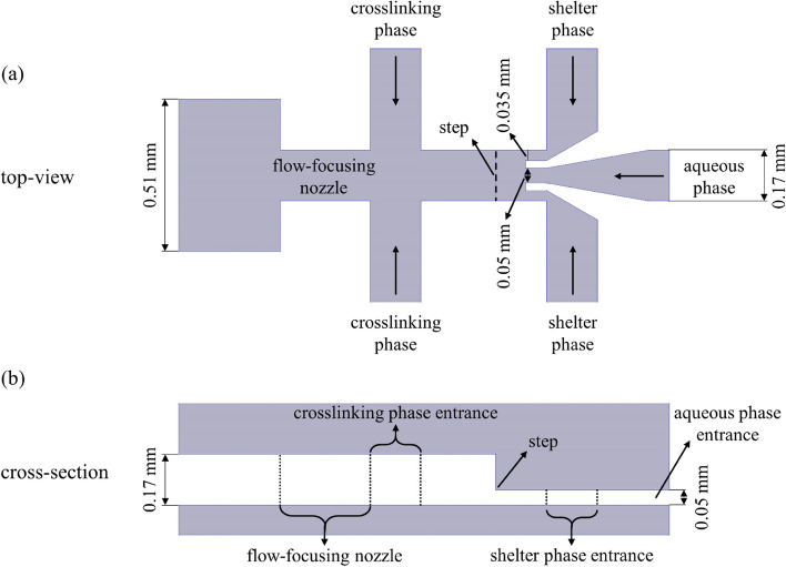
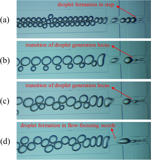
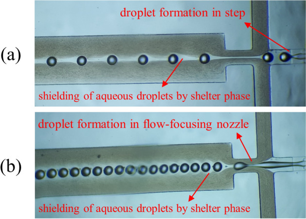
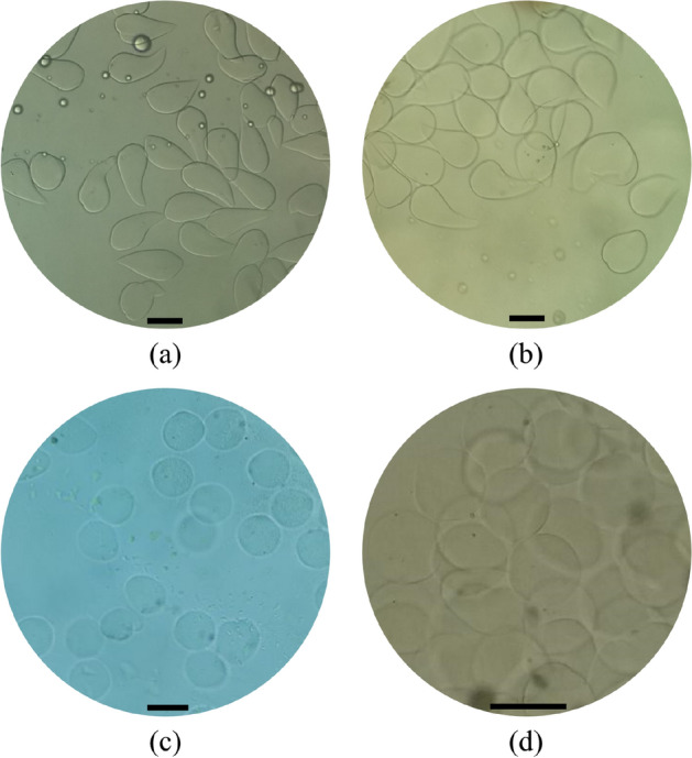
Microfluidic on-chip production of microgels using external gelation can serve numerous applications that involve encapsulation of sensitive cargos. Nevertheless, on-chip production of microgels in microfluidic devices can be challenging due to problems induced by the rapid increase in precursor solution viscosity like clogging. Here, a novel design incorporating a step, which includes a sudden increase in cross-sectional area, before a flow-focusing nozzle was proposed for microfluidic droplet generators. Besides, a shielding oil phase was utilized to avoid the occurrence of emulsification and gelation stages simultaneously. The step which was located before the flow-focusing nozzle facilitated the full shielding of the dispersed phase due to 3-dimensional fluid flow in this geometry. The results showed that the microfluidic device was capable of generating highly monodispersed spherical droplets (CV < 2% for step and CV < 5% for flow-focusing nozzle) with an average diameter in the range of 90-190 μm, both in step and flow-focusing nozzle. Moreover, it was proved that the device could adequately create a shelter for the dispersed phase regardless of the droplet formation locus. The ability of this microfluidic device in the production of microgels was validated by creating alginate microgels (with an average diameter of ~ 100 μm) through an external gelation process with on-chip calcium chloride emulsion in mineral oil.


Conflict of interest statement

The authors declare no competing interests.

Figures

Figure 1
Figure 1
Schematic illustration of the designed microfluidic system. (a) top-view. (b) cross-section.
Figure 2
Figure 2
Transition of droplet generation locus (from step (a) to flow focusing nozzle (d)) with increasing Qa and Qs at a constant ratio (α = 1) and a specific value of Qc (Qc = 30 μL/min). (a) Qa = 18 μL/min. (b) Qa = 24 μL/min. (c) Qa = 30 μL/min. (d) Qa = 36 μL/min.
Figure 3
Figure 3
Average diameter of generated droplets versus flow rate of aqueous phase. α: ■ = 1, ♦ = 2.25, and ▲ = 0.44. (a) Qc = 6 μL/min. (b) Qc = 30 μL/min. (c) Qc = 60 μL/min. Error bars indicate standard deviation for average diameter of droplets (n = 2).
Figure 4
Figure 4
Shielding of aqueous phase droplets by shelter phase when aqueous droplets were formed in (a) step and (b) flow-focusing nozzle.
Figure 5
Figure 5
Alginate microgels were synthesized by using the designed microfluidic device. Calcium chloride concentration in crosslinking phase emulsion was (a) 4 mol/L, (b) 6 mol/L, (c) 8 mol/L, and (d) 10 mol/L. Scale bars are 100 μm.
Figure 6
Figure 6
(a) Effect of calcium chloride concentration on roundness and CV of alginate microgels. (b) Size distribution of alginate microgels produced with a calcium chloride concentration of 10 mol/L.

Similar articles

Cited by

References

    1. Damiati S, Kompella UB, Damiati SA, Kodzius R. Microfluidic devices for drug delivery systems and drug screening. Genes. 2018;9:103. doi: 10.3390/genes9020103. - DOI - PMC - PubMed
    1. Chung BG, Lee K-H, Khademhosseini A, Lee S-H. Microfluidic fabrication of microengineered hydrogels and their application in tissue engineering. Lab Chip. 2012;12:45–59. doi: 10.1039/C1LC20859D. - DOI - PubMed
    1. Liu AL, García AJ. Methods for generating hydrogel particles for protein delivery. Ann. Biomed. Eng. 2016;44:1946–1958. doi: 10.1007/s10439-016-1637-z. - DOI - PMC - PubMed
    1. Li W, et al. Microfluidic fabrication of microparticles for biomedical applications. Chem. Soc. Rev. 2018;47:5646–5683. doi: 10.1039/C7CS00263G. - DOI - PMC - PubMed
    1. Theberge AB, et al. Microdroplets in microfluidics: An evolving platform for discoveries in chemistry and biology. Angew. Chem. Int. Ed. 2010;49:5846–5868. doi: 10.1002/anie.200906653. - DOI - PubMed

Publication types

LinkOut - more resources