. 2021 Jan 15;4(1):69.
doi: 10.1038/s42003-020-01614-9.

Clustered rapid induction of apoptosis limits ZIKV and DENV-2 proliferation in the midguts of Aedes aegypti

Affiliations

Clustered rapid induction of apoptosis limits ZIKV and DENV-2 proliferation in the midguts of Aedes aegypti

Jasmine B Ayers et al. Commun Biol. .

Abstract

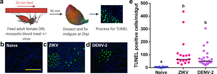
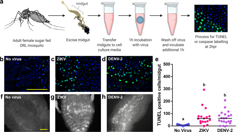
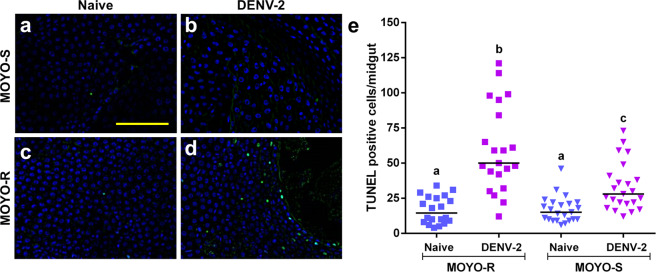
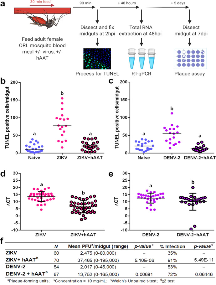
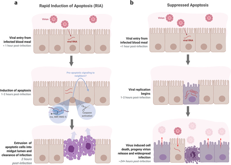
Inter-host transmission of pathogenic arboviruses such as dengue virus (DENV) and Zika virus (ZIKV) requires systemic infection of the mosquito vector. Successful systemic infection requires initial viral entry and proliferation in the midgut cells of the mosquito followed by dissemination to secondary tissues and eventual entry into salivary glands1. Lack of arbovirus proliferation in midgut cells has been observed in several Aedes aegypti strains2, but the midgut antiviral responses underlying this phenomenon are not yet fully understood. We report here that there is a rapid induction of apoptosis (RIA) in the Aedes aegypti midgut epithelium within 2 hours of infection with DENV-2 or ZIKV in both in vivo blood-feeding and ex vivo midgut infection models. Inhibition of RIA led to increased virus proliferation in the midgut, implicating RIA as an innate immune mechanism mediating midgut infection in this mosquito vector.


Conflict of interest statement

The authors declare no competing interests.

Figures

Fig. 1
Fig. 1. Exposure to a blood meal containing DENV-2 or ZIKV induces DNA fragmentation in the midgut epithelium of adult female Aedes aegypti (ORL) mosquitoes at 2hpi.
a Workflow diagram of in vivo infection. bd Representative images of DNA fragmentation visualized via TUNEL with DAPI counterstain in midguts from adult female ORL mosquitoes at 2 hours post b naive blood meal c ZIKV infected blood meal or d or DENV-2-infected blood meal. Scale bar shown is 100 µm in length and can be applied to all images. e Quantification of TUNEL-positive cells per midgut at 2 hours post in vivo infection (n (naive) = 12; n (ZIKV) = 17; n (DENV-2) = 24). The horizontal line indicates the median. Treatments without a common letter were found to be statistically significant (α = 0.05) as calculated by Kruskal–Wallis test with Mann–Whitney post hoc comparison (Kruskal–Wallis chi-squared = 32.017, df = 2, p value = 1.116e-07).
Fig. 2
Fig. 2. Exposure to a blood meal containing DENV-2 or ZIKV induces DNA fragmentation in the epithelium of ex vivo adult female Aedes aegypti (ORL) midguts at 2hpi.
a Workflow diagram of ex vivo midgut infection. bd Representative images of DNA fragmentation visualized via TUNEL at 2 hours after b ex vivo mock infection, c ZIKV infection or d DENV-2 infection. e Quantification of TUNEL-positive cells per midgut at 2 hours post ex vivo infection. (n (naive) = 21; n (ZIKV) = 17; n (DENV-2) = 18). The horizontal line indicates the median. Treatments without a common letter were found to be statistically significant (α = 0.05) as calculated by Kruskal–Wallis test with Mann–Whitney post hoc comparison (Kruskal–Wallis chi-squared = 22.285, df = 2, p value = 1.448e-05). fh Pan-caspase activation at 2 hours after f mock infection g ZIKV infection or h DENV-2 infection. Scale bars shown are 100 µm in length and can be applied to all related images.
Fig. 3
Fig. 3. Aedes aegypti MOYO-R strain mosquitoes that are partially resistant to DENV-2 have higher rates of TUNEL-positive cells in the midgut than DENV-2 susceptible MOYO-S strain mosquitoes at 2 hours post ingestion of a DENV-2-infected blood meal.
ad Representative images of TUNEL with DAPI counterstain in midguts from adult female mosquitoes at 2 hours post blood meal. a MOYO-S mosquitoes fed naive blood, b MOYO-S mosquitoes fed DENV-2-infected blood meal, c MOYO-R mosquitoes fed naïve blood or d MOYO-R mosquitoes fed DENV-2-infected blood. Scale bar = 100 µm. e Quantification of TUNEL-positive cells per midgut at 2 hours post in vivo infection (n (MOYO-R naive) = 20; n (MOYO-R DENV-2) =  21; n (MOYO-S naive) = 23; n (MOYO-S DENV-2) = 24). The horizontal line indicates the median. Treatments without a common letter were found to be statistically significant (α = 0.05) as calculated by Kruskal–Wallis test with Mann–Whitney post hoc comparison (Kruskal–Wallis chi-squared = 41.907, df = 3, p value = 4.199e-09). Scale bar shown is 100 µm in length and can be applied to all images.
Fig. 4
Fig. 4. Supplementing a DENV-2 or ZIKV blood meal with hAAT in adult female Aedes aegypti (ORL) mosquitoes inhibits the rapid induction of apoptosis in the midgut at 2hpi and increases subsequent midgut viral replication.
a Workflow of the apoptosis inhibitor co-feeding experiment, hpi=hours post infection, dpi=days post infection. bc TUNEL-positive cell counts in midguts from mosquitoes fed a b ZIKV (n (naive) = 22; n (ZIKV) = 18; n (ZIKV + hAAT) = 22) or c DENV-2-infected blood meal or a virus-infected blood meal supplemented with 10 mg/mL hAAT (n (naive) = 21; n (DENV-2) = 18; n (DENV-2 + hAAT) = 15). Treatments without a common letter were found to be statistically significant (α = 0.05) as calculated by Kruskal–Wallis test with Mann–Whitney post hoc test (panel a: Kruskal–Wallis chi-squared = 32.532, df = 2, p value = 8.624e-08; b Kruskal–Wallis chi-squared = 26.513, df = 2, p value = 1.749e-06) de ΔCT values from RT-qPCR detecting d ZIKV (n (ZIKV) = 34; n (ZIKV + hAAT) = 40) or e DENV-2 genome copy in whole mosquitoes 48 hours after an infected blood meal with or without 10 mg/mL hAAT (n (DENV-2) = 29; n (DENV-2 + hAAT) = 25). The horizontal line indicates the median. Treatments without a common letter were found to be statistically significant (α = 0.05) as calculated by Mann–Whitney U test (d W = 905, p value = 1.735e-08; e W = 466, p value = 0.006808). f Productive midgut infection quantified by midgut plaque assay at 7 days after feeding on a ZIKV or DENV-2-infected blood meal (n (ZIKV) = 34; n (ZIKV + hAAT) = 40; n (DENV-2) = 29; n (DENV-2 + hAAT) = 25)). Treatments without a common letter were found to be statistically significant (α = 0.05) as calculated by Mann–Whitney U test (ZIKV W = 608, p value = 1.102e-12; DENV-2 W = 1228.5, p value = 0.001929).
Fig. 5
Fig. 5. Hypothesized model of antiviral apoptosis in the Aedes aegypti midgut.
a RIA of infected cells in the midgut epithelium within 2 hours of an infected blood meal arrests viral replication before progeny virions can be produced and suppresses infection. The clustered appearance of apoptotic cells may be caused by pro-apoptotic signaling to neighboring cells. b At later timepoints post infection, when infection is widespread throughout the midgut and progeny virions are being produced, death of infected cells may facilitate release of virions from cells and/or from the midgut by degrading the integrity of the epithelium and basal lamina.

Similar articles

Cited by

References

    1. Franz AWE, Kantor AM, Passarelli AL, Clem RJ. Tissue barriers to arbovirus infection in mosquitoes. Viruses. 2015;7:3741–3767. doi: 10.3390/v7072795. - DOI - PMC - PubMed
    1. Caicedo PA, et al. Selection of Aedes aegypti (Diptera: Culicidae) strains that are susceptible or refractory to Dengue-2 virus. Can. Entomol. 2013;145:273–282. doi: 10.4039/tce.2012.105. - DOI
    1. Kalayanarooj S. Clinical manifestations and management of dengue/dhf/dss. Trop. Med. Health. 2011;39:83–87. doi: 10.2149/tmh.2011-S10. - DOI - PMC - PubMed
    1. Messina JP, et al. A global compendium of human dengue virus occurrence. Sci. Data. 2014;1:140004. doi: 10.1038/sdata.2014.4. - DOI - PMC - PubMed
    1. Barbi L, Coelho AVC, Alencar LCAde, Crovella S. Prevalence of Guillain-Barré syndrome among Zika virus infected cases: a systematic review and meta-analysis. Braz. J. Infect. Dis. 2018;22:137–141. doi: 10.1016/j.bjid.2018.02.005. - DOI - PMC - PubMed

Publication types